

The DOI matches its Crossref record; 23 of the 23 references it cites were found in a registry or an index.
ATM regulates ATR chromatin loading in response to DNA double-strand breaks
Open accessAbstract
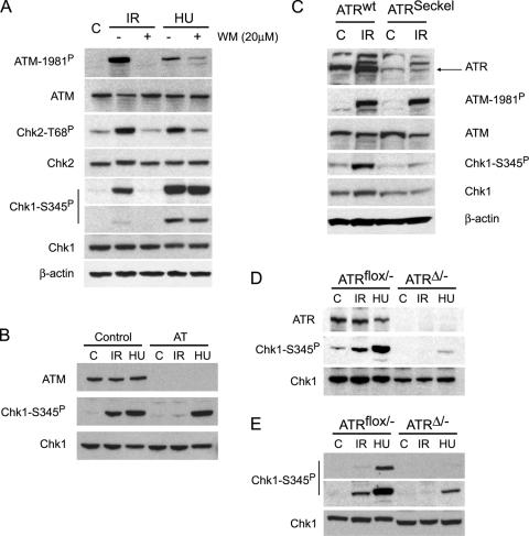
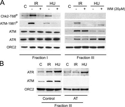
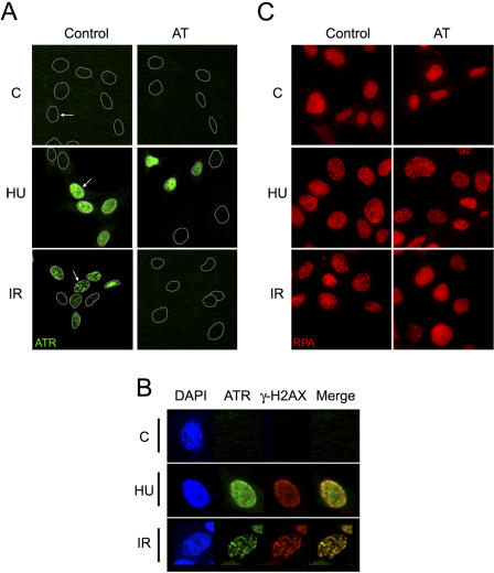
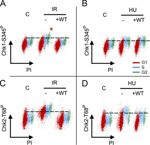
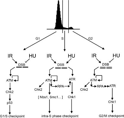
DNA double-strand breaks (DSBs) are among the most deleterious lesions that can challenge genomic integrity. Concomitant to the repair of the breaks, a rapid signaling cascade must be coordinated at the lesion site that leads to the activation of cell cycle checkpoints and/or apoptosis. In this context, ataxia telangiectasia mutated (ATM) and ATM and Rad-3–related (ATR) protein kinases are the earliest signaling molecules that are known to initiate the transduction cascade at damage sites. The current model places ATM and ATR in separate molecular routes that orchestrate distinct pathways of the checkpoint responses. Whereas ATM signals DSBs arising from ionizing radiation (IR) through a Chk2-dependent pathway, ATR is activated in a variety of replication-linked DSBs and leads to activation of the checkpoints in a Chk1 kinase–dependent manner. However, activation of the G2/M checkpoint in response to IR escapes this accepted paradigm because it is dependent on both ATM and ATR but independent of Chk2. Our data provides an explanation for this observation and places ATM activity upstream of ATR recruitment to IR-damaged chromatin. These data provide experimental evidence of an active cross talk between ATM and ATR signaling pathways in response to DNA damage.
Overview
The claims and statistics of this paper haven’t been checked yet.
Run the check — free, no account needed. It reads the open-access text and checks the sources it cites.
Integrity signals
Registry
Cites
Statistics
Figures
Verification
Claims the paper makes
The claims and statistics of this paper haven’t been checked yet.
Run the check — free, no account needed. It reads the open-access text and checks the sources it cites.
Claims other papers make about this one
Each is checked against this paper’s own text: does it say what the citing paper says it does?
In the citing paper
“ATM is essential for the recruitment of ATR to IR-damaged chromatin and Chk1 phosphorylation at Serine 345 [139].”
From Functional interplay between ATM/ATR-mediated DNA damage response and DNA repair pathways in oxidative stress (2014) · cites this paper as [139]
Self-check
Percentages, sums and reported differences re-computed from the paper’s own numbers.
The self-check needs the paper’s full text.
Citations
Cites
DNA damage activates ATM through intermolecular autophosphorylation and dimer dissociation
Bakkenist, C. J., & Kastan, M. B. (2003) · Nature · doi:10.1038/nature01368 · Cited by 3,466
Cell-cycle checkpoints and cancer
Kastan, M. B., & Bartek, J. (2004) · Nature · doi:10.1038/nature03097 · Cited by 2,836
DNA damage response as a candidate anti-cancer barrier in early human tumorigenesis
Bartkova, J., Hořejší, Z., Koed, K., Krämer, A., Tort, F., Zieger, K., Guldberg, P., Sehested, M., Nesland, J. M., Lukas, C., et al. (2005) · Nature · doi:10.1038/nature03482 · Cited by 2,688
Cell Cycle Checkpoints: Preventing an Identity Crisis
Elledge, S. J. (1996) · Science · doi:10.1126/science.274.5293.1664 · Cited by 2,050
Activation of the DNA damage checkpoint and genomic instability in human precancerous lesions
Gorgoulis, V. G., Vassiliou, L.-V. F., Karakaidos, P., Zacharatos, P., Kotsinas, A., Liloglou, T., Venere, M., DiTullio, R. A., Kastrinakis, N. G., Levy, B., et al. (2005) · Nature · doi:10.1038/nature03485 · Cited by 2,009
Cell cycle checkpoint signaling through the ATM and ATR kinases
Abraham, R. T. (2001) · Genes & Development · doi:10.1101/gad.914401 · Cited by 1,966
Liu, Q., Guntuku, S., Cui, X.-S., Matsuoka, S., Cortez, D., Tamai, K., Luo, G., Carattini-Rivera, S., DeMayo, F., Bradley, A., et al. (2000) · Genes & Development · doi:10.1101/gad.14.12.1448 · Cited by 1,625
ATM- and cell cycle-dependent regulation of ATR in response to DNA double-strand breaks
Jazayeri, A., Falck, J., Lukas, C., Bartek, J., Smith, G. C. M., Lukas, J., & Jackson, S. P. (2006) · Nature Cell Biology · doi:10.1038/ncb1337 · Cited by 1,064
ATR and ATRIP: Partners in Checkpoint Signaling
Cortez, D., Guntuku, S., Qin, J., & Elledge, S. J. (2001) · Science · doi:10.1126/science.1065521 · Cited by 985
O'Driscoll, M., Ruiz-Perez, V. L., Woods, C. G., Jeggo, P. A., & Goodship, J. A. (2003) · Nature Genetics · doi:10.1038/ng1129 · Cited by 794
Research works printed but not linked: 3
- 1998, Cancer Res., 58, 4375
- 2004, Methods Mol. Biol., 263, 67
- 2005, Nat. Cell Biol., 8, 37
Cited by
Sorted by citations
ATM, ATR, and DNA-PK: The Trinity at the Heart of the DNA Damage Response
Blackford, A. N., & Jackson, S. P. (2017) · Molecular Cell · doi:10.1016/j.molcel.2017.05.015 · Cited by 2,050
ATR: an essential regulator of genome integrity
Cimprich, K. A., & Cortez, D. (2008) · Nature Reviews Molecular Cell Biology · doi:10.1038/nrm2450 · Cited by 1,798
Human CtIP promotes DNA end resection
Sartori, A. A., Lukas, C., Coates, J., Mistrik, M., Fu, S., Bartek, J., Baer, R., Lukas, J., & Jackson, S. P. (2007) · Nature · doi:10.1038/nature06337 · Cited by 1,362
The ATM–Chk2 and ATR–Chk1 Pathways in DNA Damage Signaling and Cancer
Smith, J., Tho, L. M., Xu, N., & Gillespie, D. A. (2010) · Advances in cancer research · doi:10.1016/b978-0-12-380888-2.00003-0 · Cited by 1,331
DNA damage-induced cell death: From specific DNA lesions to the DNA damage response and apoptosis
Roos, W. P., & Kaina, B. (2013) · Cancer Letters · doi:10.1016/j.canlet.2012.01.007 · Cited by 879
Metadata
- Authors
- Cuadrado, M., Martinez-Pastor, B., Murga, M., Toledo, L. I., Gutierrez-Martinez, P., Lopez, E., & Fernandez-Capetillo, O.
- Affiliations
- Spanish National Cancer Research Centre (ES)
- Genomic Instability Group, Spanish National Cancer Center, Madrid 28029, Spain
- Venue
- The Journal of Experimental Medicine
- Publisher
- Rockefeller University Press
- Published
- 2006-02-06
- Volume
- 203
- Issue
- 2
- Pages
- 297-303
- ISSN
- 0022-1007 (ISSN-L)
- 1540-9538
- PMC
- PMC2118201
- OpenAlex
- W2149468013
- Cited by
- 232
- References
- 23
- Citations by year
- 2026: 2 · 2025: 3 · 2024: 6 · 2023: 6 · 2022: 3 · 2021: 6 · 2020: 8 · 2019: 17 · 2018: 5 · 2017: 8 · 2016: 12 · 2015: 9 · 2014: 16 · 2013: 9 · 2012: 17
- FWCI
- 9.78
- Type
- journal article
- Language
- en
- Open access
- green
- europepmc.org
- Licence
- cc-by-nc-sa
- Locations
- The Journal of Experimental Medicine (publishedVersion)
- Europe PMC (PubMed Central) (publishedVersion, cc-by-nc-sa)
- PubMed Central (publishedVersion, cc-by-nc-sa)
- PubMed (publishedVersion)
- Topics
- Life Sciences › Biochemistry, Genetics and Molecular Biology › Molecular Biology › DNA Repair Mechanisms
- Health Sciences › Medicine › Radiology, Nuclear Medicine and Imaging › Effects of Radiation Exposure
- Life Sciences › Biochemistry, Genetics and Molecular Biology › Molecular Biology › Genomics and Chromatin Dynamics
- Keywords
- ATM kinase, ATR kinase, DNA double-strand breaks, chromatin loading, ionizing radiation, G2/M checkpoint, Chk2, Chk1, DNA damage signaling, DNA repair
- MeSH terms
- Checkpoint Kinase 1, Ataxia Telangiectasia, Cell Cycle, Cell Line, Transformed, Chromatin, DNA Damage, DNA Replication, DNA-Binding Proteins, Flow Cytometry, Gamma Rays, Humans, Phosphorylation, Protein Kinases, Signal Transduction, Protein Serine-Threonine Kinases, Cell Cycle Proteins, Chromosome Breakage, Tumor Suppressor Proteins, Cell Line, Tumor, Ataxia Telangiectasia Mutated Proteins, Checkpoint Kinase 2
About these checks
Registry, retraction, Cites and statistics results are computed, not judged, with one exception: a printed reference that no index lists is read once by a model before it counts as a citation error. Claims are extracted by a model and kept only when their quote appears verbatim in the text; each cited source is then checked, with a model’s help, for whether it says what the claim relies on. Those verdicts can be wrong — read the claim’s quote and the cited source before relying on one.
Statistics and arithmetic are recomputed, not judged.
Confidence bands are shown only for fields calibrated on our annotated test set. None is calibrated yet, so no band is shown.
What counts as a claim ›How we measure confidence ›How we check this ›
Are you an author, or do you see a problem with this page? Request a review
