

The DOI matches its Crossref record; 5 of the 5 references it cites were found in a registry or an index.
B and T cells collaborate in antiviral responses via IL-6, IL-21, and transcriptional activator and coactivator, Oct2 and OBF-1
Abstract
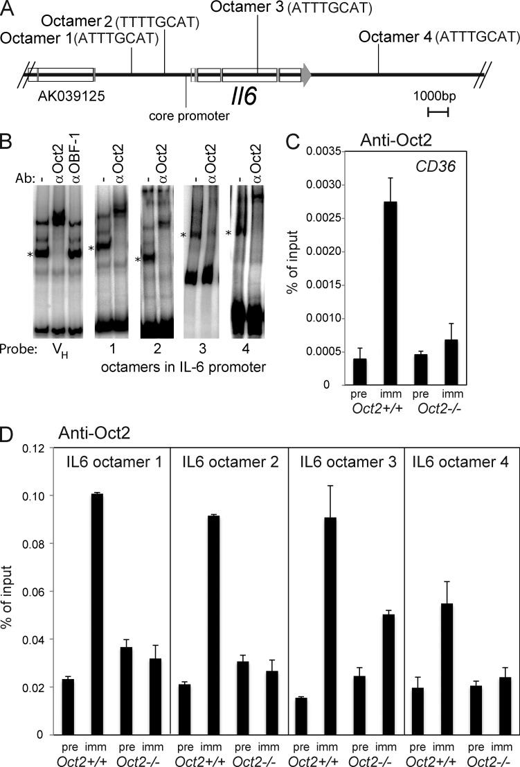
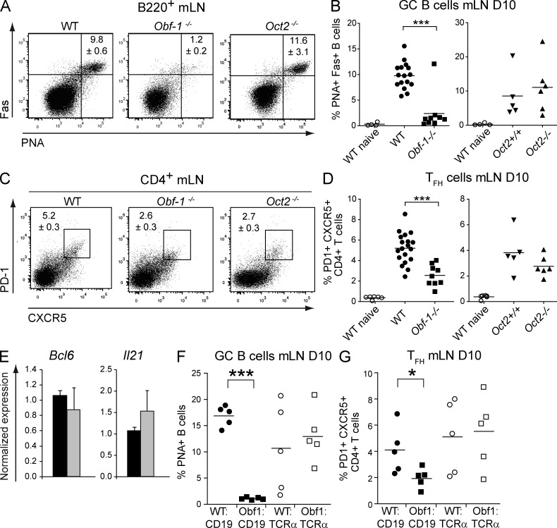
A strong humoral response to infection requires the collaboration of several hematopoietic cell types that communicate via antigen presentation, surface coreceptors and their ligands, and secreted factors. The proinflammatory cytokine IL-6 has been shown to promote the differentiation of activated CD4+ T cells into T follicular helper cells (TFH cells) during an immune response. TFH cells collaborate with B cells in the formation of germinal centers (GCs) during T cell–dependent antibody responses, in part through secretion of critical cytokines such as IL-21. In this study, we demonstrate that loss of either IL-6 or IL-21 has marginal effects on the generation of TFH cells and on the formation of GCs during the response to acute viral infection. However, mice lacking both IL-6 and IL-21 were unable to generate a robust TFH cell–dependent immune response. We found that IL-6 production in follicular B cells in the draining lymph node was an important early event during the antiviral response and that B cell–derived IL-6 was necessary and sufficient to induce IL-21 from CD4+ T cells in vitro and to support TFH cell development in vivo. Finally, the transcriptional activator Oct2 and its cofactor OBF-1 were identified as regulators of Il6 expression in B cells.
Overview
The claims and statistics of this paper haven’t been checked yet.
Run the check — free, no account needed. It reads the open-access text and checks the sources it cites.
Integrity signals
Registry
Cites
Statistics
Figures
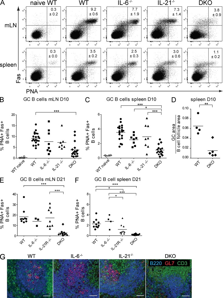
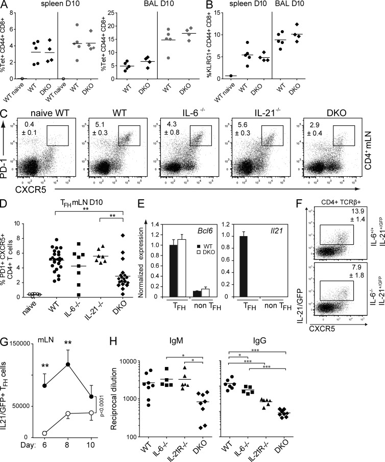
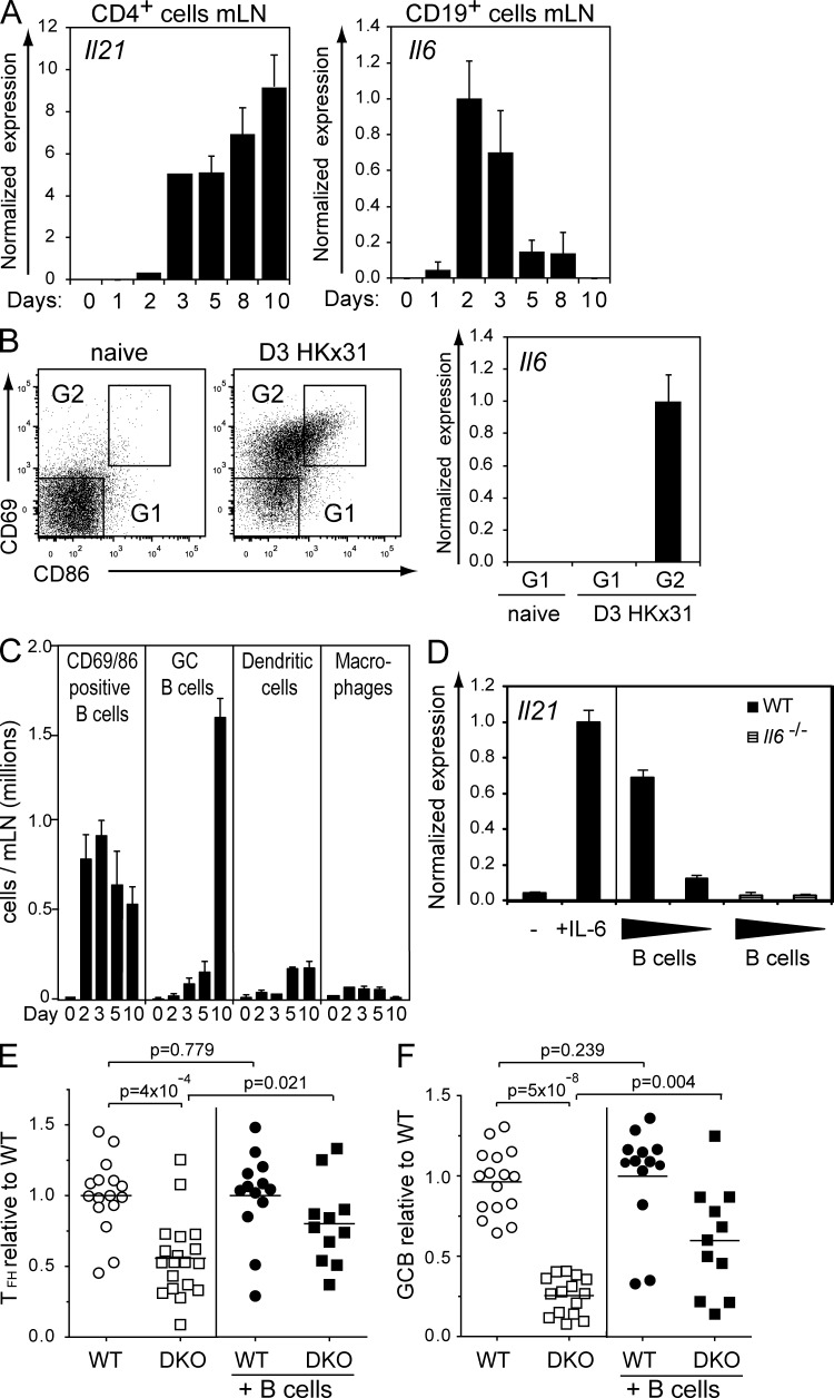
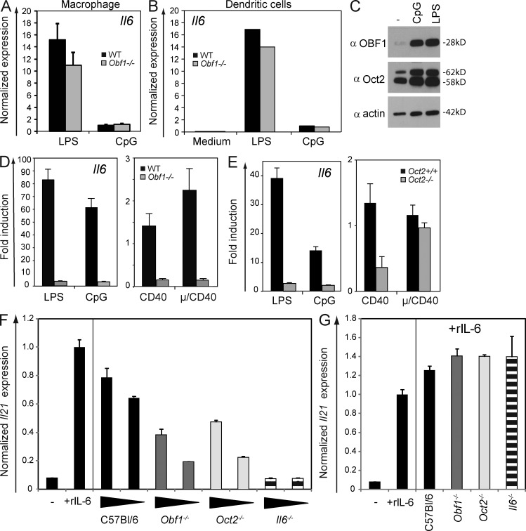
Verification
Claims the paper makes
The claims and statistics of this paper haven’t been checked yet.
Run the check — free, no account needed. It reads the open-access text and checks the sources it cites.
Claims other papers make about this one
Each is checked against this paper’s own text: does it say what the citing paper says it does?
In the citing paper
“In IL-6 knockout mice (Il6−/− mice), the frequency of Tfh cells is reduced and IL-21 is absent (Karnowski et al., 2012”
From Targeting Interleukin-6 Signaling in Clinic (2019) · cites this paper as Karnowski et al., 2012
Self-check
Percentages, sums and reported differences re-computed from the paper’s own numbers.
The self-check needs the paper’s full text.
Citations
Cites
Bcl6 mediates the development of T follicular helper cells
Nurieva, R. I., Chung, Y., Martinez, G. J., Yang, X. O., Tanaka, S., Matskevitch, T. D., Wang, Y.-H., & Dong, C. (2009) · Science · doi:10.1126/science.1176676 · Cited by 1,506
A Critical Role for IL-21 in Regulating Immunoglobulin Production
Ozaki, K., Spolski, R., Feng, C. G., Qi, C.-F., Cheng, J., Sher, A., Morse, H. C., Liu, C., Schwartzberg, P. L., & Leonard, W. J. (2002) · Science · doi:10.1126/science.1077002 · Cited by 978
Vinuesa, C. G., Cook, M. C., Angelucci, C., Athanasopoulos, V., Rui, L., Hill, K. M., Yu, D., Domaschenz, H., Whittle, B., Lambe, T., et al. (2005) · Nature · doi:10.1038/nature03555 · Cited by 862
Chtanova, T., Tangye, S. G., Newton, R., Frank, N., Hodge, M. R., Rolph, M. S., & Mackay, C. R. (2004) · The Journal of Immunology · doi:10.4049/jimmunol.173.1.68 · Cited by 722
SAP-controlled T–B cell interactions underlie germinal centre formation
Qi, H., Cannons, J. L., Klauschen, F., Schwartzberg, P. L., & Germain, R. N. (2008) · Nature · doi:10.1038/nature07345 · Cited by 637
Research works printed but not linked: 84
- Ansel, 1999, In vivo–activated CD4 T cells upregulate CXC chemokine receptor 5 and reprogram their response to lymphoid chemokines, J. Exp. Med., 190, 1123, 10.1084/jem.190.8.1123 · DOI
- Arguni, 2006, JunD/AP-1 and STAT3 are the major enhancer molecules for high Bcl6 expression in germinal center B cells, Int. Immunol., 18, 1079, 10.1093/intimm/dxl041 · DOI
- Baccam, 2003, CD40-mediated transcriptional regulation of the IL-6 gene in B lymphocytes: involvement of NF-kappa B, AP-1, and C/EBP, J. Immunol., 170, 3099, 10.4049/jimmunol.170.6.3099 · DOI
- Bartholdy, 2006, The Ets factor Spi-B is a direct critical target of the coactivator OBF-1, Proc. Natl. Acad. Sci. USA., 103, 11665, 10.1073/pnas.0509430103 · DOI
- Batten, 2010, IL-27 supports germinal center function by enhancing IL-21 production and the function of T follicular helper cells, J. Exp. Med., 207, 2895, 10.1084/jem.20100064 · DOI
- Baumjohann, 2011, Cutting Edge: Distinct waves of BCL6 expression during T follicular helper cell development, J. Immunol., 187, 2089, 10.4049/jimmunol.1101393 · DOI
- Belz, 2000, Contemporary analysis of MHC-related immunodominance hierarchies in the CD8+ T cell response to influenza A viruses, J. Immunol., 165, 2404, 10.4049/jimmunol.165.5.2404 · DOI
- Bessa, 2010, Cutting edge: IL-21 and TLR signaling regulate germinal center responses in a B cell-intrinsic manner, J. Immunol., 184, 4615, 10.4049/jimmunol.0903949 · DOI
- Blink, 2005, Early appearance of germinal center–derived memory B cells and plasma cells in blood after primary immunization, J. Exp. Med., 201, 545, 10.1084/jem.20042060 · DOI
- Brocker, 1999, CD4 T cell traffic control: in vivo evidence that ligation of OX40 on CD4 T cells by OX40-ligand expressed on dendritic cells leads to the accumulation of CD4 T cells in B follicles, Eur. J. Immunol., 29, 1610, 10.1002/(SICI)1521-4141(199905)29:05<1610::AID-IMMU1610>3.0.CO;2-8 · DOI
- Brunner, 2007, BOB.1/OBF.1 controls the balance of TH1 and TH2 immune responses, EMBO J., 26, 3191, 10.1038/sj.emboj.7601742 · DOI
- Cannons, 2010, Optimal germinal center responses require a multistage T cell:B cell adhesion process involving integrins, SLAM-associated protein, and CD84, Immunity., 32, 253, 10.1016/j.immuni.2010.01.010 · DOI
- Casey, 2007, IL-21 promotes differentiation of naive CD8 T cells to a unique effector phenotype, J. Immunol., 178, 7640, 10.4049/jimmunol.178.12.7640 · DOI
- Cepek, 1996, Sequence-specific DNA binding of the B-cell-specific coactivator OCA-B, Genes Dev., 10, 2079, 10.1101/gad.10.16.2079 · DOI
- Choi, 2011, ICOS receptor instructs T follicular helper cell versus effector cell differentiation via induction of the transcriptional repressor Bcl6, Immunity., 34, 932, 10.1016/j.immuni.2011.03.023 · DOI
- Corcoran, 1994, Oct-2 is required early in T cell-independent B cell activation for G1 progression and for proliferation, Immunity., 1, 635, 10.1016/1074-7613(94)90035-3 · DOI
- Corcoran, 1993, Oct-2, although not required for early B-cell development, is critical for later B-cell maturation and for postnatal survival, Genes Dev., 7, 570, 10.1101/gad.7.4.570 · DOI
- Corcoran, 2004, All known in vivo functions of the Oct-2 transcription factor require the C-terminal protein domain, J. Immunol., 172, 2962, 10.4049/jimmunol.172.5.2962 · DOI
- Corcoran, 2005, Differential requirement for OBF-1 during antibody-secreting cell differentiation, J. Exp. Med., 201, 1385, 10.1084/jem.20042325 · DOI
- Crotty, 2011, Follicular helper CD4 T cells (TFH), Annu. Rev. Immunol., 29, 621, 10.1146/annurev-immunol-031210-101400 · DOI · Paper page
- Cucak, 2009, Type I interferon signaling in dendritic cells stimulates the development of lymph-node-resident T follicular helper cells, Immunity., 31, 491, 10.1016/j.immuni.2009.07.005 · DOI
- Deenick, 2010, Follicular helper T cell differentiation requires continuous antigen presentation that is independent of unique B cell signaling, Immunity., 33, 241, 10.1016/j.immuni.2010.07.015 · DOI
- Deenick, 2011, Regulation of T follicular helper cell formation and function by antigen presenting cells, Curr. Opin. Immunol., 23, 111, 10.1016/j.coi.2010.10.007 · DOI
- Dienz, 2009, The induction of antibody production by IL-6 is indirectly mediated by IL-21 produced by CD4+ T cells, J. Exp. Med., 206, 69, 10.1084/jem.20081571 · DOI
- Eddahri, 2009, Interleukin-6/STAT3 signaling regulates the ability of naive T cells to acquire B-cell help capacities, Blood., 113, 2426, 10.1182/blood-2008-04-154682 · DOI
- Emslie, 2008, Oct2 enhances antibody-secreting cell differentiation through regulation of IL-5 receptor α chain expression on activated B cells, J. Exp. Med., 205, 409, 10.1084/jem.20072049 · DOI
- Engel, 1995, Abnormal B lymphocyte development, activation, and differentiation in mice that lack or overexpress the CD19 signal transduction molecule, Immunity., 3, 39, 10.1016/1074-7613(95)90157-4 · DOI
- Eto, 2011, IL-21 and IL-6 are critical for different aspects of B cell immunity and redundantly induce optimal follicular helper CD4 T cell (Tfh) differentiation, PLoS ONE., 6, e17739, 10.1371/journal.pone.0017739 · DOI
- Fazilleau, 2009, Follicular helper T cells: lineage and location, Immunity., 30, 324, 10.1016/j.immuni.2009.03.003 · DOI
- Flynn, 1998, Virus-specific CD8+ T cells in primary and secondary influenza pneumonia, Immunity., 8, 683, 10.1016/S1074-7613(00)80573-7 · DOI
- Garside, 1998, Visualization of specific B and T lymphocyte interactions in the lymph node, Science., 281, 96, 10.1126/science.281.5373.96 · DOI
- Goenka, 2011, Cutting edge: dendritic cell-restricted antigen presentation initiates the follicular helper T cell program but cannot complete ultimate effector differentiation, J. Immunol., 187, 1091, 10.4049/jimmunol.1100853 · DOI
- Gstaiger, 1996, The B cell coactivator Bob1 shows DNA sequence-dependent complex formation with Oct-1/Oct-2 factors, leading to differential promoter activation, EMBO J., 15, 2781, 10.1002/j.1460-2075.1996.tb00638.x · DOI
- Harker, 2011, Late interleukin-6 escalates T follicular helper cell responses and controls a chronic viral infection, Science., 334, 825, 10.1126/science.1208421 · DOI · Paper page
- Haynes, 2007, Role of CXCR5 and CCR7 in follicular Th cell positioning and appearance of a programmed cell death gene-1high germinal center-associated subpopulation, J. Immunol., 179, 5099, 10.4049/jimmunol.179.8.5099 · DOI
- Johnston, 2009, Bcl6 and Blimp-1 are reciprocal and antagonistic regulators of T follicular helper cell differentiation, Science., 325, 1006, 10.1126/science.1175870 · DOI · Paper page
- Kerfoot, 2011, Germinal center B cell and T follicular helper cell development initiates in the interfollicular zone, Immunity., 34, 947, 10.1016/j.immuni.2011.03.024 · DOI
- Kim, 1996, The B-cell-specific transcription coactivator OCA-B/OBF-1/Bob-1 is essential for normal production of immunoglobulin isotypes, Nature., 383, 542, 10.1038/383542a0 · DOI
- King, 2009, New insights into the differentiation and function of T follicular helper cells, Nat. Rev. Immunol., 9, 757, 10.1038/nri2644 · DOI
- King, 2008, T follicular helper (TFH) cells in normal and dysregulated immune responses, Annu. Rev. Immunol., 26, 741, 10.1146/annurev.immunol.26.021607.090344 · DOI
- Kitano, 2011, Bcl6 protein expression shapes pre-germinal center B cell dynamics and follicular helper T cell heterogeneity, Immunity., 34, 961, 10.1016/j.immuni.2011.03.025 · DOI
- König, 1995, Identification of CD36 as the first gene dependent on the B-cell differentiation factor Oct-2, Genes Dev., 9, 1598, 10.1101/gad.9.13.1598 · DOI
- Kopf, 1998, Interleukin 6 influences germinal center development and antibody production via a contribution of C3 complement component, J. Exp. Med., 188, 1895, 10.1084/jem.188.10.1895 · DOI
- Kwon, 2009, Analysis of interleukin-21-induced Prdm1 gene regulation reveals functional cooperation of STAT3 and IRF4 transcription factors, Immunity., 31, 941, 10.1016/j.immuni.2009.10.008 · DOI
- Lins, 2003, OBF1 enhances transcriptional potential of Oct1, EMBO J., 22, 2188, 10.1093/emboj/cdg199 · DOI
- Linterman, 2010, IL-21 acts directly on B cells to regulate Bcl-6 expression and germinal center responses, J. Exp. Med., 207, 353, 10.1084/jem.20091738 · DOI
- Lüthje, 2012, The development and fate of follicular helper T cells defined by an IL-21 reporter mouse, Nat. Immunol., 13, 491, 10.1038/ni.2261 · DOI
- Ma, 2009, Early commitment of naïve human CD4(+) T cells to the T follicular helper (T(FH)) cell lineage is induced by IL-12, Immunol. Cell Biol., 87, 590, 10.1038/icb.2009.64 · DOI
- Messeguer, 2002, PROMO: detection of known transcription regulatory elements using species-tailored searches, Bioinformatics., 18, 333, 10.1093/bioinformatics/18.2.333 · DOI
- Naik, 2005, Development of murine plasmacytoid dendritic cell subsets, Immunol. Cell Biol., 83, 563, 10.1111/j.1440-1711.2005.01390.x · DOI
- Novy, 2011, Intrinsic IL-21 signaling is critical for CD8 T cell survival and memory formation in response to vaccinia viral infection, J. Immunol., 186, 2729, 10.4049/jimmunol.1003009 · DOI
- Nurieva, 2008, Generation of T follicular helper cells is mediated by interleukin-21 but independent of T helper 1, 2, or 17 cell lineages, Immunity., 29, 138, 10.1016/j.immuni.2008.05.009 · DOI · Paper page
- Nutt, 2011, Germinal center B and follicular helper T cells: siblings, cousins or just good friends?, Nat. Immunol., 12, 472, 10.1038/ni.2019 · DOI
- Ozaki, 2004, Regulation of B cell differentiation and plasma cell generation by IL-21, a novel inducer of Blimp-1 and Bcl-6, J. Immunol., 173, 5361, 10.4049/jimmunol.173.9.5361 · DOI · Paper page
- Parrish-Novak, 2000, Interleukin 21 and its receptor are involved in NK cell expansion and regulation of lymphocyte function, Nature., 408, 57, 10.1038/35040504 · DOI
- Philpott, 1992, Lymphoid development in mice congenitally lacking T cell receptor alpha beta-expressing cells, Science., 256, 1448, 10.1126/science.1604321 · DOI
- Poholek, 2010, In vivo regulation of Bcl6 and T follicular helper cell development, J. Immunol., 185, 313, 10.4049/jimmunol.0904023 · DOI
- Qin, 1998, OCA-B integrates B cell antigen receptor-, CD40L- and IL 4-mediated signals for the germinal center pathway of B cell development, EMBO J., 17, 5066, 10.1093/emboj/17.17.5066 · DOI
- Rankin, 2011, IL-21 receptor is critical for the development of memory B cell responses, J. Immunol., 186, 667, 10.4049/jimmunol.0903207 · DOI
- Reinhardt, 2009, Cytokine-secreting follicular T cells shape the antibody repertoire, Nat. Immunol., 10, 385, 10.1038/ni.1715 · DOI
- Samardzic, 2002, CD22 regulates early B cell development in BOB.1/OBF.1-deficient mice, Eur. J. Immunol., 32, 2481, 10.1002/1521-4141(200209)32:9<2481::AID-IMMU2481>3.0.CO;2-C · DOI
- Sangster, 2000, Analysis of the virus-specific and nonspecific B cell response to a persistent B-lymphotropic gammaherpesvirus, J. Immunol., 164, 1820, 10.4049/jimmunol.164.4.1820 · DOI
- Sauter, 1997, The B cell-specific coactivator OBF-1 (OCA-B, Bob-1) is inducible in T cells and its expression is dispensable for IL-2 gene induction, Immunobiology., 198, 207, 10.1016/S0171-2985(97)80041-1 · DOI
- Schreiber, 1989, Rapid detection of octamer binding proteins with ‘mini-extracts’, prepared from a small number of cells, Nucleic Acids Res., 17, 6419, 10.1093/nar/17.15.6419 · DOI
- Schubart, 1996, B-cell-specific coactivator OBF-1/OCA-B/Bob1 required for immune response and germinal centre formation, Nature., 383, 538, 10.1038/383538a0 · DOI
- Schubart, 2001, B cell development and immunoglobulin gene transcription in the absence of Oct-2 and OBF-1, Nat. Immunol., 2, 69, 10.1038/83190 · DOI
- Shore, 2002, Oct-2 regulates CD36 gene expression via a consensus octamer, which excludes the co-activator OBF-1, Nucleic Acids Res., 30, 1767, 10.1093/nar/30.8.1767 · DOI
- Simon, 2003, Q-Gene: processing quantitative real-time RT-PCR data, Bioinformatics., 19, 1439, 10.1093/bioinformatics/btg157 · DOI
- Smith, 2008, Association of serum interleukin-6 concentration with a functional IL6 -6331T>C polymorphism, Clin. Chem., 54, 841, 10.1373/clinchem.2007.098608 · DOI
- Spanopoulou, 1994, Functional immunoglobulin transgenes guide ordered B-cell differentiation in Rag-1-deficient mice, Genes Dev., 8, 1030, 10.1101/gad.8.9.1030 · DOI
- Strubin, 1995, OBF-1, a novel B cell-specific coactivator that stimulates immunoglobulin promoter activity through association with octamer-binding proteins, Cell., 80, 497, 10.1016/0092-8674(95)90500-6 · DOI
- Suto, 2008, Development and characterization of IL-21–producing CD4+ T cells, J. Exp. Med., 205, 1369, 10.1084/jem.20072057 · DOI
- Vijayanand, 2012, Interleukin-4 production by follicular helper T cells requires the conserved Il4 enhancer hypersensitivity site V, Immunity., 36, 175, 10.1016/j.immuni.2011.12.014 · DOI
- Vinuesa, 2011, How T cells earn the follicular rite of passage, Immunity., 35, 671, 10.1016/j.immuni.2011.11.001 · DOI
- Vogelzang, 2008, A fundamental role for interleukin-21 in the generation of T follicular helper cells, Immunity., 29, 127, 10.1016/j.immuni.2008.06.001 · DOI
- Wu, 2009, IL-6 produced by immune complex-activated follicular dendritic cells promotes germinal center reactions, IgG responses and somatic hypermutation, Int. Immunol., 21, 745, 10.1093/intimm/dxp041 · DOI
- Yi, 1996, IFN-gamma promotes IL-6 and IgM secretion in response to CpG motifs in bacterial DNA and oligodeoxynucleotides, J. Immunol., 156, 558, 10.4049/jimmunol.156.2.558 · DOI
- Yu, 2010, The elusive identity of T follicular helper cells, Trends Immunol., 31, 377, 10.1016/j.it.2010.07.001 · DOI
- Yu, 2009, The transcriptional repressor Bcl-6 directs T follicular helper cell lineage commitment, Immunity., 31, 457, 10.1016/j.immuni.2009.07.002 · DOI
- Yusuf, 2010, Germinal center T follicular helper cell IL-4 production is dependent on signaling lymphocytic activation molecule receptor (CD150), J. Immunol., 185, 190, 10.4049/jimmunol.0903505 · DOI
- Zaretsky, 2009, T follicular helper cells differentiate from Th2 cells in response to helminth antigens, J. Exp. Med., 206, 991, 10.1084/jem.20090303 · DOI
- Zeng, 2007, The molecular basis of IL-21-mediated proliferation, Blood., 109, 4135, 10.1182/blood-2006-10-054973 · DOI
- Zotos, 2010, IL-21 regulates germinal center B cell differentiation and proliferation through a B cell–intrinsic mechanism, J. Exp. Med., 207, 365, 10.1084/jem.20091777 · DOI
- Zwilling, 1997, Inducible expression and phosphorylation of coactivator BOB.1/OBF.1 in T cells, Science., 277, 221, 10.1126/science.277.5323.221 · DOI
Cited by
Sorted by citations
T follicular helper cell differentiation, function, and roles in disease
Crotty, S. (2014) · Immunity · doi:10.1016/j.immuni.2014.10.004 · Cited by 1,847
T follicular helper cell biology: a decade of discovery and diseases
Crotty, S. (2019) · Immunity · doi:10.1016/j.immuni.2019.04.011 · Cited by 1,591
Vinuesa, C. G., Linterman, M. A., Yu, D., & MacLennan, I. C. M. (2016) · Annual Review of Immunology · doi:10.1146/annurev-immunol-041015-055605 · Cited by 1,141
Targeting Interleukin-6 Signaling in Clinic
Kang, S., Tanaka, T., Narazaki, M., & Kishimoto, T. (2019) · Immunity · doi:10.1016/j.immuni.2019.03.026 · Cited by 904
Antibody-independent functions of B cells: a focus on cytokines
Shen, P., & Fillatreau, S. (2015) · Nature Reviews Immunology · doi:10.1038/nri3857 · Cited by 508
Metadata
- Authors
- Karnowski, A., Chevrier, S., Belz, G. T., Mount, A., Emslie, D., D’Costa, K., Tarlinton, D. M., Kallies, A., & Corcoran, L. M.
- Affiliations
- Molecular Immunology Division 1 and 2
- Venue
- Journal of Experimental Medicine
- Publisher
- Rockefeller University Press
- Published
- 2012-10-08
- Volume
- 209
- Issue
- 11
- Pages
- 2049-2064
- ISSN
- 1540-9538
- 0022-1007
- PMC
- PMC3478936
- Cited by
- 206
- References
- 5
- Language
- en
About these checks
Registry, retraction, Cites and statistics results are computed, not judged, with one exception: a printed reference that no index lists is read once by a model before it counts as a citation error. Claims are extracted by a model and kept only when their quote appears verbatim in the text; each cited source is then checked, with a model’s help, for whether it says what the claim relies on. Those verdicts can be wrong — read the claim’s quote and the cited source before relying on one.
Statistics and arithmetic are recomputed, not judged.
Confidence bands are shown only for fields calibrated on our annotated test set. None is calibrated yet, so no band is shown.
What counts as a claim ›How we measure confidence ›How we check this ›
Are you an author, or do you see a problem with this page? Request a review
How to cite
Cite CoChat for the checks on this page, and the paper for its own findings.
- This page
- CoChat. (2026). Citation check: B and T cells collaborate in antiviral responses via IL-6, IL-21, and transcriptional activator and coactivator, Oct2 and OBF-1. https://app.cochat.ai/papers/10.1084/jem.20111504
- The paper
- Karnowski, A., Chevrier, S., Belz, G. T., Mount, A., Emslie, D., D’Costa, K., Tarlinton, D. M., Kallies, A., & Corcoran, L. M. (2012). B and T cells collaborate in antiviral responses via IL-6, IL-21, and transcriptional activator and coactivator, Oct2 and OBF-1. Journal of Experimental Medicine. https://doi.org/10.1084/jem.20111504
