Cited by 115

The DOI matches its Crossref record; 7 of the 7 references it cites were found in a registry or an index.

Epigenetic regulator CXXC5 recruits DNA demethylase Tet2 to regulate TLR7/9-elicited IFN response in pDCs

Ma, Shixin, Wan, Xiaoling, Deng, Zihou, Shi, Lei, Hao, Congfang, Zhou, Zhenyuan, Zhou, Chun, Fang, Yiyuan, Liu, Jinghua, Yang, Jing, Chen, Xia, Li, Tiantian, Zang, Aiping, Yin, Shigang, Li, Bin, Plumas, Joel, Chaperot, Laurence, Zhang, Xiaoming, Xu, Guoliang, Jiang, Lubin, Shen, Nan, Xiong, Sidong, Gao, Xiaoming, Zhang, Yan, Xiao, Hui·2017·Journal of Experimental Medicine

Abstract

TLR7/9 signals are capable of mounting massive interferon (IFN) response in plasmacytoid dendritic cells (pDCs) immediately after viral infection, yet the involvement of epigenetic regulation in this process has not been documented. Here, we report that zinc finger CXXC family epigenetic regulator CXXC5 is highly expressed in pDCs, where it plays a crucial role in TLR7/9- and virus-induced IFN response. Notably, genetic ablation of CXXC5 resulted in aberrant methylation of the CpG-containing island (CGI) within the Irf7 gene and impaired IRF7 expression in steady-state pDCs. Mechanistically, CXXC5 is responsible for the recruitment of DNA demethylase Tet2 to maintain the hypomethylation of a subset of CGIs, a process coincident with active histone modifications and constitutive transcription of these CGI-containing genes. Consequently, CXXC5-deficient mice had compromised early IFN response and became highly vulnerable to infection by herpes simplex virus and vesicular stomatitis virus. Together, our results identify CXXC5 as a novel epigenetic regulator for pDC-mediated antiviral response.

Overview

The claims and statistics of this paper haven’t been checked yet.

Run the check — free, no account needed. It reads the open-access text and checks the sources it cites.

A few free checks a day · papers already verified open instantly · results are public

Integrity signals

Registry

Crossref · 10.1084/jem.20161149

Cites

7 of 7 references verified0 citation errors · 0 not checkable

Statistics

Figures

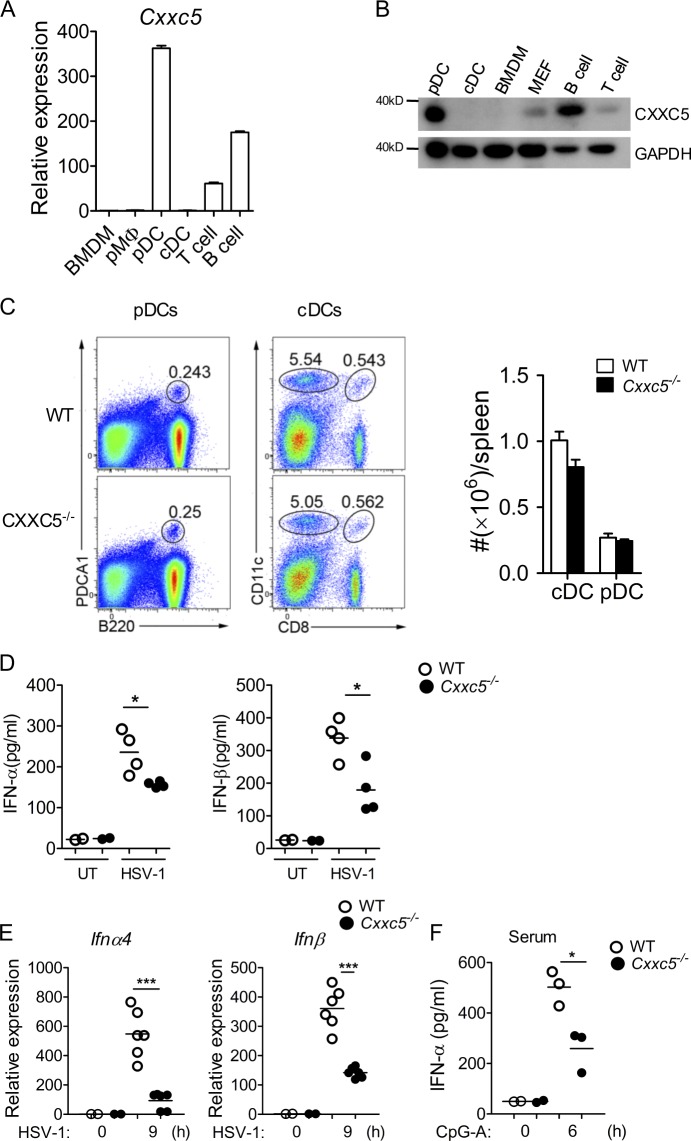
Figure 1. Expression and function of CXXC5 in pDCs. (A) Total mRNAs were extracted from BMDMs, thioglycolate-elicited peritoneal macrophages, Flt3L-differentiated pDCs, GM-CSF–differentiated BMDCs (cDCs), and splenic T and B cells isolated by anti-CD3 or anti-CD19 microbeads, respectively. The relative expression of Cxxc5 over Gapdh was measured by real-time PCR, and the data are presented as mean ± SD. This experiment was conducted twice with similar results. (B) Whole-cell lysates were prepared from Flt3L-pDCs, GM-CSF–cDCs, BMDMs, MEFs, splenic T cells, and B cells, and Western blotting was conducted with anti-CXXC5 antibody. This experiment was conducted twice with similar results. (C) Percentage of cDCs (CD11chi, gated on B220−CD3− cells) and pDCs (B220+PDCA1+CD11cint) in spleens was analyzed by flow cytometry, and total numbers of cDCs and pDCs were counted. This experiment was repeated three times, and similar results were obtained. The data are presented as mean ± SD. (D) Splenic pDCs (106 cells/ml) purified from WT and CXXC5−/− mice (n = 4) were infected with HSV-1 for 24 h. Production of IFNα/β was measured by ELISA. This experiment was repeated twice, and representative data (mean ± SD) are shown (*, P < 0.05, analyzed by Student’s t test). (E) WT and CXXC5−/− mice (n = 6) were either uninfected or i.v. infected with HSV-1 (5 × 10⁶ pfu/mouse) for 9 h, and splenocytes were sorted by FACS. Total mRNA was prepared from 105 pDCs (CD11cintB220+/PDCA1+/Siglec-Hhi) and quantified by real-time PCR. This experiment was repeated twice, and representative data are shown (***, P < 0.001, analyzed by Student’s t test). (F) WT and CXXC5−/− mice (n = 3) were either untreated or treated with i.v. injection of 20 µg CpG-A (complexed with 30 µg DOTAP) for 6 h, and sera were collected for ELISA. This experiment was performed three times with similar results (*, P < 0.05, analyzed by Student’s t test).
Figure 1. Expression and function of CXXC5 in pDCs. (A) Total mRNAs were extracted from BMDMs, thioglycolate-elicited peritoneal macrophages, Flt3L-differentiated pDCs, GM-CSF–differentiated BMDCs (cDCs), and splenic T and B cells isolated by anti-CD3 or anti-CD19 microbeads, respectively. The relative expression of Cxxc5 over Gapdh was measured by real-time PCR, and the data are presented as mean ± SD. This experiment was conducted twice with similar results. (B) Whole-cell lysates were prepared from Flt3L-pDCs, GM-CSF–cDCs, BMDMs, MEFs, splenic T cells, and B cells, and Western blotting was conducted with anti-CXXC5 antibody. This experiment was conducted twice with similar results. (C) Percentage of cDCs (CD11chi, gated on B220−CD3− cells) and pDCs (B220+PDCA1+CD11cint) in spleens was analyzed by flow cytometry, and total numbers of cDCs and pDCs were counted. This experiment was repeated three times, and similar results were obtained. The data are presented as mean ± SD. (D) Splenic pDCs (106 cells/ml) purified from WT and CXXC5−/− mice (n = 4) were infected with HSV-1 for 24 h. Production of IFNα/β was measured by ELISA. This experiment was repeated twice, and representative data (mean ± SD) are shown (*, P < 0.05, analyzed by Student’s t test). (E) WT and CXXC5−/− mice (n = 6) were either uninfected or i.v. infected with HSV-1 (5 × 10⁶ pfu/mouse) for 9 h, and splenocytes were sorted by FACS. Total mRNA was prepared from 105 pDCs (CD11cintB220+/PDCA1+/Siglec-Hhi) and quantified by real-time PCR. This experiment was repeated twice, and representative data are shown (***, P < 0.001, analyzed by Student’s t test). (F) WT and CXXC5−/− mice (n = 3) were either untreated or treated with i.v. injection of 20 µg CpG-A (complexed with 30 µg DOTAP) for 6 h, and sera were collected for ELISA. This experiment was performed three times with similar results (*, P < 0.05, analyzed by Student’s t test).CC BY-NC-SA · PubMed Central
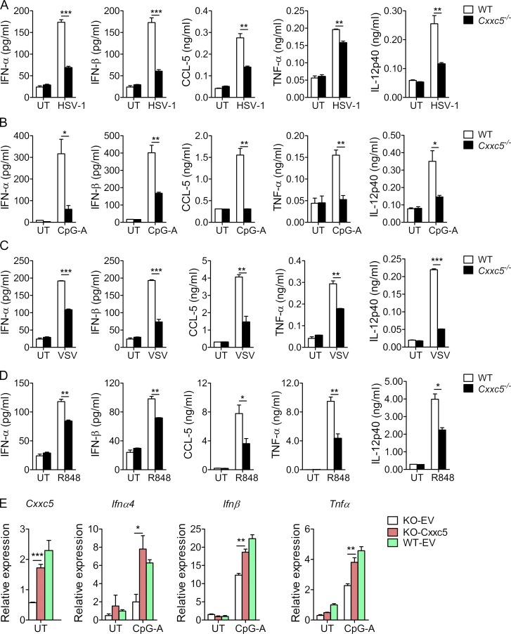
Figure 2. Virus- and TLR7/9-induced gene expression in WT and CXXC5−/− pDCs. (A–D) 2 × 10⁵ Flt3L-differentiated BM-derived pDCs (CD11cintB220+CD11b−) sorted by FACS were either untreated (UT) or treated with HSV-1 (MOI: 1; A), CpG-A (2 µg/ml; B), VSV (MOI: 1; C), or R848 (1 µg/ml; D) for 24 h, and the supernatants (n = 3) were collected for ELISA. These experiments were conducted three times, and representative data (mean ± SD) are shown (*, P < 0.05; **, P < 0.01; ***, P < 0.001, analyzed by Student’s t test). (E) WT and CXXC5−/− (KO) Flt3L-pDCs transduced by lentiviral vector or CXXC5-expressing lentiviruses were sorted as GFP-positive cells by FACS and treated with CpG-A (2 µg/ml) for 6 h. Expression levels of Ifnα/β and Tnfα mRNAs were quantified by real-time PCR, and the data are presented as mean ± SD. This experiment was repeated twice with similar results (*, P < 0.05; **, P < 0.01; ***, P < 0.001, analyzed by Student’s t test).
Figure 2. Virus- and TLR7/9-induced gene expression in WT and CXXC5−/− pDCs. (A–D) 2 × 10⁵ Flt3L-differentiated BM-derived pDCs (CD11cintB220+CD11b−) sorted by FACS were either untreated (UT) or treated with HSV-1 (MOI: 1; A), CpG-A (2 µg/ml; B), VSV (MOI: 1; C), or R848 (1 µg/ml; D) for 24 h, and the supernatants (n = 3) were collected for ELISA. These experiments were conducted three times, and representative data (mean ± SD) are shown (*, P < 0.05; **, P < 0.01; ***, P < 0.001, analyzed by Student’s t test). (E) WT and CXXC5−/− (KO) Flt3L-pDCs transduced by lentiviral vector or CXXC5-expressing lentiviruses were sorted as GFP-positive cells by FACS and treated with CpG-A (2 µg/ml) for 6 h. Expression levels of Ifnα/β and Tnfα mRNAs were quantified by real-time PCR, and the data are presented as mean ± SD. This experiment was repeated twice with similar results (*, P < 0.05; **, P < 0.01; ***, P < 0.001, analyzed by Student’s t test).CC BY-NC-SA · PubMed Central
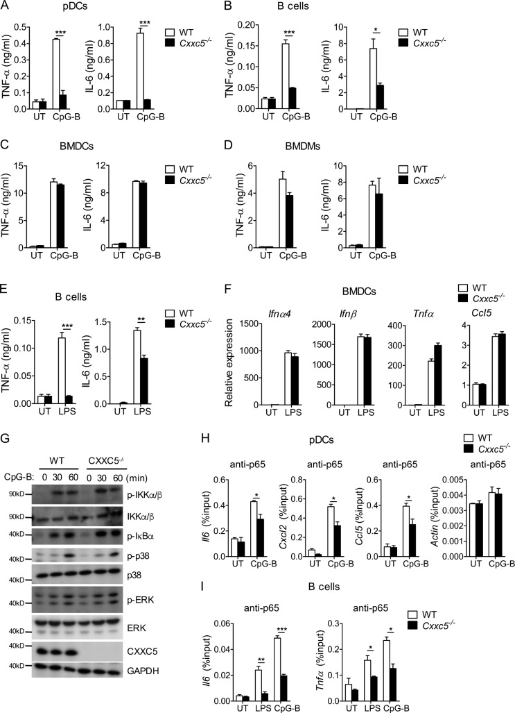
Figure 3. Effects of CXXC5 on CpG-B– and LPS-induced signaling in various immune cell populations. (A–D) Flt3L-pDCs (106/ml), splenic B cells (107/ml, purified by anti-CD19 microbeads), GM-CSF–BMDCs (cDCs, 106/ml), and BMDMs (106/ml) were stimulated by CpG-B (100 nM) for 24 h, and supernatants (n = 3) were collected for ELISA. These experiments were conducted three times, and the data are presented as mean ± SD (*, P < 0.05; ***, P < 0.001, by Student’s t test). (E) Splenic B cells (107/ml) were either untreated (UT) or treated with LPS (5 µg/ml) for 48 h, and secreted cytokines were measured by ELISA. This experiment was conducted twice with similar results, and the data are presented as mean ± SD (**, P < 0.01; ***, P < 0.001, by Student’s t test). (F) BMDCs (cDCs; 106/ml) were either untreated or treated with LPS (100 ng/ml) for 4 h, and the induction of Ifnα/β, chemokine, and cytokine mRNAs was analyzed by real-time PCR. This experiment was repeated once, and the data are presented as mean ± SD. (G) Flt3L-differentiated pDCs from WT and CXXC5−/− mice were stimulated with CpG-B (100 nM) for various times. Cell lysates were resolved by 10% SDS-PAGE and probed with the indicated antibodies. This experiment was repeated twice. (H and I) Flt3L-pDCs were stimulated with CpG-B (100 nM) for 2 h, and splenic B cells were treated with LPS (5 µg/ml) or CpG-B (150 nM) for 2 h. After cross-linking and FACS sorting, cell lysates were prepared and immunoprecipitated with 2 µg anti-p65. Immunoprecipitated promoters of proinflammatory genes were measured by real-time PCR and quantified over respective inputs (n = 4). These experiments were repeated twice, and representative data (mean ± SD) are shown (*, P < 0.05; **, P < 0.01; ***, P < 0.001, analyzed by Student’s t test).
Figure 3. Effects of CXXC5 on CpG-B– and LPS-induced signaling in various immune cell populations. (A–D) Flt3L-pDCs (106/ml), splenic B cells (107/ml, purified by anti-CD19 microbeads), GM-CSF–BMDCs (cDCs, 106/ml), and BMDMs (106/ml) were stimulated by CpG-B (100 nM) for 24 h, and supernatants (n = 3) were collected for ELISA. These experiments were conducted three times, and the data are presented as mean ± SD (*, P < 0.05; ***, P < 0.001, by Student’s t test). (E) Splenic B cells (107/ml) were either untreated (UT) or treated with LPS (5 µg/ml) for 48 h, and secreted cytokines were measured by ELISA. This experiment was conducted twice with similar results, and the data are presented as mean ± SD (**, P < 0.01; ***, P < 0.001, by Student’s t test). (F) BMDCs (cDCs; 106/ml) were either untreated or treated with LPS (100 ng/ml) for 4 h, and the induction of Ifnα/β, chemokine, and cytokine mRNAs was analyzed by real-time PCR. This experiment was repeated once, and the data are presented as mean ± SD. (G) Flt3L-differentiated pDCs from WT and CXXC5−/− mice were stimulated with CpG-B (100 nM) for various times. Cell lysates were resolved by 10% SDS-PAGE and probed with the indicated antibodies. This experiment was repeated twice. (H and I) Flt3L-pDCs were stimulated with CpG-B (100 nM) for 2 h, and splenic B cells were treated with LPS (5 µg/ml) or CpG-B (150 nM) for 2 h. After cross-linking and FACS sorting, cell lysates were prepared and immunoprecipitated with 2 µg anti-p65. Immunoprecipitated promoters of proinflammatory genes were measured by real-time PCR and quantified over respective inputs (n = 4). These experiments were repeated twice, and representative data (mean ± SD) are shown (*, P < 0.05; **, P < 0.01; ***, P < 0.001, analyzed by Student’s t test).CC BY-NC-SA · PubMed Central
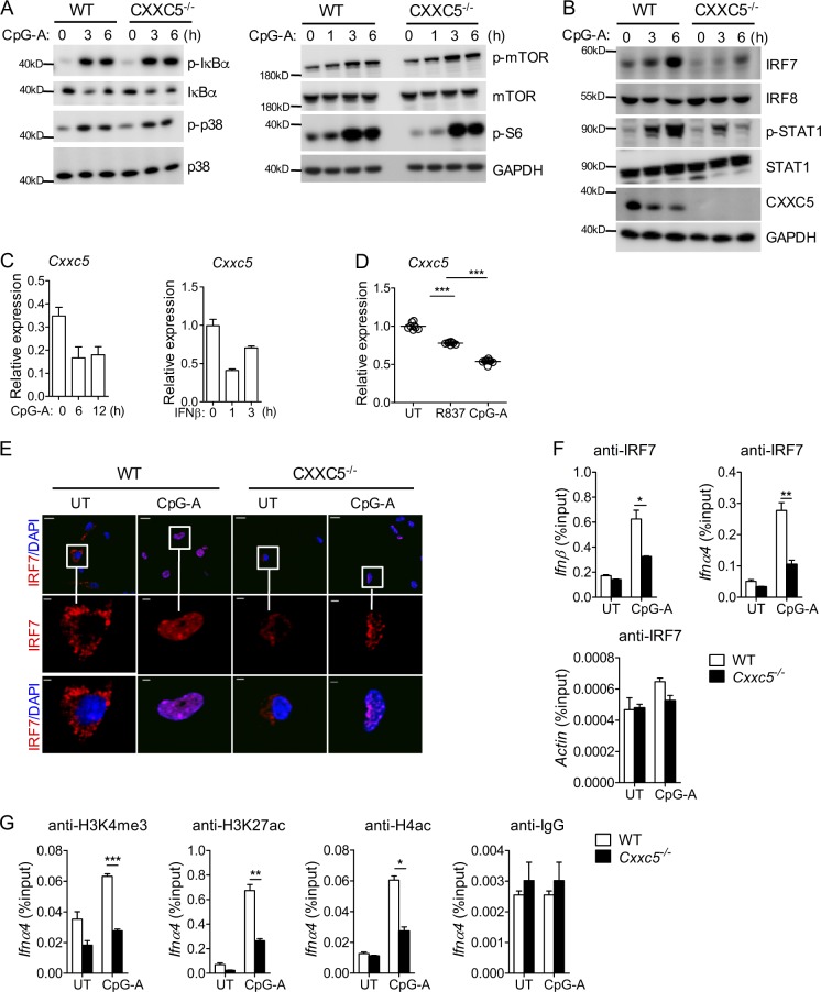
Figure 4. Impact of CXXC5 on CpG-A–induced TLR9 signaling in pDCs. (A and B) Flt3L-differentiated pDCs from WT and CXXC5−/− mice were stimulated with CpG-A (2 µg/ml) for various times. Cell lysates were resolved by 10% SDS-PAGE and probed with the indicated antibodies. These experiments were repeated twice with similar results. (C and D) Flt3L-differentiated pDCs were stimulated with CpG-A (ODN1585; 2 µg/ml) or IFNβ (100 U/ml) for various times (C), and human pDCs sorted from PBMCs of eight healthy individuals were treated with R837 (20 µg/ml) or CpG-A (ODN2216; 1 µM) for 24 h (D). These experiments were repeated twice, and expression levels of Cxxc5 were analyzed by real-time PCR (mean ± SD; ***, P < 0.001). (E) Flt3L-differentiated pDCs were either untreated (UT) or treated with CpG-A (2 µg/ml) for 5 h. Cells were fixed with paraformaldehyde and stained with anti-IRF7 antibodies (red) and DAPI (blue) sequentially. This experiment was repeated twice, and images were collected by a laser capture confocal microscope. Bars: (top) 10 µm; (bottom) 2 µm. (F and G) Flt3L-differentiated pDCs were either untreated or treated with CpG-A (2 µg/ml) for 2 h and cross-linked with paraformaldehyde. pDCs sorted by FACS were lysed for ChIP by anti-IRF7 (3 µg; F) or anti-H4ac, anti-H3K4me3, and anti-H3K27ac (1 µg; G). Immunoprecipitated Ifnα4 and Ifnβ promoter DNA fragments were measured by real-time PCR and quantified over respective inputs. These experiments were repeated twice, and data (n = 3, mean ± SD) were analyzed by Student’s t test (*, P < 0.05; **, P < 0.01; ***, P < 0.001).
Figure 4. Impact of CXXC5 on CpG-A–induced TLR9 signaling in pDCs. (A and B) Flt3L-differentiated pDCs from WT and CXXC5−/− mice were stimulated with CpG-A (2 µg/ml) for various times. Cell lysates were resolved by 10% SDS-PAGE and probed with the indicated antibodies. These experiments were repeated twice with similar results. (C and D) Flt3L-differentiated pDCs were stimulated with CpG-A (ODN1585; 2 µg/ml) or IFNβ (100 U/ml) for various times (C), and human pDCs sorted from PBMCs of eight healthy individuals were treated with R837 (20 µg/ml) or CpG-A (ODN2216; 1 µM) for 24 h (D). These experiments were repeated twice, and expression levels of Cxxc5 were analyzed by real-time PCR (mean ± SD; ***, P < 0.001). (E) Flt3L-differentiated pDCs were either untreated (UT) or treated with CpG-A (2 µg/ml) for 5 h. Cells were fixed with paraformaldehyde and stained with anti-IRF7 antibodies (red) and DAPI (blue) sequentially. This experiment was repeated twice, and images were collected by a laser capture confocal microscope. Bars: (top) 10 µm; (bottom) 2 µm. (F and G) Flt3L-differentiated pDCs were either untreated or treated with CpG-A (2 µg/ml) for 2 h and cross-linked with paraformaldehyde. pDCs sorted by FACS were lysed for ChIP by anti-IRF7 (3 µg; F) or anti-H4ac, anti-H3K4me3, and anti-H3K27ac (1 µg; G). Immunoprecipitated Ifnα4 and Ifnβ promoter DNA fragments were measured by real-time PCR and quantified over respective inputs. These experiments were repeated twice, and data (n = 3, mean ± SD) were analyzed by Student’s t test (*, P < 0.05; **, P < 0.01; ***, P < 0.001).CC BY-NC-SA · PubMed Central
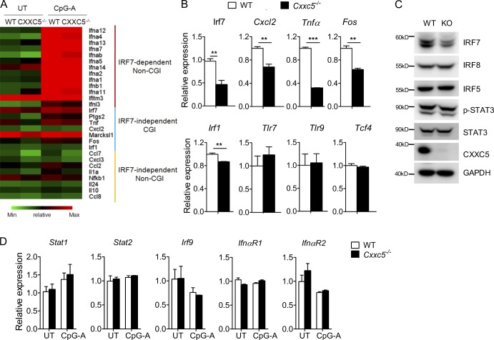
Figure 5. CXXC5-regulated gene expression in pDCs. (A) Graphic presentation of three groups of genes that were down-regulated >1.5-fold in CXXC5−/− pDCs compared with WT pDCs by microarray analysis. The relative expression of each gene in four different groups is presented as log2 value. (B) Validation of subsets of genes differentially expressed in WT and CXXC5−/− pDCs by real-time PCR. This experiment was conducted twice, and data (mean ± SD) were analyzed by Student’s t test (**, P < 0.01; ***, P < 0.001). (C) Cell lysates were prepared from Flt3L-differentiated WT and CXXC5−/− (KO) pDCs, and Western blotting was conducted to detect the expression of transcriptional factors. This experiment was repeated twice with similar results. (D) Flt3-pDCs were either untreated (UT) or treated with CpG-A (2 µg/ml) for 6 h, and gene expression was analyzed by real-time PCR. This experiment was conducted twice (n = 3), and the data are presented as mean ± SD.
Figure 5. CXXC5-regulated gene expression in pDCs. (A) Graphic presentation of three groups of genes that were down-regulated >1.5-fold in CXXC5−/− pDCs compared with WT pDCs by microarray analysis. The relative expression of each gene in four different groups is presented as log2 value. (B) Validation of subsets of genes differentially expressed in WT and CXXC5−/− pDCs by real-time PCR. This experiment was conducted twice, and data (mean ± SD) were analyzed by Student’s t test (**, P < 0.01; ***, P < 0.001). (C) Cell lysates were prepared from Flt3L-differentiated WT and CXXC5−/− (KO) pDCs, and Western blotting was conducted to detect the expression of transcriptional factors. This experiment was repeated twice with similar results. (D) Flt3-pDCs were either untreated (UT) or treated with CpG-A (2 µg/ml) for 6 h, and gene expression was analyzed by real-time PCR. This experiment was conducted twice (n = 3), and the data are presented as mean ± SD.CC BY-NC-SA · PubMed Central
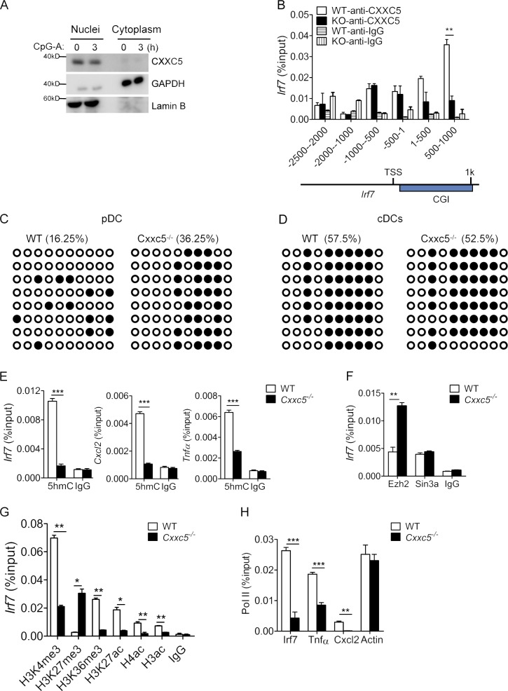
Figure 6. Epigenetic modifications on the CpG island of Irf7 gene. (A) Flt3-pDCs were either untreated or treated with CpG-A (2 µg/ml) for 3 h, and nuclear versus cytoplasmic distribution of CXXC5 was analyzed by Western blotting. This experiment was repeated twice with similar results. (B) Cell lysates from Flt3L-differentiated WT and CXXC5−/− (KO) pDCs were cross-linked and immunoprecipitated with anti-CXXC5. The enrichment of various regions of Irf7 promoter was measured by real-time PCR and quantified over respective inputs. This experiment was repeated three times, and data (n = 4, mean ± SD) were analyzed by Student’s t test (**, P < 0.01). TSS, transcription start site. (C and D) 500 ng genomic DNA from Flt3L-pDCs (C) and GM-CSF–cDCs (D) generated from WT and CXXC5−/− mice were treated with sodium bisulfite, and the CGI region of Irf7 (500–1,000) was amplified by PCR and sequenced (n = 10). This experiment was conducted twice. (E) Flt3L-differentiated pDCs were cross-linked and sonicated, and chromatin fragments were immunoprecipitated with 5hmC antibodies. 5hmC levels at the promoter region of Irf7, Cxcl2, and Tnfα were quantified by real-time PCR (n = 3). This experiment was repeated once, and the data are presented as mean ± SD (***, P < 0.001). (F–H) Enrichment of Ezh2 and Sin3a and histone modifications in Irf7 CGI (500–1,000; F and G) and enrichment of pol II at the promoter region of Irf7, Tnfα, and Cxcl2 (H) in pDCs were quantified by real-time PCR (n = 3). These experiments were repeated twice with similar results (data were analyzed by Student’s t test and are presented as mean ± SD; *, P < 0.05; **, P < 0.01; ***, P < 0.001).
Figure 6. Epigenetic modifications on the CpG island of Irf7 gene. (A) Flt3-pDCs were either untreated or treated with CpG-A (2 µg/ml) for 3 h, and nuclear versus cytoplasmic distribution of CXXC5 was analyzed by Western blotting. This experiment was repeated twice with similar results. (B) Cell lysates from Flt3L-differentiated WT and CXXC5−/− (KO) pDCs were cross-linked and immunoprecipitated with anti-CXXC5. The enrichment of various regions of Irf7 promoter was measured by real-time PCR and quantified over respective inputs. This experiment was repeated three times, and data (n = 4, mean ± SD) were analyzed by Student’s t test (**, P < 0.01). TSS, transcription start site. (C and D) 500 ng genomic DNA from Flt3L-pDCs (C) and GM-CSF–cDCs (D) generated from WT and CXXC5−/− mice were treated with sodium bisulfite, and the CGI region of Irf7 (500–1,000) was amplified by PCR and sequenced (n = 10). This experiment was conducted twice. (E) Flt3L-differentiated pDCs were cross-linked and sonicated, and chromatin fragments were immunoprecipitated with 5hmC antibodies. 5hmC levels at the promoter region of Irf7, Cxcl2, and Tnfα were quantified by real-time PCR (n = 3). This experiment was repeated once, and the data are presented as mean ± SD (***, P < 0.001). (F–H) Enrichment of Ezh2 and Sin3a and histone modifications in Irf7 CGI (500–1,000; F and G) and enrichment of pol II at the promoter region of Irf7, Tnfα, and Cxcl2 (H) in pDCs were quantified by real-time PCR (n = 3). These experiments were repeated twice with similar results (data were analyzed by Student’s t test and are presented as mean ± SD; *, P < 0.05; **, P < 0.01; ***, P < 0.001).CC BY-NC-SA · PubMed Central
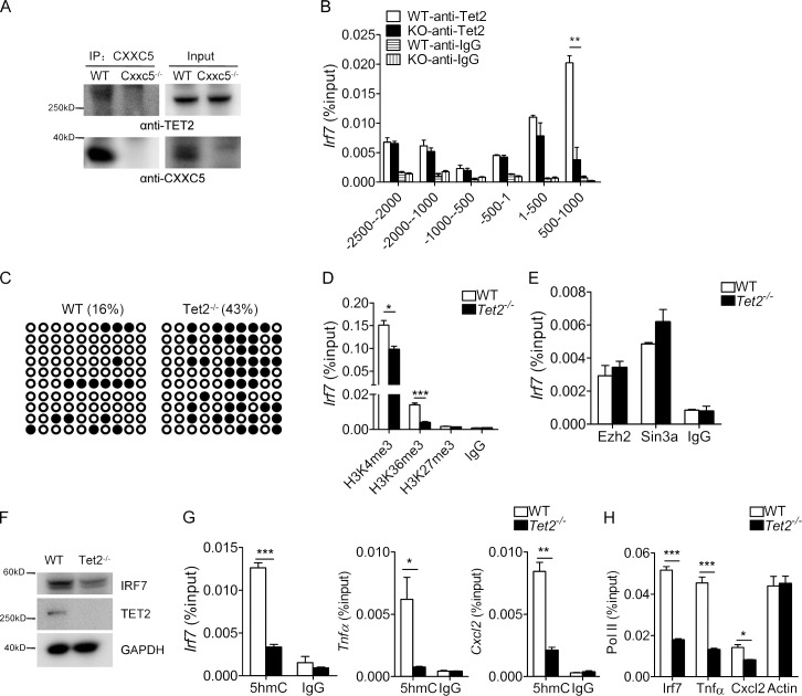
Figure 7. CXXC5 recruits Tet2 to regulate the methylation of Irf7 CGI in pDCs. (A) Cell lysates were prepared from WT and CXXC5−/− Flt3L-pDCs and immunoprecipitated (IP) with anti-CXXC5 antibody. The immunoprecipitates and inputs were blotted by anti-Tet2 and anti-CXXC5, respectively. This experiment was repeated once. (B) Whole-cell lysates from Flt3L-differentiated WT and CXXC5−/− (KO) pDCs were cross-linked and immunoprecipitated by control IgG or anti-Tet2. Enrichment of Tet2 on various regions of Irf7 promoter was determined by real-time PCR (n = 3). This experiment was repeated three times, and data (mean ± SD) were analyzed by Student’s t test (**, P < 0.01). (C) Genomic DNA from WT and Tet2−/− Flt3L-pDCs were treated with sodium bisulfite, and the CGI region of Irf7 (500–1,000) was amplified by PCR and sequenced (n = 10). This experiment was conducted twice. (D and E) Flt3L-differentiated pDCs were cross-linked and subjected to ChIP with the indicated antibodies. Histone modifications (D) and enrichment of Ezh2 and Sin3a (E) in Irf7 CGI (500–1,000) were quantified by real-time PCR (n = 3). These experiments were repeated twice, and data were analyzed by Student’s t test and are presented as mean ± SD (*, P < 0.05; ***, P < 0.001). (F) Whole-cell lysates from Flt3L-differentiated WT and Tet2−/− pDCs were resolved by 10% SDS-PAGE and probed with anti-IRF7 and anti-Tet2, respectively. This experiment was repeated once with similar results. (G and H) Flt3L-differentiated pDCs were cross-linked and sonicated, and chromatin fragments were immunoprecipitated with the indicated antibodies. 5hmC levels (G) and pol II enrichment (H) at the promoter regions of Irf7, Cxcl2, and Tnfα were quantified by real-time PCR (n = 3). These experiments were repeated twice with similar results (*, P < 0.05; **, P < 0.01; ***, P < 0.001, analyzed by Student’s t test and are presented as mean ± SD).
Figure 7. CXXC5 recruits Tet2 to regulate the methylation of Irf7 CGI in pDCs. (A) Cell lysates were prepared from WT and CXXC5−/− Flt3L-pDCs and immunoprecipitated (IP) with anti-CXXC5 antibody. The immunoprecipitates and inputs were blotted by anti-Tet2 and anti-CXXC5, respectively. This experiment was repeated once. (B) Whole-cell lysates from Flt3L-differentiated WT and CXXC5−/− (KO) pDCs were cross-linked and immunoprecipitated by control IgG or anti-Tet2. Enrichment of Tet2 on various regions of Irf7 promoter was determined by real-time PCR (n = 3). This experiment was repeated three times, and data (mean ± SD) were analyzed by Student’s t test (**, P < 0.01). (C) Genomic DNA from WT and Tet2−/− Flt3L-pDCs were treated with sodium bisulfite, and the CGI region of Irf7 (500–1,000) was amplified by PCR and sequenced (n = 10). This experiment was conducted twice. (D and E) Flt3L-differentiated pDCs were cross-linked and subjected to ChIP with the indicated antibodies. Histone modifications (D) and enrichment of Ezh2 and Sin3a (E) in Irf7 CGI (500–1,000) were quantified by real-time PCR (n = 3). These experiments were repeated twice, and data were analyzed by Student’s t test and are presented as mean ± SD (*, P < 0.05; ***, P < 0.001). (F) Whole-cell lysates from Flt3L-differentiated WT and Tet2−/− pDCs were resolved by 10% SDS-PAGE and probed with anti-IRF7 and anti-Tet2, respectively. This experiment was repeated once with similar results. (G and H) Flt3L-differentiated pDCs were cross-linked and sonicated, and chromatin fragments were immunoprecipitated with the indicated antibodies. 5hmC levels (G) and pol II enrichment (H) at the promoter regions of Irf7, Cxcl2, and Tnfα were quantified by real-time PCR (n = 3). These experiments were repeated twice with similar results (*, P < 0.05; **, P < 0.01; ***, P < 0.001, analyzed by Student’s t test and are presented as mean ± SD).CC BY-NC-SA · PubMed Central
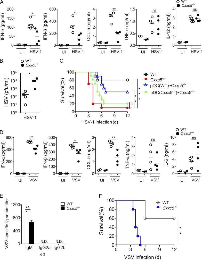
Figure 8. CXXC5−/− mice are vulnerable to viral infection in vivo. (A and B) 6–8-wk-old WT and CXXC5−/− littermates (n = 4) were intravenously infected with HSV-1 (5 × 10⁶ pfu/mouse). The sera were collected at 6 h for ELISA (A) or at 24 h for plaque assay (B). These experiments were repeated three times, and data were analyzed by Student’s t test and are presented as mean ± SD (*, P < 0.05; **, P < 0.01; ns, not significant). (C) 6–8-wk-old WT and CXXC5−/− littermates (n = 10) or CXXC5−/− mice (n = 10) adoptively transferred with 1.2 × 10⁶ Flt3L-pDCs were i.v. infected with HSV-1 (5 × 10⁶ pfu/mouse) and monitored every 12 h for survival (**, P < 0.01; ***, P < 0.01; ns, not significant, log-rank [Mantel–Cox] test). (D–F) 6–8-wk-old WT and CXXC5−/− littermates (n = 5) were intravenously infected with VSV (5 × 10⁶ pfu/mouse). Sera were collected 6 h (D) or 3 d (E) after infection for ELISA, and survival was monitored every 12 h (F). These experiments were repeated once with similar results, and data were analyzed by Student’s t test and are presented as mean ± SD (D and E) or log-rank (Mantel-Cox) test (F; *, P < 0.05; **, P < 0.01; ns, not significant).
Figure 8. CXXC5−/− mice are vulnerable to viral infection in vivo. (A and B) 6–8-wk-old WT and CXXC5−/− littermates (n = 4) were intravenously infected with HSV-1 (5 × 10⁶ pfu/mouse). The sera were collected at 6 h for ELISA (A) or at 24 h for plaque assay (B). These experiments were repeated three times, and data were analyzed by Student’s t test and are presented as mean ± SD (*, P < 0.05; **, P < 0.01; ns, not significant). (C) 6–8-wk-old WT and CXXC5−/− littermates (n = 10) or CXXC5−/− mice (n = 10) adoptively transferred with 1.2 × 10⁶ Flt3L-pDCs were i.v. infected with HSV-1 (5 × 10⁶ pfu/mouse) and monitored every 12 h for survival (**, P < 0.01; ***, P < 0.01; ns, not significant, log-rank [Mantel–Cox] test). (D–F) 6–8-wk-old WT and CXXC5−/− littermates (n = 5) were intravenously infected with VSV (5 × 10⁶ pfu/mouse). Sera were collected 6 h (D) or 3 d (E) after infection for ELISA, and survival was monitored every 12 h (F). These experiments were repeated once with similar results, and data were analyzed by Student’s t test and are presented as mean ± SD (D and E) or log-rank (Mantel-Cox) test (F; *, P < 0.05; **, P < 0.01; ns, not significant).CC BY-NC-SA · PubMed Central
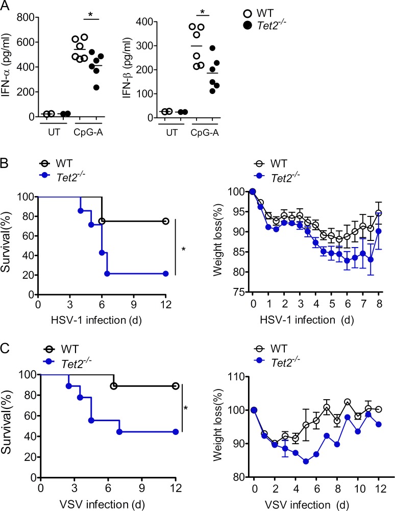
Figure 9. Tet2−/− mice are susceptible to viral infection in vivo. (A) 105 splenic pDCs sorted from WT and Tet2−/− mice were stimulated by CpG-A (2 µg/ml) for 48 h, and supernatants were collected for ELISA (n = 6). This experiment was conducted three times, and data were analyzed by Student’s t test and are presented as mean ± SD (*, P < 0.05). UT, untreated. (B) 8-wk-old WT (n = 8) and Tet2−/− (n = 14) littermates were intravenously infected with HSV-1 (5 × 10⁶ pfu) and monitored every 12 h for mortality and morbidity. This experiment was conducted twice, and log-rank (Mantel-Cox) test was used for statistical analysis (*, P < 0.05). (C) 8-wk-old WT and Tet2−/− littermates (n = 9) were intravenously infected with VSV (5 × 10⁶ pfu) and monitored every 12 h for survival and weight loss. This experiment was conducted twice, and log-rank (Mantel-Cox) test was used for statistical analysis (data are presented as mean ± SD; *, P < 0.05).
Figure 9. Tet2−/− mice are susceptible to viral infection in vivo. (A) 105 splenic pDCs sorted from WT and Tet2−/− mice were stimulated by CpG-A (2 µg/ml) for 48 h, and supernatants were collected for ELISA (n = 6). This experiment was conducted three times, and data were analyzed by Student’s t test and are presented as mean ± SD (*, P < 0.05). UT, untreated. (B) 8-wk-old WT (n = 8) and Tet2−/− (n = 14) littermates were intravenously infected with HSV-1 (5 × 10⁶ pfu) and monitored every 12 h for mortality and morbidity. This experiment was conducted twice, and log-rank (Mantel-Cox) test was used for statistical analysis (*, P < 0.05). (C) 8-wk-old WT and Tet2−/− littermates (n = 9) were intravenously infected with VSV (5 × 10⁶ pfu) and monitored every 12 h for survival and weight loss. This experiment was conducted twice, and log-rank (Mantel-Cox) test was used for statistical analysis (data are presented as mean ± SD; *, P < 0.05).CC BY-NC-SA · PubMed Central
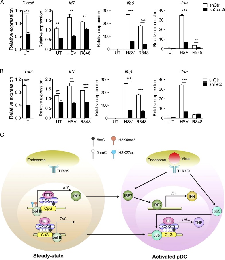
Figure 10. CXXC5 and Tet2 regulate IFN production in human pDCs. (A and B) Control and CXXC5- or Tet2-knockdown Gen2.2 cells were stimulated by HSV-1 (MOI: 2) or R848 (1 µg/ml) for 4 h, and expression of IRF7 and IFNα/β was analyzed by real-time PCR (n = 4). These experiments were repeated twice, and data shown as mean ± SD were analyzed by Student’s t test (**, P < 0.01; ***, P < 0.001). UT, untreated. (C) In light of our data presented here, we propose that CXXC5 may function as an epigenetic reader for hypomethylated CpG islands and work in concert with Tet2 to regulate gene expression in pDCs. By impacting DNA methylation and the ensuing histone modifications, CXXC5 not only promotes constitutive expression of the CGI-containing transcriptional factors (such as IRF7), but also opens up the proinflammatory gene promoters (such as Tnfα), thereby priming pDCs for TLR7/9-induced IFN and proinflammatory responses.
Figure 10. CXXC5 and Tet2 regulate IFN production in human pDCs. (A and B) Control and CXXC5- or Tet2-knockdown Gen2.2 cells were stimulated by HSV-1 (MOI: 2) or R848 (1 µg/ml) for 4 h, and expression of IRF7 and IFNα/β was analyzed by real-time PCR (n = 4). These experiments were repeated twice, and data shown as mean ± SD were analyzed by Student’s t test (**, P < 0.01; ***, P < 0.001). UT, untreated. (C) In light of our data presented here, we propose that CXXC5 may function as an epigenetic reader for hypomethylated CpG islands and work in concert with Tet2 to regulate gene expression in pDCs. By impacting DNA methylation and the ensuing histone modifications, CXXC5 not only promotes constitutive expression of the CGI-containing transcriptional factors (such as IRF7), but also opens up the proinflammatory gene promoters (such as Tnfα), thereby priming pDCs for TLR7/9-induced IFN and proinflammatory responses.CC BY-NC-SA · PubMed Central

Verification

Claims the paper makes

The claims and statistics of this paper haven’t been checked yet.

Run the check — free, no account needed. It reads the open-access text and checks the sources it cites.

A few free checks a day · papers already verified open instantly · results are public

Claims other papers make about this one

Each is checked against this paper’s own text: does it say what the citing paper says it does?

Self-check

Percentages, sums and reported differences re-computed from the paper’s own numbers.

The self-check needs the paper’s full text.

Citations

Cites

Research works printed but not linked: 70

  1. Andersson, 2009, CXXC5 is a novel BMP4-regulated modulator of Wnt signaling in neural stem cells, J. Biol. Chem., 284, 3672, 10.1074/jbc.M808119200 · DOI
  2. Bach, 2007, Vesicular stomatitis virus glycoprotein displaying retrovirus-like particles induce a type I IFN receptor-dependent switch to neutralizing IgG antibodies, J. Immunol., 178, 5839, 10.4049/jimmunol.178.9.5839 · DOI
  3. Barozzi, 2014, Coregulation of transcription factor binding and nucleosome occupancy through DNA features of mammalian enhancers, Mol. Cell., 54, 844, 10.1016/j.molcel.2014.04.006 · DOI · Paper page
  4. Blackledge, 2011, CpG island chromatin: A platform for gene regulation, Epigenetics., 6, 147, 10.4161/epi.6.2.13640 · DOI
  5. Blackledge, 2013, CpG island chromatin is shaped by recruitment of ZF-CxxC proteins, Cold Spring Harb. Perspect. Biol., 5, a018648, 10.1101/cshperspect.a018648 · DOI
  6. Blasius, 2012, Slc15a4, a gene required for pDC sensing of TLR ligands, is required to control persistent viral infection, PLoS Pathog., 8, e1002915, 10.1371/journal.ppat.1002915 · DOI
  7. Cao, 2008, Toll-like receptor-mediated induction of type I interferon in plasmacytoid dendritic cells requires the rapamycin-sensitive PI(3)K-mTOR-p70S6K pathway, Nat. Immunol., 9, 1157, 10.1038/ni.1645 · DOI
  8. Cervantes-Barragan, 2012, Plasmacytoid dendritic cells control T-cell response to chronic viral infection, Proc. Natl. Acad. Sci. USA., 109, 3012, 10.1073/pnas.1117359109 · DOI
  9. Chaperot, 2006, Virus or TLR agonists induce TRAIL-mediated cytotoxic activity of plasmacytoid dendritic cells, J. Immunol., 176, 248, 10.4049/jimmunol.176.1.248 · DOI
  10. Chehimi, 2002, Persistent decreases in blood plasmacytoid dendritic cell number and function despite effective highly active antiretroviral therapy and increased blood myeloid dendritic cells in HIV-infected individuals, J. Immunol., 168, 4796, 10.4049/jimmunol.168.9.4796 · DOI
  11. Ciancanelli, 2015, Infectious disease. Life-threatening influenza and impaired interferon amplification in human IRF7 deficiency, Science., 348, 448, 10.1126/science.aaa1578 · DOI
  12. Decalf, 2007, Plasmacytoid dendritic cells initiate a complex chemokine and cytokine network and are a viable drug target in chronic HCV patients, J. Exp. Med., 204, 2423, 10.1084/jem.20070814 · DOI
  13. Deng, 2015, Tyrosine phosphatase SHP-2 mediates C-type lectin receptor-induced activation of the kinase Syk and anti-fungal TH17 responses, Nat. Immunol., 16, 642, 10.1038/ni.3155 · DOI
  14. Di Domizio, 2009, TLR7 stimulation in human plasmacytoid dendritic cells leads to the induction of early IFN-inducible genes in the absence of type I IFN, Blood., 114, 1794, 10.1182/blood-2009-04-216770 · DOI
  15. Duan, 2004, Decreased frequency and function of circulating plasmocytoid dendritic cells (pDC) in hepatitis B virus infected humans, J. Clin. Immunol., 24, 637, 10.1007/s10875-004-6249-y · DOI
  16. Farcas, 2012, KDM2B links the Polycomb Repressive Complex 1 (PRC1) to recognition of CpG islands, eLife., 1, e00205, 10.7554/eLife.00205 · DOI
  17. Ganguly, 2009, Self-RNA-antimicrobial peptide complexes activate human dendritic cells through TLR7 and TLR8, J. Exp. Med., 206, 1983, 10.1084/jem.20090480 · DOI
  18. Ganguly, 2013, The role of dendritic cells in autoimmunity, Nat. Rev. Immunol., 13, 566, 10.1038/nri3477 · DOI
  19. Ghosh, 2010, Continuous expression of the transcription factor e2-2 maintains the cell fate of mature plasmacytoid dendritic cells, Immunity., 33, 905, 10.1016/j.immuni.2010.11.023 · DOI
  20. Gu, 2011, The role of Tet3 DNA dioxygenase in epigenetic reprogramming by oocytes, Nature., 477, 606, 10.1038/nature10443 · DOI
  21. He, 2015, mTOR regulates TLR-induced c-fos and Th1 responses to HBV and HCV vaccines, Virol. Sin., 30, 174, 10.1007/s12250-015-3606-3 · DOI
  22. Honda, 2005, IRF-7 is the master regulator of type-I interferon-dependent immune responses, Nature., 434, 772, 10.1038/nature03464 · DOI
  23. Hoshino, 2006, IkappaB kinase-α is critical for interferon-α production induced by Toll-like receptors 7 and 9, Nature., 440, 949, 10.1038/nature04641 · DOI
  24. Hu, 2014, Tet and TDG mediate DNA demethylation essential for mesenchymal-to-epithelial transition in somatic cell reprogramming, Cell Stem Cell., 14, 512, 10.1016/j.stem.2014.01.001 · DOI
  25. Ichiyama, 2015, The methylcytosine dioxygenase Tet2 promotes DNA demethylation and activation of cytokine gene expression in T cells, Immunity., 42, 613, 10.1016/j.immuni.2015.03.005 · DOI · Paper page
  26. Iurlaro, 2013, A screen for hydroxymethylcytosine and formylcytosine binding proteins suggests functions in transcription and chromatin regulation, Genome Biol., 14, R119, 10.1186/gb-2013-14-10-r119 · DOI
  27. Izaguirre, 2003, Comparative analysis of IRF and IFN-α expression in human plasmacytoid and monocyte-derived dendritic cells, J. Leukoc. Biol., 74, 1125, 10.1189/jlb.0603255 · DOI
  28. Karrich, 2014, The plasmacytoid dendritic cell as the Swiss army knife of the immune system: Molecular regulation of its multifaceted functions, J. Immunol., 193, 5772, 10.4049/jimmunol.1401541 · DOI
  29. Kawai, 2004, Interferon-α induction through Toll-like receptors involves a direct interaction of IRF7 with MyD88 and TRAF6, Nat. Immunol., 5, 1061, 10.1038/ni1118 · DOI
  30. Kim, 2015, CXXC5 is a negative-feedback regulator of the Wnt/β-catenin pathway involved in osteoblast differentiation, Cell Death Differ., 22, 912, 10.1038/cdd.2014.238 · DOI
  31. Kim, 2010, A novel Wilms tumor 1 (WT1) target gene negatively regulates the WNT signaling pathway, J. Biol. Chem., 285, 14585, 10.1074/jbc.M109.094334 · DOI
  32. Kim, 2016, CXXC5 plays a role as a transcription activator for myelin genes on oligodendrocyte differentiation, Glia., 64, 350, 10.1002/glia.22932 · DOI
  33. Ko, 2013, Modulation of TET2 expression and 5-methylcytosine oxidation by the CXXC domain protein IDAX, Nature., 497, 122, 10.1038/nature12052 · DOI · Paper page
  34. Ko, 2015, TET proteins and 5-methylcytosine oxidation in hematological cancers, Immunol. Rev., 263, 6, 10.1111/imr.12239 · DOI
  35. Laouar, 2003, STAT3 is required for Flt3L-dependent dendritic cell differentiation, Immunity., 19, 903, 10.1016/S1074-7613(03)00332-7 · DOI · Paper page
  36. Lee, 2015, The Dishevelled-binding protein CXXC5 negatively regulates cutaneous wound healing, J. Exp. Med., 212, 1061, 10.1084/jem.20141601 · DOI
  37. Lewis, 2012, Dendritic cells: Arbiters of immunity and immunological tolerance, Cold Spring Harb. Perspect. Biol., 4, a007401, 10.1101/cshperspect.a007401 · DOI
  38. Li, 2014, DNA methylation in mammals, Cold Spring Harb. Perspect. Biol., 6, a019133, 10.1101/cshperspect.a019133 · DOI
  39. Li, 2014, Plasmacytoid dendritic cells suppress HIV-1 replication but contribute to HIV-1 induced immunopathogenesis in humanized mice, PLoS Pathog., 10, e1004291, 10.1371/journal.ppat.1004291 · DOI
  40. Li, 2012, The signal transducers STAT5 and STAT3 control expression of Id2 and E2-2 during dendritic cell development, Blood., 120, 4363, 10.1182/blood-2012-07-441311 · DOI
  41. Long, 2013, ZF-CxxC domain-containing proteins, CpG islands and the chromatin connection, Biochem. Soc. Trans., 41, 727, 10.1042/BST20130028 · DOI
  42. Lu, 2000, Regulation of the promoter activity of interferon regulatory factor-7 gene. Activation by interferon and silencing by hypermethylation, J. Biol. Chem., 275, 31805, 10.1074/jbc.M005288200 · DOI
  43. Lund, 2006, Cutting edge: Plasmacytoid dendritic cells provide innate immune protection against mucosal viral infection in situ, J. Immunol., 177, 7510, 10.4049/jimmunol.177.11.7510 · DOI
  44. Mildner, 2014, Development and function of dendritic cell subsets, Immunity., 40, 642, 10.1016/j.immuni.2014.04.016 · DOI
  45. Miller, 2012, Deciphering the transcriptional network of the dendritic cell lineage, Nat. Immunol., 13, 888, 10.1038/ni.2370 · DOI
  46. Ng, 2013, Networking at the level of host immunity: Immune cell interactions during persistent viral infections, Cell Host Microbe., 13, 652, 10.1016/j.chom.2013.05.014 · DOI
  47. Ning, 2011, IRF7: Activation, regulation, modification and function, Genes Immun., 12, 399, 10.1038/gene.2011.21 · DOI
  48. Pendino, 2009, Functional involvement of RINF, retinoid-inducible nuclear factor (CXXC5), in normal and tumoral human myelopoiesis, Blood., 113, 3172, 10.1182/blood-2008-07-170035 · DOI
  49. Ramirez-Carrozzi, 2009, A unifying model for the selective regulation of inducible transcription by CpG islands and nucleosome remodeling, Cell., 138, 114, 10.1016/j.cell.2009.04.020 · DOI
  50. Reizis, 2011, Plasmacytoid dendritic cells: Recent progress and open questions, Annu. Rev. Immunol., 29, 163, 10.1146/annurev-immunol-031210-101345 · DOI
  51. Reizis, 2011, Plasmacytoid dendritic cells: One-trick ponies or workhorses of the immune system?, Nat. Rev. Immunol., 11, 558, 10.1038/nri3027 · DOI
  52. Robbins, 2008, Novel insights into the relationships between dendritic cell subsets in human and mouse revealed by genome-wide expression profiling, Genome Biol., 9, R17, 10.1186/gb-2008-9-1-r17 · DOI
  53. Rowland, 2014, Early, transient depletion of plasmacytoid dendritic cells ameliorates autoimmunity in a lupus model, J. Exp. Med., 211, 1977, 10.1084/jem.20132620 · DOI
  54. Sato, 2000, Distinct and essential roles of transcription factors IRF-3 and IRF-7 in response to viruses for IFN-α/β gene induction, Immunity., 13, 539, 10.1016/S1074-7613(00)00053-4 · DOI
  55. Sisirak, 2014, Genetic evidence for the role of plasmacytoid dendritic cells in systemic lupus erythematosus, J. Exp. Med., 211, 1969, 10.1084/jem.20132522 · DOI
  56. Smale, 2014, Chromatin contributions to the regulation of innate immunity, Annu. Rev. Immunol., 32, 489, 10.1146/annurev-immunol-031210-101303 · DOI
  57. Smit, 2006, Plasmacytoid dendritic cells inhibit pulmonary immunopathology and promote clearance of respiratory syncytial virus, J. Exp. Med., 203, 1153, 10.1084/jem.20052359 · DOI
  58. Swiecki, 2015, The multifaceted biology of plasmacytoid dendritic cells, Nat. Rev. Immunol., 15, 471, 10.1038/nri3865 · DOI
  59. Swiecki, 2010, Plasmacytoid dendritic cell ablation impacts early interferon responses and antiviral NK and CD8(+) T cell accrual, Immunity., 33, 955, 10.1016/j.immuni.2010.11.020 · DOI
  60. Swiecki, 2013, Plasmacytoid dendritic cells contribute to systemic but not local antiviral responses to HSV infections, PLoS Pathog., 9, e1003728, 10.1371/journal.ppat.1003728 · DOI
  61. Takagi, 2011, Plasmacytoid dendritic cells are crucial for the initiation of inflammation and T cell immunity in vivo, Immunity., 35, 958, 10.1016/j.immuni.2011.10.014 · DOI
  62. Treppendahl, 2013, Downregulation but lack of promoter hypermethylation or somatic mutations of the potential tumor suppressor CXXC5 in MDS and AML with deletion 5q, Eur. J. Haematol., 90, 259, 10.1111/ejh.12045 · DOI
  63. Tsuchiya, 2016, ThPOK represses CXXC5, which induces methylation of histone H3 lysine 9 in Cd40lg promoter by association with SUV39H1: Implications in repression of CD40L expression in CD8+ cytotoxic T cells, J. Leukoc. Biol., 100, 327, 10.1189/jlb.1A0915-396RR · DOI
  64. Vremec, 2000, CD4 and CD8 expression by dendritic cell subtypes in mouse thymus and spleen, J. Immunol., 164, 2978, 10.4049/jimmunol.164.6.2978 · DOI
  65. Webster, 2016, Cell-cell sensing of viral infection by plasmacytoid dendritic cells, J. Virol., 90, 10050, 10.1128/JVI.01692-16 · DOI
  66. Xu, 2011, Genome-wide regulation of 5hmC, 5mC, and gene expression by Tet1 hydroxylase in mouse embryonic stem cells, Mol. Cell., 42, 451, 10.1016/j.molcel.2011.04.005 · DOI
  67. Xu, 2012, Tet3 CXXC domain and dioxygenase activity cooperatively regulate key genes for Xenopus eye and neural development, Cell., 151, 1200, 10.1016/j.cell.2012.11.014 · DOI · Paper page
  68. Yang, 2015, Hydrogen sulfide promotes Tet1- and Tet2-mediated Foxp3 demethylation to drive regulatory T cell differentiation and maintain immune homeostasis, Immunity., 43, 251, 10.1016/j.immuni.2015.07.017 · DOI
  69. Zhang, 2009, The CXXC finger 5 protein is required for DNA damage-induced p53 activation, Sci. China C Life Sci., 52, 528, 10.1007/s11427-009-0083-7 · DOI
  70. Zhang, 2015, Plasmacytoid dendritic cells promote HIV-1-induced group 3 innate lymphoid cell depletion, J. Clin. Invest., 125, 3692, 10.1172/JCI82124 · DOI

Cited by

See all 115

Metadata

Authors
Ma, S., Wan, X., Deng, Z., Shi, L., Hao, C., Zhou, Z., Zhou, C., Fang, Y., Liu, J., Yang, J., Chen, X., Li, T., Zang, A., Yin, S., Li, B., Plumas, J., Chaperot, L., Zhang, X., Xu, G., Jiang, L., Shen, N., Xiong, S., Gao, X., Zhang, Y., & Xiao, H.
Affiliations
  • CAS Key Laboratory of Molecular Virology and Immunology, Institut Pasteur of Shanghai; CAS Center for Excellence in Molecular Cell Sciences; University of Chinese Academy of Sciences, Chinese Academy of Sciences, Shanghai 200031, China 1
  • CAS Key Laboratory of Molecular Virology and Immunology, Institut Pasteur of Shanghai, Chinese Academy of Sciences, Shanghai 200031, China 3
  • Shanghai Institute of Rheumatology, Renji Hospital, Shanghai Jiao Tong University School of Medicine, Shanghai 200240, China 5
  • Institute for Advanced Biosciences (IAB), Team Immunobiology and Immunotherapy in Chronic Diseases, Institut National de la Santé et de la Recherche Médicale U1209, Centre National de la Recherche Scientifique UMR5309, Université Grenoble Alpes, Etablissement Français du Sang-Rhone-Alpes, F-38700 Grenoble, France 6
  • State Key Laboratory of Molecular Biology, CAS Excellence Center in Molecular Cell Sciences, Institute of Biochemistry and Cell Biology, Shanghai Institutes for Biological Sciences, Chinese Academy of Sciences, Shanghai 200031, China 4
  • Institute of Biology and Medical Sciences, Soochow University, Soochow, Jiangsu 215006, China 2
Venue
Journal of Experimental Medicine
Published
2017-04-17
Volume
214
Issue
5
Pages
1471-1491
ISSN
  • 0022-1007
  • 1540-9538
Cited by
115
References
7
Language
en
Funders
  • Ministry of Science and Technology of the People's Republic of China (2016YFA0502102, 2014CB541902)
  • National Natural Science Foundation of China (31470847, 91542206)
  • Chinese Academy of Sciences (XDB19000000)

About these checks

Registry, retraction, Cites and statistics results are computed, not judged, with one exception: a printed reference that no index lists is read once by a model before it counts as a citation error. Claims are extracted by a model and kept only when their quote appears verbatim in the text; each cited source is then checked, with a model’s help, for whether it says what the claim relies on. Those verdicts can be wrong — read the claim’s quote and the cited source before relying on one.

Statistics and arithmetic are recomputed, not judged.

Confidence bands are shown only for fields calibrated on our annotated test set. None is calibrated yet, so no band is shown.

What counts as a claim ›How we measure confidence ›How we check this ›

Are you an author, or do you see a problem with this page? Request a review