

The DOI matches its Crossref record; 71 of the 71 references it cites were found in a registry or an index.
Human neural stem cells enhance structural plasticity and axonal transport in the ischaemic brain
Open accessAbstract
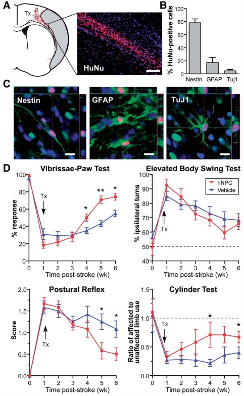
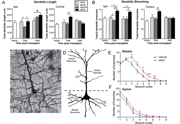
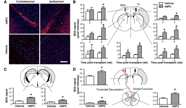
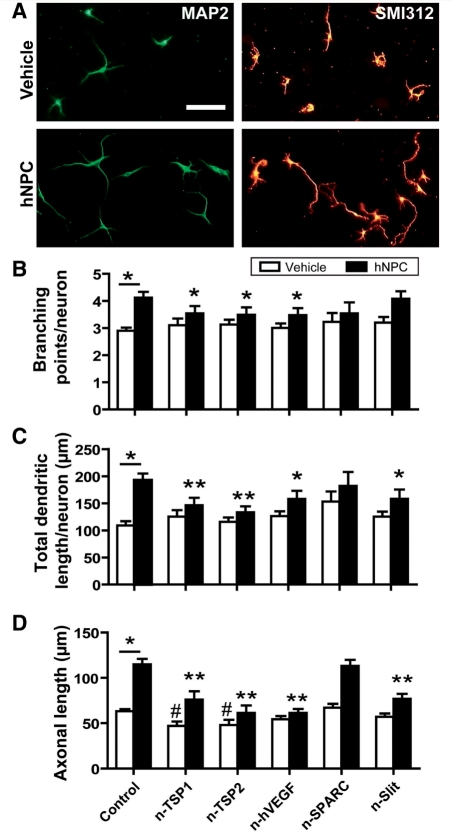
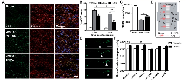
Stem cell transplantation promises new hope for the treatment of stroke although significant questions remain about how the grafted cells elicit their effects. One hypothesis is that transplanted stem cells enhance endogenous repair mechanisms activated after cerebral ischaemia. Recognizing that bilateral reorganization of surviving circuits is associated with recovery after stroke, we investigated the ability of transplanted human neural progenitor cells to enhance this structural plasticity. Our results show the first evidence that human neural progenitor cell treatment can significantly increase dendritic plasticity in both the ipsi- and contralesional cortex and this coincides with stem cell-induced functional recovery. Moreover, stem cell-grafted rats demonstrated increased corticocortical, corticostriatal, corticothalamic and corticospinal axonal rewiring from the contralesional side; with the transcallosal and corticospinal axonal sprouting correlating with functional recovery. Furthermore, we demonstrate that axonal transport, which is critical for both proper axonal function and axonal sprouting, is inhibited by stroke and that this is rescued by the stem cell treatment, thus identifying another novel potential mechanism of action of transplanted cells. Finally, we established in vitro co-culture assays in which these stem cells mimicked the effects observed in vivo. Through immunodepletion studies, we identified vascular endothelial growth factor, thrombospondins 1 and 2, and slit as mediators partially responsible for stem cell-induced effects on dendritic sprouting, axonal plasticity and axonal transport in vitro. Thus, we postulate that human neural progenitor cells aid recovery after stroke through secretion of factors that enhance brain repair and plasticity.
Overview
The claims and statistics of this paper haven’t been checked yet.
Run the check — free, no account needed. It reads the open-access text and checks the sources it cites.
Integrity signals
Registry
Cites
Statistics
Figures
Verification
Claims the paper makes
The claims and statistics of this paper haven’t been checked yet.
Run the check — free, no account needed. It reads the open-access text and checks the sources it cites.
Claims other papers make about this one
Each is checked against this paper’s own text: does it say what the citing paper says it does?
The check wasn’t confident either way, so it gives no verdict — never disputed.
In the citing paper
“With simple growth factor withdrawal for three weeks, most of these iPS-NPCs develop into neurons expressing Tuj1 or MAP250”
From GDF10 is a signal for axonal sprouting and functional recovery after stroke (2015) · cites this paper as [50]
Experimental — accuracy not yet measuredIn the citing paper
“In rodents, beneficial effects were reported even when treatments were initiated days, weeks, or months after stroke.14–20”
From Promoting brain remodelling and plasticity for stroke recovery: therapeutic promise and potential pitfalls of clinical translation (2012) · cites this paper as [15]
In the citing paper
“By contrast with neuroprotection, which requires an acute intervention, treatments promoting neurological recovery by remodelling of brain tissue can be instituted during the stroke recovery phase.10–15”
From Promoting brain remodelling and plasticity for stroke recovery: therapeutic promise and potential pitfalls of clinical translation (2012) · cites this paper as [15]
In the citing paper
“By sharp contrast with neuroprotection, the efficacy of neuro restorative therapies does not depend on the successful reperfusion of ischaemic brain tissue.11,12,15,17,21”
From Promoting brain remodelling and plasticity for stroke recovery: therapeutic promise and potential pitfalls of clinical translation (2012) · cites this paper as [15]
In the citing paper
“The finding that neural precursor cells prevent ischaemic injury and promote neurological recovery could be attributed to bystander effects related to the interaction of neuroblasts with the microvasculature in the vicinity of the ischaemic lesion, which creates an environment that promotes brain remodelling (figure 2).14,15,37,45”
From Promoting brain remodelling and plasticity for stroke recovery: therapeutic promise and potential pitfalls of clinical translation (2012) · cites this paper as [15]
In the citing paper
“Meanwhile, evidence that neuro genesis contributes to functional neurological recovery has been obtained in studies with transgenic ablation of neural precursor cells expressing the marker doublecortin43 and in studies with exogenous delivery of adult neural precursor cells,14,15,44 which reported impaired or enhanced neurological recovery, respectively.”
From Promoting brain remodelling and plasticity for stroke recovery: therapeutic promise and potential pitfalls of clinical translation (2012) · cites this paper as [15]
In the citing paper
“Instead, homologous fibres originating from the contralesional motor cortex grow out across the midline to reach denervated neurons in target structures of the lesioned pyramidal tract (figure 1).10–12,15,17,19,24”
From Promoting brain remodelling and plasticity for stroke recovery: therapeutic promise and potential pitfalls of clinical translation (2012) · cites this paper as [15]
In the citing paper
“Contralesional plasticity as a basis for neurological recovery occurs in several species (eg, in rats,11,15,17 mice,12,19,24 and macaques32).”
From Promoting brain remodelling and plasticity for stroke recovery: therapeutic promise and potential pitfalls of clinical translation (2012) · cites this paper as [15]
In the citing paper
“This lesion-remote plasticity involves the midline crossing of fibres from the contralesional to the ipsilesional hemisphere at the level of the red nucleus10,17,19,24 and the re-entry of fibres into the contralesional hemicord at the level of the cervical spinal cord (figure 1).11,12,15”
From Promoting brain remodelling and plasticity for stroke recovery: therapeutic promise and potential pitfalls of clinical translation (2012) · cites this paper as [15]
In the citing paper
“Microvessels and neuroblasts mutually support each other via release of trophic factors, resulting in a tight interplay that stimulates brain remodelling processes, reducing delayed neuronal degeneration,14,45 promoting neuronal plasticity,15 modulating glial responses,14,46 and attenuating brain inflammation14,46 (figure 2).”
From Promoting brain remodelling and plasticity for stroke recovery: therapeutic promise and potential pitfalls of clinical translation (2012) · cites this paper as [15]
Self-check
Percentages, sums and reported differences re-computed from the paper’s own numbers.
The self-check needs the paper’s full text.
Citations
Cites
Neuronal replacement from endogenous precursors in the adult brain after stroke
Arvidsson, A., Collin, T., Kirik, D., Kokaia, Z., & Lindvall, O. (2002) · Nature Medicine · doi:10.1038/nm747 · Cited by 2,799
Bederson, J. B., Pitts, L. H., Tsuji, M., Nishimura, M. C., Davis, R. L., & Bartkowski, H. (1986) · Stroke · doi:10.1161/01.str.17.3.472 · Cited by 2,681
Discovery and development of bevacizumab, an anti-VEGF antibody for treating cancer
Ferrara, N., Hillan, K. J., Gerber, H.-P., & Novotny, W. (2004) · Nature Reviews Drug Discovery · doi:10.1038/nrd1381 · Cited by 2,625
Plasticity during stroke recovery: from synapse to behaviour
Murphy, T. H., & Corbett, D. (2009) · Nature Reviews Neuroscience · doi:10.1038/nrn2735 · Cited by 1,938
Pyramidal neurons: dendritic structure and synaptic integration
Spruston, N. (2008) · Nature Reviews Neuroscience · doi:10.1038/nrn2286 · Cited by 1,688
Schallert, T. J., Fleming, S. M., Leasure, J. L., Tillerson, J. L., & Bland, S. T. (2000) · Neuropharmacology · doi:10.1016/s0028-3908(00)00005-8 · Cited by 1,373
Chen, J., Sanberg, P. R., Li, Y., Wang, L., Lü, M., Willing, A. E., Sanchez‐Ramos, J., & Chopp, M. (2001) · Stroke · doi:10.1161/hs1101.098367 · Cited by 1,347
A microfluidic culture platform for CNS axonal injury, regeneration and transport
Taylor, A. M., Blurton-Jones, M. M., Rhee, S. W., Cribbs, D. H., Cotman, C. W., & Jeon, N. L. (2005) · Nature Methods · doi:10.1038/nmeth777 · Cited by 1,172
Autologous mesenchymal stem cell transplantation in stroke patients
Bang, O. Y., Lee, J. S., Lee, P. H., & Lee, G. (2005) · Annals of Neurology · doi:10.1002/ana.20501 · Cited by 1,101
Axon degeneration mechanisms: Commonality amid diversity
Coleman, M. (2005) · Nature reviews. Neuroscience · doi:10.1038/nrn1788 · Cited by 819
Cited by
Sorted by citations
Graham, H. K., Rosenbaum, P., Paneth, N., Dan, B., Lin, J., Damiano, D. L., Becher, J. G., Gaebler-Spira, D. J., Colver, A., Reddihough, D. S., et al. (2016) · Nature Reviews Disease Primers · doi:10.1038/nrdp.2015.82 · Cited by 1,099
Microglia and Monocytes/Macrophages Polarization Reveal Novel Therapeutic Mechanism against Stroke
Kanazawa, M., Ninomiya, I., Hatakeyama, M., Takahashi, T., & Shimohata, T. (2017) · International Journal of Molecular Sciences · doi:10.3390/ijms18102135 · Cited by 405
Novel Stroke Therapeutics: Unraveling Stroke Pathophysiology and Its Impact on Clinical Treatments
George, P. M., & Steinberg, G. K. (2015) · Neuron · doi:10.1016/j.neuron.2015.05.041 · Cited by 404
Self-citationThe stem cell secretome and its role in brain repair
Drago, D., Cossetti, C., Iraci, N., Gaude, E., Musco, G., Bachi, A., & Pluchino, S. (2013) · Biochimie · doi:10.1016/j.biochi.2013.06.020 · Cited by 354
Hermann, D. M., & Chopp, M. (2012) · The Lancet Neurology · doi:10.1016/s1474-4422(12)70039-x · Cited by 333
Oki, K., Tatarishvili, J., Wood, J. C., Koch, P., Wattananit, S., Mine, Y., Monni, E., Tornero, D., Ahlenius, H., Ladewig, J., et al. (2012) · Stem Cells · doi:10.1002/stem.1104 · Cited by 299
Tornero, D., Wattananit, S., Grønning Madsen, M., Koch, P., Wood, J., Tatarishvili, J., Mine, Y., Ge, R., Monni, E., Devaraju, K., et al. (2013) · Brain · doi:10.1093/brain/awt278 · Cited by 241
Stem Cell-Based Therapies for Ischemic Stroke
Hao, L., Zou, Z., Tian, H. Y., Zhang, Y., Zhou, H., & Liu, L. (2014) · BioMed Research International · doi:10.1155/2014/468748 · Cited by 219
Advances in tissue engineering through stem cell-based co-culture
Paschos, N. K., Brown, W. E., Eswaramoorthy, R., Hu, J. C. Y., & Athanasiou, K. A. (2014) · Journal of Tissue Engineering and Regenerative Medicine · doi:10.1002/term.1870 · Cited by 212
Borlongan, C. V., Glover, L. E., Tajiri, N., Kaneko, Y., & Freeman, T. B. (2011) · Progress in Neurobiology · doi:10.1016/j.pneurobio.2011.08.005 · Cited by 209
Neural stem cell therapy for subacute and chronic ischemic stroke
Boese, A. C., Le, Q.-S. E., Pham, D., Hamblin, M. H., & Lee, J.-P. (2018) · Stem Cell Research & Therapy · doi:10.1186/s13287-018-0913-2 · Cited by 205
De Feo, D., Merlini, A., Laterza, C., & Martino, G. (2012) · Current Opinion in Neurology · doi:10.1097/wco.0b013e328352ec45 · Cited by 181
GDF10 is a signal for axonal sprouting and functional recovery after stroke
Li, S., Nie, E. H., Yin, Y., Benowitz, L. I., Tung, S., Vinters, H. V., Bahjat, F. R., Stenzel-Poore, M. P., Kawaguchi, R., Coppola, G., et al. (2015) · Nature Neuroscience · doi:10.1038/nn.4146 · Cited by 181
“Axons of primary cortical neurons and iPS-NPCs were stained with anti-SMI-312 monoclonal antibody 51 .”
Cell based therapies for ischemic stroke: From basic science to bedside
Liu, X., Ye, R., Yan, T., Yu, S. P., Wei, L., Xu, G., Fan, X., Jiang, Y., Stetler, R. A., Liu, C., et al. (2013) · Progress in Neurobiology · doi:10.1016/j.pneurobio.2013.11.007 · Cited by 179
The dark side of the force – constraints and complications of cell therapies for stroke
Boltze, J. (2015) · Frontiers in Neurology · doi:10.3389/fneur.2015.00155 · Cited by 175
A Review of Exercise-Induced Neuroplasticity in Ischemic Stroke: Pathology and Mechanisms
Xing, Y., & Bai, Y. (2020) · Molecular Neurobiology · doi:10.1007/s12035-020-02021-1 · Cited by 179
Zăgrean, A., Hermann, D. M., Opriș, I. C., Zăgrean, L., & Popa‐Wagner, A. (2018) · Frontiers in Neuroscience · doi:10.3389/fnins.2018.00811 · Cited by 173
Human neural stem cells alleviate Alzheimer-like pathology in a mouse model
Lee, I.-S., Jung, K., Kim, I., Lee, H.-J., Kim, M., Yun, S., Hwang, K., Shin, J. E., & Park, K. I. (2015) · Molecular Neurodegeneration · doi:10.1186/s13024-015-0035-6 · Cited by 170
Baloh, R. H., Johnson, J. P., Avalos, P., Allred, P., Svendsen, S. P., Gowing, G., Roxas, K., Wu, A., Donahue, B., Osborne, S., et al. (2022) · Nature Medicine · doi:10.1038/s41591-022-01956-3 · Cited by 157
Self-citationSmith, E. J., Stroemer, R. P., Gorenkova, N., Nakajima, M., Crum, W. R., Tang, E., Stevanato, L., Sinden, J. D., & Modo, M. (2011) · Stem Cells · doi:10.1002/stem.1024 · Cited by 154
Stem Cells as an Emerging Paradigm in Stroke 3
Savitz, S. I., Cramer, S. C., Wechsler, L. R., Aronowski, J. A., Boltze, J., Borlongan, C. V., Case, C., Chase, T. N., Chopp, M., Carmichael, S. T., et al. (2013) · Stroke · doi:10.1161/strokeaha.113.003379 · Cited by 157
Self-citationMine, Y., Tatarishvili, J., Oki, K., Monni, E., Kokaia, Z., & Lindvall, O. (2012) · Neurobiology of Disease · doi:10.1016/j.nbd.2012.12.006 · Cited by 127
Enhancing Brain Plasticity to Promote Stroke Recovery
Su, F., & Xu, W. (2020) · Frontiers in Neurology · doi:10.3389/fneur.2020.554089 · Cited by 137
Rosell, A., Morancho, A., Navarro-Sobrino, M., Martínez-Sáez, E. A., Hernández‐Guillamón, M., Lope‐Piedrafita, S., Barceló, V., Borràs, F. E., Peñalba, A., García‐Bonilla, L., et al. (2013) · PLoS ONE · doi:10.1371/journal.pone.0073244 · Cited by 122
Chen, D.-C., Lin, S., Fan, J.-R., Lin, C.-H., Lee, W., Lin, C.-C., Liu, Y., Tsai, C., Chen, J., Cho, D.-Y., et al. (2014) · Cell Transplantation · doi:10.3727/096368914x678562 · Cited by 119
Neural stem cell therapy for stroke: A multimechanistic approach to restoring neurological function
Baker, E. W., Kinder, H. A., & West, F. D. (2019) · Brain and Behavior · doi:10.1002/brb3.1214 · Cited by 119
Interleukin 6-preconditioned neural stem cells reduce ischaemic injury in stroke mice
Sakata, H., Narasimhan, P., Niizuma, K., Maier, C. M., Wakai, T., & Chan, P. H. (2012) · Brain · doi:10.1093/brain/aws259 · Cited by 113
Understanding the Mechanisms of Recovery and/or Compensation following Injury
Hylin, M. J., Kerr, A. L., & Holden, R. (2017) · Neural Plasticity · doi:10.1155/2017/7125057 · Cited by 113
Electrical preconditioning of stem cells with a conductive polymer scaffold enhances stroke recovery
George, P. M., Bliss, T. M., Hua, T., Lee, A. G., Oh, B., Levinson, A., Mehta, S. S., Sun, G.-H., & Steinberg, G. K. (2017) · Biomaterials · doi:10.1016/j.biomaterials.2017.07.020 · Cited by 110
Self-citationSareen, D., Gowing, G., Sahabian, A., Staggenborg, K., Paradis, R., Avalos, P., Latter, J., Ornelas, L., Garcia, L., & Svendsen, C. N. (2014) · The Journal of Comparative Neurology · doi:10.1002/cne.23578 · Cited by 110
Self-citationBacigaluppi, M., Russo, G. L., Peruzzotti‐Jametti, L., Rossi, S., Sandrone, S., Butti, E., Ceglia, R. D., Bergamaschi, A., Motta, C., Gallizioli, M., et al. (2016) · Journal of Neuroscience · doi:10.1523/jneurosci.1643-16.2016 · Cited by 109
Microglia preconditioned by oxygen-glucose deprivation promote functional recovery in ischemic rats
Kanazawa, M., Miura, M., Toriyabe, M., Koyama, M., Hatakeyama, M., Ishikawa, M., Nakajima, T., Onodera, O., Takahashi, T., Nishizawa, M., et al. (2017) · Scientific Reports · doi:10.1038/srep42582 · Cited by 106
Ishizaka, S., Horie, N., Satoh, K., Fukuda, Y., Nishida, N., & Nagata, I. (2013) · Stroke · doi:10.1161/strokeaha.112.677328 · Cited by 106
Self-citationRewiring the connectome: Evidence and effects
Bennett, S. H., Kirby, A. J., & Finnerty, G. T. (2018) · Neuroscience & Biobehavioral Reviews · doi:10.1016/j.neubiorev.2018.03.001 · Cited by 105
Endothelial Progenitor Cells: Therapeutic Perspective for Ischemic Stroke
Zhao, Y., Yuan, B., Chen, J., Feng, D., Zhao, B., Qin, C., & Chen, Y. (2012) · CNS Neuroscience & Therapeutics · doi:10.1111/cns.12040 · Cited by 100
Brain repair: cell therapy in stroke
Kalladka, D., & Muir, K. W. (2014) · Stem Cells and Cloning Advances and Applications · doi:10.2147/sccaa.s38003 · Cited by 101
Li, W., Guan, T., Zhang, X., Wang, Z., Wang, M., Zhong, W., Feng, H., Xing, M. M. Q., & Kong, J. (2014) · ACS Applied Materials & Interfaces · doi:10.1021/am504456t · Cited by 101
Stem Cell–Based Tissue Replacement After Stroke
Janowski, M., Wagner, D., & Boltze, J. (2015) · Stroke · doi:10.1161/strokeaha.114.007803 · Cited by 93
Role of neural precursor cells in promoting repair following stroke
Dibajnia, P., & Morshead, C. M. (2012) · Acta Pharmacologica Sinica · doi:10.1038/aps.2012.107 · Cited by 98
Optimizing Stem Cell Therapy after Ischemic Brain Injury
Zhang, S., Lachance, B. B., Moiz, B., & Jia, X. (2020) · Journal of Stroke · doi:10.5853/jos.2019.03048 · Cited by 89
Giusto, E., Donegà, M., Cossetti, C., & Pluchino, S. (2013) · Experimental Neurology · doi:10.1016/j.expneurol.2013.03.009 · Cited by 92
Shah, A. M., Ishizaka, S., Cheng, M., Wang, E. H.-J., Bautista, A. R., Levy, S. L., Smerin, D., Sun, G.-H., & Steinberg, G. K. (2017) · Scientific Reports · doi:10.1038/srep46612 · Cited by 89
Self-citationFunctional Assessment of Long-Term Deficits in Rodent models of Traumatic Brain Injury
Gold, E. M., Su, D., López-Velázquez, L., Haus, D. L., Perez, H. D., Lacuesta, G. A., Anderson, A. J., & Cummings, B. J. (2013) · Regenerative Medicine · doi:10.2217/rme.13.41 · Cited by 90
The 3 Rs of Stroke Biology: Radial, Relayed, and Regenerative
Carmichael, S. T. (2015) · Neurotherapeutics · doi:10.1007/s13311-015-0408-0 · Cited by 99
Tajiri, N., Kaneko, Y., Shinozuka, K., Ishikawa, H., Yankee, E. W., McGrogan, M. P., Case, C., & Borlongan, C. V. (2013) · PLoS ONE · doi:10.1371/journal.pone.0074857 · Cited by 85
Hermann, D. M., Peruzzotti‐Jametti, L., Schlechter, J., Bernstock, J. D., Doeppner, T. R., & Pluchino, S. (2014) · Frontiers in Cellular Neuroscience · doi:10.3389/fncel.2014.00291 · Cited by 84
Paracrine effects of stem cells in wound healing and cancer progression
Dittmer, J., & Leyh, B. (2014) · International Journal of Oncology · doi:10.3892/ijo.2014.2385 · Cited by 82
Rosenblum, S., Smith, T. N., Wang, N. N., Chua, J. Y., Westbroek, E. M., Wang, K. M., & Guzman, R. G. (2014) · Cell Transplantation · doi:10.3727/096368914x679354 · Cited by 81
Doeppner, T. R., Ewert, T. A. S., Tönges, L., Herz, J., Zechariah, A., ElAli, A., Ludwig, A., Giebel, B., Nagel, F., Dietz, G. P. H., et al. (2012) · Stem Cells · doi:10.1002/stem.1098 · Cited by 81
Sinden, J. D., Hicks, C. A., Stroemer, P., Vishnubhatla, I., & Corteling, R. L. (2017) · Stem Cells and Development · doi:10.1089/scd.2017.0009 · Cited by 79
Bălşeanu, T. A., Buga, A.-M., Cătălin, B., Wagner, D., Boltze, J., Zăgrean, A., Reymann, K. G., Schaebitz, W., & Popa‐Wagner, A. (2014) · Frontiers in Aging Neuroscience · doi:10.3389/fnagi.2014.00130 · Cited by 76
Opportunities and Challenges: Stem Cell‐Based Therapy for the Treatment of Ischemic Stroke
Tang, Y., Ma, Y., Zhang, Z., Wang, Y., & Yang, G. (2015) · CNS Neuroscience & Therapeutics · doi:10.1111/cns.12386 · Cited by 77
Neural Stem Cells Therapy for Ischemic Stroke: Progress and Challenges
Zhao, T., Zhu, T., Xie, L., Li, Y., Xie, R., Xu, F., Tang, H., & Zhu, J. (2022) · Translational Stroke Research · doi:10.1007/s12975-022-00984-y · Cited by 74
Spaethling, J. M., Na, Y.-J., Lee, J., Ulyanova, A. V., Baltuch, G. H., Bell, T. J., Brem, S. S., Chen, H. I., Dueck, H., Fisher, S. A., et al. (2017) · Cell Reports · doi:10.1016/j.celrep.2016.12.066 · Cited by 72
Liu, J., Jiao, Y., Liu, Y.-W., & Chen, W.-G. (2020) · Neural Regeneration Research · doi:10.4103/1673-5374.286955 · Cited by 76
Human induced pluripotent stem cells improve recovery in stroke-injured aged rats
Tatarishvili, J., Oki, K., Monni, E., Koch, P., Memanishvili, T., Buga, A.-M., Verma, V., Popa‐Wagner, A., Brüstle, O., Lindvall, O., et al. (2014) · Restorative Neurology and Neuroscience · doi:10.3233/rnn-140404 · Cited by 71
Ex vivo gene therapy for the treatment of neurological disorders
Gowing, G., Svendsen, S. P., & Svendsen, C. N. (2017) · Progress in brain research · doi:10.1016/bs.pbr.2016.11.003 · Cited by 74
Self-citationDonega, V., van Velthoven, C. T. J., Nijboer, C. H. A., Kavelaars, A., & Heijnen, C. J. (2013) · Journal of Cerebral Blood Flow & Metabolism · doi:10.1038/jcbfm.2013.3 · Cited by 70
Parambi, D. G. T., Alharbi, K. S., Kumar, R., Harilal, S., Batiha, G. E., Martins, N. A., Hendawy, O. M., Musa, A., Panda, D. S., & Mathew, B. (2021) · Molecular Neurobiology · doi:10.1007/s12035-021-02555-y · Cited by 76
The Influence of Oxidative Stress Markers in Patients with Ischemic Stroke
Pawluk, H., Tafelska‐Kaczmarek, A., Sopońska, M., Porzych, M., Modrzejewska, M., Pawluk, M., Kurhaluk, N., Tkaczenko, H., & Kołodziejska, R. (2024) · Biomolecules · doi:10.3390/biom14091130 · Cited by 73
The Rise of Cell Therapy Trials for Stroke: Review of Published and Registered Studies
Rosado-de-Castro, P. H., Pimentel‐Coelho, P. M., da Fonseca, L. M. B., de Freitas, G. R., & Méndez‐Otero, R. (2013) · Stem Cells and Development · doi:10.1089/scd.2013.0089 · Cited by 68
Zhang, K., Zhang, Q., Deng, J., Li, J., Li, J., Wen, L., Ma, J., & Li, C. (2019) · Cell Death and Disease · doi:10.1038/s41419-019-1596-z · Cited by 67
Stem‐Cell Challenges in the Treatment of Alzheimer's Disease: A Long Way from Bench to Bedside
Fan, X., Sun, D., Tang, X., Cai, Y., Yin, Z. Q., & Xu, H. (2014) · Medicinal Research Reviews · doi:10.1002/med.21309 · Cited by 66
Nagpal, A., Kremer, K. L., Hamilton‐Bruce, M. A., Kaidonis, X., Milton, A. G., Levi, C. R., Shi, S., Carey, L. M., Hillier, S., Rose, M. L., et al. (2016) · International Journal of Stroke · doi:10.1177/1747493016641111 · Cited by 65
Stem cell therapies in preclinical models of stroke associated with aging
Popa‐Wagner, A., Buga, A.-M., Doeppner, T. R., & Hermann, D. M. (2014) · Frontiers in Cellular Neuroscience · doi:10.3389/fncel.2014.00347 · Cited by 66
Sasaki, Y., Sasaki, M., Kataoka-Sasaki, Y., Nakazaki, M., Nagahama, H., Suzuki, J., Tateyama, D., Oka, S., Namioka, T., Namioka, A., et al. (2016) · Physical Therapy · doi:10.2522/ptj.20150504 · Cited by 64
Herz, J., Reitmeir, R., Hagen, S. I., Reinboth, B. S., Guo, Z., Zechariah, A., ElAli, A., Doeppner, T. R., Bacigaluppi, M., Pluchino, S., et al. (2012) · Neurobiology of Disease · doi:10.1016/j.nbd.2011.12.026 · Cited by 62
Amyloid-β oligomers regulate the properties of human neural stem cells through GSK-3β signaling
Lee, I.-S., Jung, K., Kim, I., & Park, K. I. (2013) · Experimental & Molecular Medicine · doi:10.1038/emm.2013.125 · Cited by 63
Stem cell-paved biobridge facilitates neural repair in traumatic brain injury
Tajiri, N., Duncan, K., Antoine, A., Pabon, M. M., Acosta, S. A., de la Peña, I., Hernandez‐Ontiveros, D. G., Shinozuka, K., Ishikawa, H., Kaneko, Y., et al. (2014) · Frontiers in Systems Neuroscience · doi:10.3389/fnsys.2014.00116 · Cited by 61
Imaging Neural Stem Cell Graft-Induced Structural Repair in Stroke
Daadi, M. M., Hu, S., Klausner, J. Q., Li, Z., Sofilos, M. C., Sun, G.-H., Wu, J. C., & Steinberg, G. K. (2013) · Cell Transplantation · doi:10.3727/096368912x656144 · Cited by 61
Self-citationUsing Dental Pulp Stem Cells for Stroke Therapy
Gancheva, M. R., Kremer, K. L., Gronthos, S., & Koblar, S. A. (2019) · Frontiers in Neurology · doi:10.3389/fneur.2019.00422 · Cited by 62
Carenza, E., Barceló, V., Morancho, A., Levander, L., Boada, C., Laromaine, A., Roig, A., Montaner, J., & Rosell, A. (2013) · Nanomedicine Nanotechnology Biology and Medicine · doi:10.1016/j.nano.2013.06.005 · Cited by 60
RetractedConcise Review: Can Stem Cells be Used to Treat or Model Alzheimer's Disease?
Chen, W. W., & Blurton-Jones, M. M. (2012) · Stem Cells · doi:10.1002/stem.1240 · Cited by 62
Tambalo, S., Peruzzotti‐Jametti, L., Rigolio, R., Fiorini, S., Bontempi, P., Mallucci, G., Balzarotti, B., Marmiroli, P., Sbarbati, A., Cavaletti, G., et al. (2015) · Journal of Neuroscience · doi:10.1523/jneurosci.0540-15.2015 · Cited by 59
Neural Stem Cells for Early Ischemic Stroke
Hamblin, M. H., & Lee, J. (2021) · International Journal of Molecular Sciences · doi:10.3390/ijms22147703 · Cited by 61
Neurogenic and non-neurogenic functions of endogenous neural stem cells
Butti, E., Cusimano, M., Bacigaluppi, M., & Martino, G. (2014) · Frontiers in Neuroscience · doi:10.3389/fnins.2014.00092 · Cited by 58
Neural stem cell therapy for brain disease
Zhao, L., Liu, J.-W., Shi, H., & Ma, Y. (2021) · World Journal of Stem Cells · doi:10.4252/wjsc.v13.i9.1278 · Cited by 57
Getting Closer to an Effective Intervention of Ischemic Stroke: The Big Promise of Stem Cell
Sarmah, D., Kaur, H., Saraf, J., Pravalika, K., Goswami, A., Kalia, K., Borah, A., Wang, X., Dave, K. R., Yavagal, D. R., et al. (2017) · Translational Stroke Research · doi:10.1007/s12975-017-0580-0 · Cited by 56
Influence of the extracellular matrix on endogenous and transplanted stem cells after brain damage
Roll, L., & Faissner, A. (2014) · Frontiers in Cellular Neuroscience · doi:10.3389/fncel.2014.00219 · Cited by 54
Stem cell therapy for acute cerebral injury
Lemmens, R., & Steinberg, G. K. (2013) · Current Opinion in Neurology · doi:10.1097/wco.0000000000000023 · Cited by 55
Self-citationNeural Stem Cell Plasticity: Advantages in Therapy for the Injured Central Nervous System
Ottoboni, L., Merlini, A., & Martino, G. (2017) · Frontiers in Cell and Developmental Biology · doi:10.3389/fcell.2017.00052 · Cited by 56
Stem cells for brain repair in neonatal hypoxia–ischemia
Chicha, L., Smith, T. N., & Guzman, R. G. (2013) · Child s Nervous System · doi:10.1007/s00381-013-2304-4 · Cited by 53
Method parameters’ impact on mortality and variability in rat stroke experiments: a meta-analysis
Ström, J. O., Ingberg, E., Theodorsson, A., & Theodorsson, E. (2013) · BMC Neuroscience · doi:10.1186/1471-2202-14-41 · Cited by 53
Fernández-García, L., Pérez‐Rigueiro, J., Martínez‐Murillo, R., Panetsos, F., Ramos, M., Guinea, G. V., & González‐Nieto, D. (2018) · Frontiers in Cellular Neuroscience · doi:10.3389/fncel.2018.00296 · Cited by 54
Ma, Y., Wang, K., Pan, J., Fan, Z., Tian, C.-H., Deng, X., Ma, K.-M., Xia, X.-H., Huang, Y.-L., & Zheng, J.-L. C. (2018) · Neurobiology of Disease · doi:10.1016/j.nbd.2018.12.003 · Cited by 53
Trophic factors and cell therapy to stimulate brain repair after ischaemic stroke
Gutiérrez‐Fernández, M., Fuentes, B., Rodríguez‐Frutos, B., Ramos‐Cejudo, J., Vallejo‐Cremades, M. T., & Díez‐Tejedor, E. (2012) · Journal of Cellular and Molecular Medicine · doi:10.1111/j.1582-4934.2012.01575.x · Cited by 52
Cell therapy for neurological disorders
Svendsen, S. P., & Svendsen, C. N. (2024) · Nature Medicine · doi:10.1038/s41591-024-03281-3 · Cited by 52
Self-citationSanto, S. D., Seiler, S., Fuchs, A.-L., Staudigl, J., & Widmer, H. R. (2014) · PLoS ONE · doi:10.1371/journal.pone.0095731 · Cited by 50
Abeysinghe, H. C. S., Bokhari, L., Quigley, A. F., Choolani, M. A., Chan, J. K. Y., Dusting, G. J., Crook, J. M., Kobayashi, N., & Roulston, C. L. (2015) · Stem Cell Research & Therapy · doi:10.1186/s13287-015-0175-1 · Cited by 50
Detante, O., Jaillard, A. S., Moisan, A., Barbieux, M., Favre, I., Garambois, K., Hommel, M., & Rémy, C. (2014) · Revue Neurologique · doi:10.1016/j.neurol.2014.10.005 · Cited by 53
Cui, J., Cui, C., Cui, Y., Li, R., Sheng, H., Jiang, X., Tian, Y., Wang, R. C., & Gao, J. (2017) · Cellular Physiology and Biochemistry · doi:10.1159/000477122 · Cited by 49
m6A methylation: Critical roles in aging and neurological diseases
Fan, Y., Lv, X., Chen, Z., Peng, Y., & Zhang, M. (2023) · Frontiers in Molecular Neuroscience · doi:10.3389/fnmol.2023.1102147 · Cited by 50
Controlling properties of human neural progenitor cells using 2D and 3D conductive polymer scaffolds
Song, S., Amores, D., Chen, C., McConnell, K. W., Oh, B., Poon, A. S. Y., & George, P. M. (2019) · Scientific Reports · doi:10.1038/s41598-019-56021-w · Cited by 49
Zhao, H., Yun, W., Zhang, Q., Cai, X., Li, X., Hui, G., Zhou, X., & Ni, J. (2016) · Journal of Stroke and Cerebrovascular Diseases · doi:10.1016/j.jstrokecerebrovasdis.2015.11.018 · Cited by 47
Kim, M., Jung, K., Kim, I., Lee, I.-S., Ko, Y., Shin, J. E., & Park, K. I. (2018) · Experimental & Molecular Medicine · doi:10.1038/s12276-018-0033-1 · Cited by 47
Williamson, M. R., Le, S. P., Franzen, R. L., Donlan, N. A., Rosow, J. L., Nicot-Cartsonis, M. S., Cervantes, A., Deneen, B., Dunn, A. K., Jones, T. A., et al. (2023) · Nature Communications · doi:10.1038/s41467-023-42138-0 · Cited by 48
Cell-based therapy for ischemic stroke
Zhang, J., & Chopp, M. (2013) · Expert Opinion on Biological Therapy · doi:10.1517/14712598.2013.804507 · Cited by 48
Drury-Stewart, D., Song, M., Mohamad, O., Guo, Y., Gu, X., Chen, D., & Wei, L. (2013) · Stem Cell Research & Therapy · doi:10.1186/scrt292 · Cited by 46
Peng, J., Sha, R., Li, M., Chen, L., Han, X., Guo, F., Chen, H., & Huang, X. (2018) · Experimental Neurology · doi:10.1016/j.expneurol.2018.12.002 · Cited by 46
Gowing, G., Shelley, B., Staggenborg, K., Hurley, A. M., Avalos, P., Victoroff, J., Latter, J., Garcia, L., & Svendsen, C. N. (2013) · Neuroreport · doi:10.1097/wnr.0000000000000092 · Cited by 45
Self-citation
Metadata
- Authors
- Andres, R. H., Horie, N., Slikker, W., Keren-Gill, H., Zhan, K., Sun, G., Manley, N. C., Pereira, M. P., Sheikh, L. A., McMillan, E. L., Schaar, B. T., Svendsen, C. N., Bliss, T. M., & Steinberg, G. K.
- Affiliations
- Stanford Medicine (US)
- Stanford University (US)
- University of Wisconsin–Madison (US)
- Cedars-Sinai Medical Center (US)
- Corresponding author
- Bliss, Tonya M.
- Steinberg, Gary K.
- ORCID
- Venue
- Brain
- Publisher
- Oxford University Press (OUP)
- Published
- 2011-05-24
- Volume
- 134
- Issue
- 6
- Pages
- 1777-1789
- ISSN
- 0006-8950 (ISSN-L)
- 1460-2156
- PMC
- PMC3102243
- OpenAlex
- W2009703235
- Cited by
- 329
- References
- 71
- Citations by year
- 2026: 4 · 2025: 9 · 2024: 18 · 2023: 18 · 2022: 14 · 2021: 22 · 2020: 20 · 2019: 16 · 2018: 23 · 2017: 30 · 2016: 18 · 2015: 39 · 2014: 28 · 2013: 33 · 2012: 24
- FWCI
- 10.19
- Type
- journal article
- Language
- en
- Open access
- hybrid
- doi.org
- Licence
- cc-by-nc
- Locations
- Brain (publishedVersion, cc-by-nc)
- PubMed Central (publishedVersion, cc-by-nc)
- PubMed (publishedVersion)
- Bern Open Repository and Information System (University of Bern) (submittedVersion, other-oa)
- Open Access CRIS of the University of Bern
- Funders
- National Science Foundation
- Hearst Foundations
- Schweizerischer Nationalfonds zur Förderung der Wissenschaftlichen Forschung (123221, 117034, PASMP3-123221/1)
- National Institutes of Health
- National Institute of Neurological Disorders and Stroke (R01 NS27292, R01NS058784, P01 NS 37520, P01NS057778, P01NS037520, R01NS027292)
- Topics
- Life Sciences › Neuroscience › Developmental Neuroscience › Neurogenesis and neuroplasticity mechanisms
- Health Sciences › Medicine › Rehabilitation › Stroke Rehabilitation and Recovery
- Life Sciences › Neuroscience › Cellular and Molecular Neuroscience › Nerve injury and regeneration
- Keywords
- neural stem cell transplantation, neural progenitor cells, stroke, cerebral ischemia, structural plasticity, dendritic sprouting, axonal transport, corticospinal axons, functional recovery, vascular endothelial growth factor, thrombospondin, slits, co-culture, brain repair
- MeSH terms
- Analysis of Variance, Animals, Axonal Transport, Biotin, Cell Survival, Cells, Cultured, Cerebral Cortex, Brain Ischemia, Corpus Callosum, Dendrites, Dextrans, Disease Models, Animal, Fetus, Gene Expression Regulation, Humans, Male, Nerve Tissue Proteins, Neuronal Plasticity, Psychomotor Performance, Rats, Nude, RNA, Messenger, Time Factors, Vibrissae, Rats, Sprague-Dawley, Brain Infarction, Rats, Neural Stem Cells
About these checks
Registry, retraction, Cites and statistics results are computed, not judged, with one exception: a printed reference that no index lists is read once by a model before it counts as a citation error. Claims are extracted by a model and kept only when their quote appears verbatim in the text; each cited source is then checked, with a model’s help, for whether it says what the claim relies on. Those verdicts can be wrong — read the claim’s quote and the cited source before relying on one.
Statistics and arithmetic are recomputed, not judged.
Confidence bands are shown only for fields calibrated on our annotated test set. None is calibrated yet, so no band is shown.
What counts as a claim ›How we measure confidence ›How we check this ›
Are you an author, or do you see a problem with this page? Request a review
