

The DOI matches its Crossref record; 67 of the 68 references it cites were found in a registry or an index.
Loss of ‘homeostatic’ microglia and patterns of their activation in active multiple sclerosis
Open accessAbstract
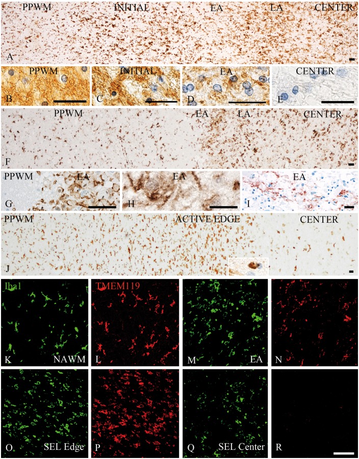
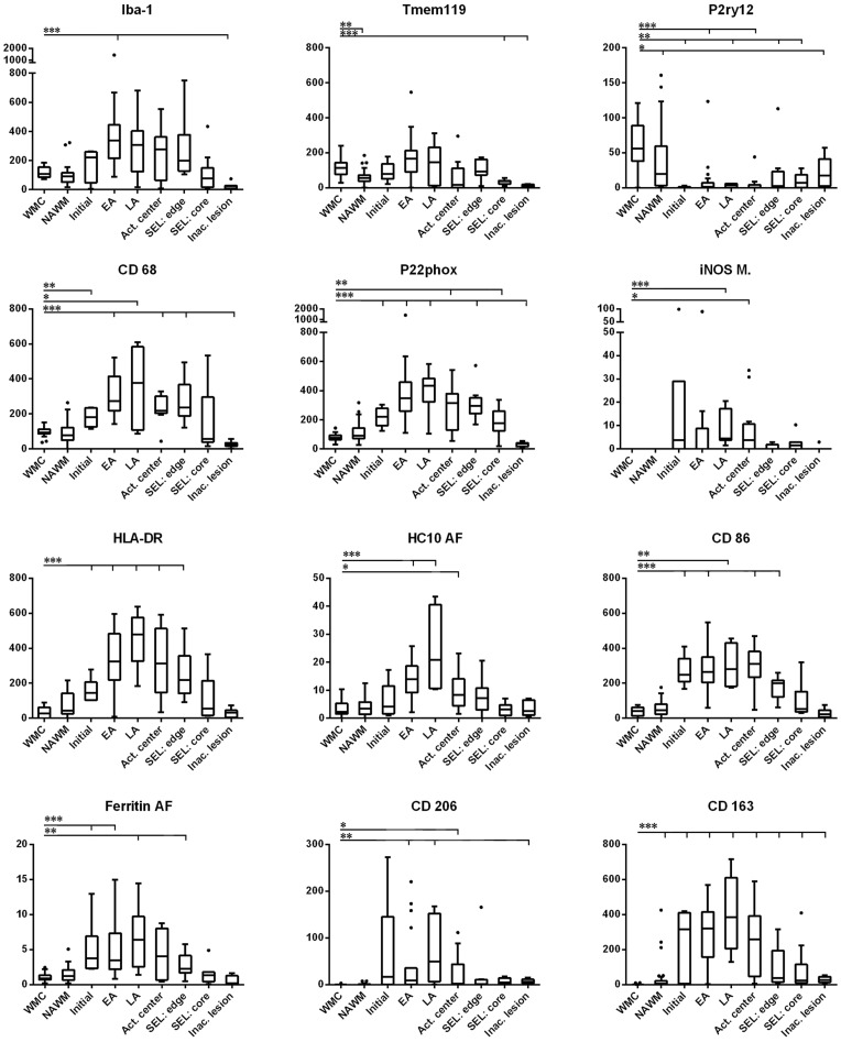
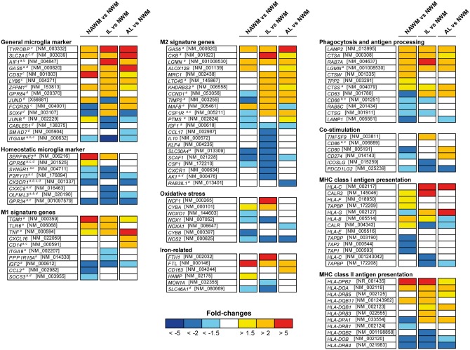
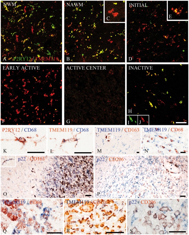
Microglia and macrophages accumulate at the sites of active demyelination and neurodegeneration in the multiple sclerosis brain and are thought to play a central role in the disease process. We used recently described markers to characterize the origin and functional states of microglia/macrophages in acute, relapsing and progressive multiple sclerosis. We found microglia activation in normal white matter of controls and that the degree of activation increased with age. This microglia activation was more pronounced in the normal-appearing white matter of patients in comparison to controls and increased with disease duration. In contrast to controls, the normal-appearing white matter of patients with multiple sclerosis showed a significant reduction of P2RY12, a marker expressed in homeostatic microglia in rodents, which was completely lost in active and slowly expanding lesions. Early stages of demyelination and neurodegeneration in active lesions contained microglia with a pro-inflammatory phenotype, which expressed molecules involved in phagocytosis, oxidative injury, antigen presentation and T cell co-stimulation. In later stages, the microglia and macrophages in active lesions changed to a phenotype that was intermediate between pro- and anti-inflammatory activation. In inactive lesions, the density of microglia/macrophages was significantly reduced and microglia in part converted to a P2RY12+ phenotype. Analysis of TMEM119, which is expressed on microglia but not on recruited macrophages, demonstrated that on average 45% of the macrophage-like cells in active lesions were derived from the resident microglia pool. Our study demonstrates the loss of the homeostatic microglial signature in active multiple sclerosis with restoration associated with disease inactivity.
Overview
The claims and statistics of this paper haven’t been checked yet.
Run the check — free, no account needed. It reads the open-access text and checks the sources it cites.
Integrity signals
Registry
Cites
Statistics
Figures
Verification
Claims the paper makes
The claims and statistics of this paper haven’t been checked yet.
Run the check — free, no account needed. It reads the open-access text and checks the sources it cites.
Claims other papers make about this one
Each is checked against this paper’s own text: does it say what the citing paper says it does?
The cited source doesn’t support the claim as written.
In the citing paper
“Similarly, microglia in active MS lesions lack Tmem119 (Satoh et al., 2016; Zrzavy et al., 2017).”
From Activated microglia drive demyelination via CSF1R signaling (2021) · cites this paper as Zrzavy et al., 2017
Checked by a reasoning model — may be wrong.
Experimental — accuracy not yet measuredThe source backs part of the claim, not all of it — or backs it for a different population, condition or comparison.
In the citing paper
“The loss of homeostatic microglial proteins is visible in immunofluorescence double-staining of IBA1 and TMEM119 in terminally sick mice and mock-infected healthy control brains, and shows the reciprocal regulation of both proteins in prion disease (Figure 5A) [55,73,88,89,90,91].”
From Loss of Homeostatic Microglia Signature in Prion Diseases (2022) · cites this paper as [90]
Experimental — accuracy not yet measuredThe source backs part of the claim, not all of it — or backs it for a different population, condition or comparison.
In the citing paper
“Microglial cells within active white matter lesions are characterized by the absence of homeostatic markers such as P2RY12 and an upregulation of CXCR4, indicating a shift to a disease-associated state (31, 41, 42).”
From The role of microglia in multiple sclerosis: implications for treatment with Bruton’s tyrosine kinase inhibitors (2025) · cites this paper as [41]
Experimental — accuracy not yet measuredThe source backs part of the claim, not all of it — or backs it for a different population, condition or comparison.
In the citing paper
“TMEM119 is one of the main homeostatic microglia markers that is not expressed on monocyte-derived macrophages and has been shown to be downregulated in active MS lesions and other inflammatory conditions of the brain47–49.”
From CHIT1 at diagnosis predicts faster disability progression and reflects early microglial activation in multiple sclerosis (2024) · cites this paper as [47]
Experimental — accuracy not yet measuredThe source backs part of the claim, not all of it — or backs it for a different population, condition or comparison.
In the citing paper
“Interestingly, the phenotype of microglia we observed in HDLS is similar to that found in an SOD1 ALS mouse model and in multiple sclerosis patients, in which the molecules associated with the homeostatic microglial phenotype (e.g., P2RY12, CSF-1R) are down-regulated and inflammatory markers are up-regulated [24, 57].”
From Loss of homeostatic microglial phenotype in CSF1R-related Leukoencephalopathy (2020) · cites this paper as [57]
Experimental — accuracy not yet measured
The source backs part of the claim, not all of it — or backs it for a different population, condition or comparison.
In the citing paper
“As reported previously (Zrzavy et al., 2017), P2RY12+ cells expressing TSPO were substantially reduced in active lesions (Fig. 2E–H).”
From A quantitative neuropathological assessment of translocator protein expression in multiple sclerosis (2019) · cites this paper as Zrzavy et al., 2017
Experimental — accuracy not yet measuredThe source backs part of the claim, not all of it — or backs it for a different population, condition or comparison.
In the citing paper
“Thus, in contrast to the pattern of TSPO expression in rodent models, it may not be so specifically associated with pro-inflammatory microglia in multiple sclerosis (Zrzavy et al., 2017).”
From A quantitative neuropathological assessment of translocator protein expression in multiple sclerosis (2019) · cites this paper as Zrzavy et al., 2017
Experimental — accuracy not yet measuredThe cited source says what the claim attributes to it.
In the citing paper
“Indeed, most phagocytes initially present in active MS lesions are microglia, and with lesion maturation, more macrophages will infiltrate from the blood47.”
From CHIT1 at diagnosis predicts faster disability progression and reflects early microglial activation in multiple sclerosis (2024) · cites this paper as [47]
The cited source says what the claim attributes to it.
In the citing paper
“TMEM119 is one of the main homeostatic microglia markers that is not expressed on monocyte-derived macrophages and has been shown to be downregulated in active MS lesions and other inflammatory conditions of the brain47–49.”
From CHIT1 at diagnosis predicts faster disability progression and reflects early microglial activation in multiple sclerosis (2024) · cites this paper as [47]
In the citing paper
“since its expression is maintained by resident human microglia in MS lesions and is not expressed by infiltrating immune cells (Zrzavy et al., 2017).”
From Complex cell-state changes revealed by single cell RNA sequencing of 76,149 microglia throughout the mouse lifespan and in the injured brain (2018) · cites this paper as Zrzavy et al., 2017
In the citing paper
“Several of these microglial genes were also observed in human microglia, including P2RY12 and TMEM119 (REFs25,33–36).”
From Microglial signatures and their role in health and disease (2018) · cites this paper as [36]
In the citing paper
“In human MS, microglia lose the expression of the P2RY12 homeostatic microglial marker and adopt a pro-inflammatory phenotype that includes the expression of phagocytic-related markers (macrosialin (also known as CD68)), antigen presentation markers (MHC class I and II molecules and T lymphocyte activation antigen CD86) and proteins involved in the production of reactive oxygen species (cytochrome”
From Microglial signatures and their role in health and disease (2018) · cites this paper as [36]
In the citing paper
“and appears to exhibit stable expression even in response to a variety of inflammatory conditions (9, 96).”
From The Transcriptional Landscape of Microglial Genes in Aging and Neurodegenerative Disease (2019) · cites this paper as [96]
In the citing paper
“Studies have shown that microglial migration, proliferation, and activation are involved in the onset and progression of disease in acute, relapsing, and progressive forms of MS in humans (Zrzavy et al. 2017).”
From MiR-124 and MiR-155 as Therapeutic Targets in Microglia-Mediated Inflammation in Multiple Sclerosis (2025) · cites this paper as Zrzavy et al. 2017
In the citing paper
“However, activation conditions are variable and microglial phenotype changes with disease progression (Zrzavy et al. 2017).”
From MiR-124 and MiR-155 as Therapeutic Targets in Microglia-Mediated Inflammation in Multiple Sclerosis (2025) · cites this paper as Zrzavy et al. 2017
Self-check
Percentages, sums and reported differences re-computed from the paper’s own numbers.
The self-check needs the paper’s full text.
Citations
Cites
Fate Mapping Analysis Reveals That Adult Microglia Derive from Primitive Macrophages
Ginhoux, F., Greter, M., Leboeuf, M., Nandi, S., See, P., Gokhan, S., Mehler, M. F., Conway, S. J., Ng, L. G., Stanley, E. R., et al. (2010) · Science · doi:10.1126/science.1194637 · Cited by 5,175
The M1 and M2 paradigm of macrophage activation: Time for reassessment
Martinez, F. O., & Gordon, S. (2014) · F1000Prime Reports · doi:10.12703/p6-13 · Cited by 4,689
Kettenmann, H., Hanisch, U.-K., Noda, M., & Verkhratsky, A. (2011) · Physiological Reviews · doi:10.1152/physrev.00011.2010 · Cited by 3,547
Identification of a unique TGF-β–dependent molecular and functional signature in microglia
Butovsky, O., Jedrychowski, M. P., Moore, C. S., Cialic, R., Lanser, A. J., Gabriely, G., Koeglsperger, T., Dake, B., Wu, P. M., Doykan, C. E., et al. (2014) · Nature Neuroscience · doi:10.1038/nn.3599 · Cited by 2,543
Self-citationthe Immunological Genome Consortium, Gautier, E. L., Shay, T., Miller, J., Greter, M., Jakubzick, C., Ivanov, S., Helft, J., Chow, A., Elpek, K. G., et al. (2012) · Nature Immunology · doi:10.1038/ni.2419 · Cited by 1,972 · cited 1×
“…to be microglia-specific in comparison to other brain cells (Chiu et al., 2013; Hickman et al., 2013) or in comparison to peripheral myeloid cells (Gautier et al., 2012; Hickman et al., 2013; Butovsky et al., 2014) or to be involved in pro- or anti-inflammatory microglia/macrophage function such…”
BackgroundNew tools for studying microglia in the mouse and human CNS
Bennett, M. L., Bennett, F. C., Liddelow, S. A., Ajami, B., Zamanian, J. L., Fernhoff, N. B., Mulinyawe, S. B., Bohlen, C. J., Adil, A., Tucker, A., et al. (2016) · Proceedings of the National Academy of Sciences · doi:10.1073/pnas.1525528113 · Cited by 1,804
M2 microglia and macrophages drive oligodendrocyte differentiation during CNS remyelination
Miron, V. E., Boyd, A., Zhao, J.-W., Yuen, T. J., Ruckh, J. M., Shadrach, J. L., van Wijngaarden, P., Wagers, A. J., Williams, A., Franklin, R. J. M., et al. (2013) · Nature Neuroscience · doi:10.1038/nn.3469 · Cited by 1,777 · cited 1×
“It has been suggested to stimulate remyelination (Butovsky et al., 2006; Miron et al., 2013) and early stages of remyelination within multiple sclerosis lesions are mainly seen in the core of active lesions (Raine et al., 1981; Prineas et al., 1993; Raine, 2017).”
A polarizing question: do M1 and M2 microglia exist?
Ransohoff, R. M. (2016) · Nature Neuroscience · doi:10.1038/nn.4338 · Cited by 1,573
Microglia in neurodegenerative disease
Perry, V. H., Nicoll, J. A. R., & Holmes, C. (2010) · Nature Reviews Neurology · doi:10.1038/nrneurol.2010.17 · Cited by 1,543
Axonal damage in acute multiple sclerosis lesions
Ferguson, B. (1997) · Brain · doi:10.1093/brain/120.3.393 · Cited by 1,532
Research works printed but not linked: 2
- Babinski, 1885, Recherches sur l’anatomie pathologique de la sclerose en plaque et étude comparative des diverses variétés de la scleroses de la moelle, Arch Physiol, 5, 186
- Marburg, 1906, Die sogenannte akute multiple Sklerose, J Psychiatric Neurol, 27, 1
Cited by
Sorted by citations
Items 501–600 of 698
Müller, M., Pavlović, I., Wiberg, A., & Burman, J. (2025) · Frontiers in Immunology · doi:10.3389/fimmu.2025.1601223 · Cited by 3
Zhang, Q.-M., Chen, Y., Xing, Y.-Y., Yang, M., Li, N., Jiang, X., Gao, H., Lu, S., & Yao, J. (2025) · Molecular Psychiatry · doi:10.1038/s41380-025-03139-1 · Cited by 4
Prajjwal, P., Patel, P. V., Patel, Z. V., Inban, P., Patel, D., Mahato, P., Shamim, L., Godinho, N. J. S., & Tekuru, Y. (2025) · Disease-a-Month · doi:10.1016/j.disamonth.2025.102012 · Cited by 3
Victoriano, G. M., Zhu, Y., Kostoglou, Y., Walker, A. K., Middleton, F. A., Massa, P. T., Eggen, B. J. L., Webster, M. J., Sommer, I. E., & Weickert, C. S. (2026) · Brain Behavior and Immunity · doi:10.1016/j.bbi.2026.106276 · Cited by 5
Kuo, R., Carlson, M. L., Reyes, S. T., Nagy, S. C., Kalita, M., Alam, I. S., Malik, N., Jackson, I. M., Acosta, C., Falk, I. N., et al. (2026) · Nature Communications · doi:10.1038/s41467-025-67907-x · Cited by 3
Candia, Y. A., Aladyeva, E., Boyden, W., Bollman, B., Rogerson‐Wood, L., Davies, D., Affleck, A. J., Buckland, M. E., Harari, O., Piccio, L. M., et al. (2026) · Acta Neuropathologica Communications · doi:10.1186/s40478-026-02241-x · Cited by 3
Leung, W. L., Dill, L. K., Perucca, P., O'Brien, T. J., Casillas-Espinosa, P. M., & Semple, B. D. (2023) · Journal of Neurotrauma · doi:10.1089/neu.2022.0463 · Cited by 3
Peripherally derived myeloid cells induce disease-dependent phenotypic changes in microglia
Thougaard, E., Carney, B. N., Wlodarczyk, A., Brambilla, R., & Lambertsen, K. (2023) · Frontiers in Cellular Neuroscience · doi:10.3389/fncel.2023.1295840 · Cited by 3
Dissecting surveying behavior of reactive microglia under chronic neurodegeneration
Subhramanian, S., Bocharova, O. V., Makarava, N., Safadi, T., & Baskakov, I. V. (2026) · eLife · doi:10.7554/elife.107650.3 · Cited by 3
Multiple Sclerosis - Genetics, Disease Mechanisms and Clinical Developments
Kishore, U., & Shastri, A. (2022) · IntechOpen eBooks · doi:10.5772/intechopen.98110 · Cited by 3
Pistolesi, A., Ranieri, G., Calvani, M., Guasti, D., Chiarugi, A., & Buonvicino, D. (2024) · Experimental Neurology · doi:10.1016/j.expneurol.2024.115095 · Cited by 4
Neuroinflammation: 2021 update
Lassmann, H. (2020) · Free Neuropathology · doi:10.17879/freeneuropathology-2021-3166 · Cited by 3
Self-citationLes vésicules extracellulaires
Leroux, É., Perbet, R., Buée, L., & Colin, M. (2021) · médecine/sciences · doi:10.1051/medsci/2021205 · Cited by 4
Coyle, P. K. (2019) · Current clinical neurology · doi:10.1007/978-3-030-24436-1_3 · Cited by 3
Fransson, J., Bachelin, C., Deknuydt, F., Ichou, F., Guillot‐Noël, L., Ponnaiah, M., Gloaguen, A., Maillart, É., Stankoff, B., Tenenhaus, A., et al. (2021) · bioRxiv (Cold Spring Harbor Laboratory) · doi:10.1101/2021.01.13.426327 · Cited by 3
Berki, P., Cserép, C., Pósfai, B., Szabadits, E., Környei, Z., Kellermayer, A., Nyerges, M., Wei, X., Módy, I., Araki, K., et al. (2022) · bioRxiv (Cold Spring Harbor Laboratory) · doi:10.1101/2022.04.12.487998 · Cited by 3
Makarava, N., Safadi, T., Bocharova, O. V., Mychko, O., Pandit, N. P., Molesworth, K., Eyo, U. B., & Baskakov, I. V. (2025) · bioRxiv (Cold Spring Harbor Laboratory) · doi:10.1101/2025.04.07.647619 · Cited by 3
Innate Immunity and Parkinson's DiseasePreprint
Oludipe, D. B., Du, X., Akter, S., Zhang, C., Mosley, R. L., Gendelman, H. E., & Sil, S. (2025) · Cold Spring Harbor Perspectives in Medicine · doi:10.1101/cshperspect.a041640 · Cited by 3
Mitochondrial complex I: the key to sustained microglia activation and neuroinflammation maintenance
Wang, H., Yu, S.-Y., Nielsen, S., Wang, X., & Zhao, W.-W. (2024) · Military Medical Research · doi:10.1186/s40779-024-00554-3 · Cited by 2
Microglia-derived sEV: Friend or foe in the pathogenesis of cognitive impairment
Chen, L., & Wang, W. (2025) · Progress in Neuro-Psychopharmacology and Biological Psychiatry · doi:10.1016/j.pnpbp.2025.111287 · Cited by 3
Alatawi, Z. (2025) · Developmental Neurobiology · doi:10.1002/dneu.23014 · Cited by 2
Multiple sclerosis: Glial cell activation – Biomarker advances and their translational significance
Zeng, W., Wu, H., & Huang, Y. (2025) · Science Progress · doi:10.1177/00368504251390710 · Cited by 2
Laloli, K. J., Rentsch, P., Stayte, S., & Vissel, B. (2025) · Frontiers in Pharmacology · doi:10.3389/fphar.2025.1682979 · Cited by 3
Su, L., Tian, M., Hao, Y., Zhang, X., Liu, X., Liu, L., & Yao, E. (2026) · Progress in Neurobiology · doi:10.1016/j.pneurobio.2026.102911 · Cited by 2
Fowler, D. K., Savage, T. M., & Mackie, D. I. (2026) · Journal of Cannabis Research · doi:10.1186/s42238-026-00445-5 · Cited by 3
Microglial physiology and function
Verkhratsky, A., & Butt, A. M. (2023) · Elsevier eBooks · doi:10.1016/b978-0-12-821565-4.00002-x · Cited by 2
Microglia: a potential target for neurological disorder
Parajuli, B., & Koizumi, S. (2023) · Folia Pharmacologica Japonica · doi:10.1254/fpj.23005 · Cited by 2
Gushchina, S. V., Bo, K., Балашов, В. П., Pankov, V. G., & Balashov, A. V. (2018) · MORPHOLOGICAL NEWSLETTER · doi:10.20340/mv-mn.18(26).02.8-11 · Cited by 2
Role of A1 Adenosinergic System in Multiple Sclerosis and Possible Therapeutic Strategy
Justin, A., Basavaraj, M., Murugan, D., Rao, G. N., & Jeyabalan, J. B. (2022) · Research Journal of Pharmacy and Technology · doi:10.52711/0974-360x.2022.00505 · Cited by 2
Biswas, B. K., & Goswami, R. (2021) · Meta Gene · doi:10.1016/j.mgene.2021.100886 · Cited by 2
Davalos, D., Akassoglou, K., & Cardona, A. E. (2020) · Elsevier eBooks · doi:10.1016/b978-0-12-814405-3.00041-2 · Cited by 3
Hwang, D., Seyedsadr, M., Ishikawa, L. L. W., Boehm, A., Sahin, Z., Casella, G., Jang, S., Gonzalez, M. V., Garifallou, J. P., Hakonarson, H. H., et al. (2020) · bioRxiv (Cold Spring Harbor Laboratory) · doi:10.1101/2020.10.23.352534 · Cited by 2
Das, R. R., & Chinnathambi, S. (2021) · SSRN Electronic Journal · doi:10.2139/ssrn.3919950 · Cited by 2
Aberrant iron deposition in the multiple sclerosis spinal cord relates to neurodegenerationPreprint
Pisa, M., Lockhart, A., Angell, T., Avery, A., Waldman, A. D., Rodriguez, C., Duarte, T. L., Pansieri, J., Dhari, Z., Bailey, M. A., et al. (2024) · bioRxiv (Cold Spring Harbor Laboratory) · doi:10.1101/2024.10.25.619794 · Cited by 2
Self-citationInsights and challenges on animal models for progressive multiple sclerosis
Nalepa, I. F., Haahr, N. O., Bekiaris, V., Jakobsen, M. R., Asuni, A. A., & Benmamar-Badel, A. (2026) · Journal of Neuroinflammation · doi:10.1186/s12974-026-03805-3 · Cited by 1
“Of CNS resident cells, both activated microglia and astrocytes have been strongly implicated in MS pathology, even though astrocytes are less extensively studied [41–44].”
Specificities of Living Human Microglial Cells
Milior, G., Chiano, M. D., & Morin‐Brureau, M. (2024) · Advances in neurobiology · doi:10.1007/978-3-031-55529-9_31 · Cited by 1
The role of vitamin D in multiple sclerosis and its mechanisms of action
Kowalczyk, A., Kiełbasa, J., Przestrzelska, M., Rykucka, A., Wąs, M., Zozula, N., Tomczewska, Z., Bil, K., Ślesicka, I., & Latała, A. (2024) · Quality in Sport · doi:10.12775/qs.2024.19.54188 · Cited by 1
Knockdown of BMP7 induced oligodendrocyte apoptosis, demyelination and motor function loss
Wei, X., Liu, S., Chen, K., Wang, M., Wang, Y., Zou, D., & Xiao, Y. (2024) · Molecular and Cellular Neuroscience · doi:10.1016/j.mcn.2024.103973 · Cited by 1
Zhou, L., Wu, Z., Yi, X., Xie, D., Wang, J., & Wu, W. (2025) · Biochimica et Biophysica Acta (BBA) - Molecular Cell Research · doi:10.1016/j.bbamcr.2025.119905 · Cited by 1
Demyelination and Remyelination: General Principles
Niu, J., Verkhratsky, A., Butt, A. M., & Yi, C. (2025) · Advances in neurobiology · doi:10.1007/978-3-031-87919-7_9 · Cited by 2
Sun, L., Feng, L., Zhang, Y., Shan, M., Tong, C., Zhang, K., Zheng, J., & Fu, X. (2025) · Brain Research Bulletin · doi:10.1016/j.brainresbull.2025.111564 · Cited by 1
Innate Immunity in the Pathogenesis of Selected Autoimmune Neurological Diseases
Rudnicka-Czerwiec, J., & Bartosik-Psujek, H. (2025) · Journal of Clinical Medicine · doi:10.3390/jcm14207235 · Cited by 1
Chang, Y., Wu, C.-C., Hsiao, F.-T., Lin, Y.-S., Lai, T., Huang, K.-H., Tang, S., Lee, C., Chou, S., & Hsu, L.-C. (2025) · Journal of Neuroinflammation · doi:10.1186/s12974-025-03550-z · Cited by 1
Modulation of axon excitable domains by astrocytes and microglia
Garrido, J. J. (2026) · Frontiers in Cellular Neuroscience · doi:10.3389/fncel.2025.1749755 · Cited by 1
Wang, Y., Li, X., Li, W., Wang, C., Xu, G., & Yu, S. (2026) · Experimental Neurology · doi:10.1016/j.expneurol.2026.115662 · Cited by 1
Otálora-Alcaraz, A., Sun, M. C., Hofman, N., Costelloe, L., KEARNEY, H. F., Prenderville, J. A., & Downer, E. J. (2026) · Multiple Sclerosis and Related Disorders · doi:10.1016/j.msard.2026.107040 · Cited by 1
Microglial Senescence in Neurodegenerative Diseases: Mechanisms and Therapeutic Perspectives
Liu, Y., Hu, Z., Xiao, Y., Han, X., Cheng, X., & Ye, T. (2026) · ACS Chemical Neuroscience · doi:10.1021/acschemneuro.6c00016 · Cited by 1
Viscidi, R. P., Huang, J., Li, Y., Severance, E. G., & Xiao, J. (2026) · Scientific Reports · doi:10.1038/s41598-026-49283-8 · Cited by 1
McMahan, R. H., Evans, M. R., Najarro, K. M., Frank, D. N., Basak, J. M., & Kovacs, E. J. (2026) · Alcohol · doi:10.1016/j.alcohol.2026.04.007 · Cited by 1
Disease-associated RNA and protein signatures in iPSC-derived microglia model of Alzheimer’s disease
Wu, W., Choi, E. S., Liu, L., Thamilselvan, V., LI, L., Rippee‐Brooks, M. D., Khatkar, K., Li, D.-Y., McGrath, D., Manning, A. C., et al. (2026) · Frontiers in Neuroscience · doi:10.3389/fnins.2026.1799542 · Cited by 1
Koyya, P., Alavilli, H., Pandrangi, S. L., & Manthari, R. K. (2026) · Immunological Investigations · doi:10.1080/08820139.2026.2702083 · Cited by 1
Nanomedicine Strategies to Ameliorate Neuroinflammation and Prevent Secondary Neural Tissue Damage
Hegab, R. A., & Peppas, N. A. (2026) · ACS Nano Medicine · doi:10.1021/acsnanomed.6c00120 · Cited by 2
Immune-mediated excitotoxicity in brain disorders
Shin, J., Vogelaar, C. F., Bittner, S., & Zipp, F. (2026) · Nature reviews. Immunology · doi:10.1038/s41577-026-01348-5 · Cited by 1
Intranasal HSV‑1 infection drives region‑specific interferon‑dominant microglial remodeling
Frietze, S. E., Lunn, C., Oldham, D., Boyd, J. R., Bubak, A. N., Bustillos, A., Nagel, M. A., Restrepo, D., Bruce, K. D., & Niemeyer, C. S. (2026) · PLoS Pathogens · doi:10.1371/journal.ppat.1014684 · Cited by 1
NEURO PROTECTIVE EFFECTS OF WHEAT GERM OIL ON THE BRAIN INFLAMMATION INDUCED BY METHOTREXATE IN MICE
Hussein, R., & Radwan, O. K. (2019) · International Research Journal of Pharmacy · doi:10.7897/2230-8407.1012330 · Cited by 1
DAMPs in Organ-Specific Autoimmune Diseases
Land, W. G. (2023) · Springer eBooks · doi:10.1007/978-3-031-21776-0_8 · Cited by 1
Kasarełło, K., Seta, M., Sulejczak, D., Snarski, E., & Cudnoch‐Jędrzejewska, A. (2023) · Archivum Immunologiae et Therapiae Experimentalis · doi:10.1007/s00005-023-00675-y · Cited by 1
Jakimovski, D., Weinstock‐Guttman, B., & Zivadinov, R. (2023) · Elsevier eBooks · doi:10.1016/b978-0-443-18578-6.00010-6 · Cited by 1
Al-Hroub, H., Musawi, H. A. M. A., Abdelrahman, A., Namasivayam, V., Corbel, M., Petit, F., Aksiyote, G., Beau, F., Jan, C., Bemelmans, A., et al. (2025) · Journal of Medicinal Chemistry · doi:10.1021/acs.jmedchem.5c00213 · Cited by 1
Pincemail, E., Peyronneau, M., Denis, C., Bensalah, H., Corbel, M., Goutal, S., Goislard, M., Al-Hroub, H., Théry, M., Beuché, S., et al. (2025) · ACS Chemical Neuroscience · doi:10.1021/acschemneuro.5c00187 · Cited by 1
Myeloid Cells in Multiple Sclerosis
Wang, M., Caryotakis, S. E., Kumar, N., Nguyen, A. V., & Soulika, A. M. (2019) · IntechOpen eBooks · doi:10.5772/intechopen.86285 · Cited by 2
Epstein–Barr virus and multiple sclerosis
Skliar, A. I., Torianyk, I., Осолодченко, Т. П., & Ponomarenko, S. V. (2020) · Pathologia · doi:10.14739/2310-1237.2020.3.221870 · Cited by 3
Microglia and border-associated macrophages in the central nervous system
Zanluqui, N. G., Polonio, C. M., de Oliveira, M. G., de Oliveira, L. G., Faria, L. C., & Peron, J. P. S. (2022) · Elsevier eBooks · doi:10.1016/b978-0-12-821385-8.00013-5 · Cited by 1
Glial Cells in Neuroinflammation in Various Disease States
Barthels, D., & Das, H. (2022) · Springer eBooks · doi:10.1007/978-981-19-2655-6_39 · Cited by 1
Ramos, K. A., Soares, I. G. M., Oliveira, L. M. A., Bragaa, M. A., Soares, P. P. C., Scherrer, E. C., Silva, F. D. S., Lima, N. M., de Castro, S. B. R., Alves, C. C. D. S., et al. (2023) · SSRN Electronic Journal · doi:10.2139/ssrn.4564796 · Cited by 1
Mehrabani-Tabari, A. A., Hegdekar, N., Herb, B. R., Kachi, S. A., Sarkar, C., Thapa, S., Nguyen, D. P., Morel, Y., Weldemariam, M. M., Muller, L., et al. (2025) · bioRxiv (Cold Spring Harbor Laboratory) · doi:10.1101/2025.09.04.674092 · Cited by 2
Neuronal activity promotes repair by microglial reprogramming following demyelinationPreprint
Perrot, C., Ronzano, R., Li, Z., Aigrot, M., Pantazou, V., Stheneur, P., Lejeune, F., Zalc, B., Quintana, F. J., Stankoff, B., et al. (2025) · bioRxiv (Cold Spring Harbor Laboratory) · doi:10.1101/2025.11.19.689194 · Cited by 2
Modeling the Multiple Sclerosis Brain Disease Using Agents: What Works and What Doesn't?Preprint
Muqaddas, A., & Niazi, M. A. (2017) · arXiv (Cornell University) · doi:10.48550/arxiv.1712.00190
On the role of macrophages, microglia and the extracellular matrix in remyelination
Wang, P. (2018) · University of Groningen research database (University of Groningen / Centre for Information Technology)
Much, if not all, of the cortical damage in MS can be attributed to the microglial cell – Commentary
Spencer, J. I., & DeLuca, G. C. (2018) · Multiple Sclerosis Journal · doi:10.1177/1352458518775549
Anuradha, S., Satyanarayana, C., & Srinivas, Y. (2018) · International Journal of Image Graphics and Signal Processing · doi:10.5815/ijigsp.2018.08.06
Kurt Jellinger 90: his contribution to neuroimmunology
Dal‐Bianco, A., Höftberger, R., Lassmann, H., & Berger, T. (2021) · Journal of Neural Transmission · doi:10.1007/s00702-021-02358-4
Self-citationHwang, D., Seyedsadr, M., Ishikawa, L. L. W., Boehm, A., Sahin, Z., Casella, G., Jang, S., Gonzalez, M. V., Garifallo, J. P., Hakonarson, H. H., et al. (2022) · The Journal of Immunology · doi:10.4049/jimmunol.208.supp.60.14
Neuroinflammation: 2022 update
Lassmann, H. (2022) · Free Neuropathology · doi:10.17879/freeneuropathology-2022-3790 · Cited by 1
Self-citationHe, X., Zhao, Y., Li, G., Luo, J., Xu, J., Zheng, H., Zhang, L., & Hu, X. (2022) · SSRN Electronic Journal · doi:10.2139/ssrn.4072048
Timmerman, R. (2022) · Utrecht University Repository (Utrecht University) · doi:10.33540/1603
Mikroglia-/Makrophagenphänotypen bei humanen Entmarkungserkrankungen (MS, NMO und PML)
Nau-Gietz, C. A. (2022) · eDiss (Georg-August-Universität Göttingen) · doi:10.53846/goediss-9627
Immunopathology and Pathogenesis
Zettl, U. K., & Patejdl, R. (2023) · Springer eBooks · doi:10.1007/978-3-031-13498-2_4
Exercise Improved P2Y12-Regulated Microglial Dynamics During Stroke Via Endocannabinoid SignalingPreprint
He, X., Zhao, Y., Li, G., Luo, J., Xu, J., Zheng, H., Zhang, L., & Hu, X. (2023) · bioRxiv (Cold Spring Harbor Laboratory) · doi:10.1101/2023.04.24.538065
(2023) · Advances in magnetic resonance technology and applications · doi:10.1016/b978-0-323-91771-1.00006-x
Innate Immunopathological Mechanisms in Multiple Sclerosis
Shastri, A., Singh, I., & Kishore, U. (2023) · IntechOpen eBooks · doi:10.5772/intechopen.110626
Effect of Tinospora cordifolia on neuroinflammation
Dhote, V. V., Kilor, V. A., Raja, M. K. M. M., Singhai, A., Mandloi, A. S., & Upaganlawar, A. B. (2023) · Elsevier eBooks · doi:10.1016/b978-0-323-90052-2.00019-6
Imagem PET de processos relacionados à esclerose múltipla: estudo clínico
Pitombeira, M. S. (2022) · Digital Library of Theses and Dissertations (Universidade de São Paulo) · doi:10.11606/t.5.2022.tde-20042023-115057
Magnani, L., Fusar‐Poli, L., Parise, A., Nguyen, K. D., Saverino, D., & Costanza, A. (2023) · Medical Hypotheses · doi:10.1016/j.mehy.2023.111158
Research Progress on the Dual Role of Microglia in Neurological Diseases
赵, 璐. (2023) · Advances in Clinical Medicine · doi:10.12677/acm.2023.13102295
Etebar, F. (2023) · Queensland University of Technology · doi:10.5204/thesis.eprints.244774
Additional file 11 of Epigenetic regulation of innate immune memory in microglia
Zhang, X., Kracht, L., Lerário, A. M., Dubbelaar, M. L., Brouwer, N., Wesseling, E. M., Boddeke, E., Eggen, B. J. L., & Kooistra, S. M. (2022) · Figshare · doi:10.6084/m9.figshare.19768066
Additional file 6 of Epigenetic regulation of innate immune memory in microglia
Zhang, X., Kracht, L., Lerário, A. M., Dubbelaar, M. L., Brouwer, N., Wesseling, E. M., Boddeke, E., Eggen, B. J. L., & Kooistra, S. M. (2022) · Figshare · doi:10.6084/m9.figshare.19768093
Additional file 13 of Epigenetic regulation of innate immune memory in microglia
Zhang, X., Kracht, L., Lerário, A. M., Dubbelaar, M. L., Brouwer, N., Wesseling, E. M., Boddeke, E., Eggen, B. J. L., & Kooistra, S. M. (2022) · Figshare · doi:10.6084/m9.figshare.19768072
Additional file 14 of Epigenetic regulation of innate immune memory in microglia
Zhang, X., Kracht, L., Lerário, A. M., Dubbelaar, M. L., Brouwer, N., Wesseling, E. M., Boddeke, E., Eggen, B. J. L., & Kooistra, S. M. (2022) · Figshare · doi:10.6084/m9.figshare.19768075
Li, S., Sakurai, K., Ohgidani, M., Kato, T. A., & Hikida, T. (2023) · Figshare · doi:10.6084/m9.figshare.23672669
Additional file 8 of Epigenetic regulation of innate immune memory in microglia
Zhang, X., Kracht, L., Lerário, A. M., Dubbelaar, M. L., Brouwer, N., Wesseling, E. M., Boddeke, E., Eggen, B. J. L., & Kooistra, S. M. (2022) · Figshare · doi:10.6084/m9.figshare.19768099
Li, S., Sakurai, K., Ohgidani, M., Kato, T. A., & Hikida, T. (2023) · Figshare · doi:10.6084/m9.figshare.23672672
Additional file 10 of Epigenetic regulation of innate immune memory in microglia
Zhang, X., Kracht, L., Lerário, A. M., Dubbelaar, M. L., Brouwer, N., Wesseling, E. M., Boddeke, E., Eggen, B. J. L., & Kooistra, S. M. (2022) · Figshare · doi:10.6084/m9.figshare.19768063
Additional file 9 of Epigenetic regulation of innate immune memory in microglia
Zhang, X., Kracht, L., Lerário, A. M., Dubbelaar, M. L., Brouwer, N., Wesseling, E. M., Boddeke, E., Eggen, B. J. L., & Kooistra, S. M. (2022) · Figshare · doi:10.6084/m9.figshare.19768102
Additional file 7 of Epigenetic regulation of innate immune memory in microglia
Zhang, X., Kracht, L., Lerário, A. M., Dubbelaar, M. L., Brouwer, N., Wesseling, E. M., Boddeke, E., Eggen, B. J. L., & Kooistra, S. M. (2022) · Figshare · doi:10.6084/m9.figshare.19768096
Additional file 5 of Epigenetic regulation of innate immune memory in microglia
Zhang, X., Kracht, L., Lerário, A. M., Dubbelaar, M. L., Brouwer, N., Wesseling, E. M., Boddeke, E., Eggen, B. J. L., & Kooistra, S. M. (2022) · Figshare · doi:10.6084/m9.figshare.19768090
Additional file 12 of Epigenetic regulation of innate immune memory in microglia
Zhang, X., Kracht, L., Lerário, A. M., Dubbelaar, M. L., Brouwer, N., Wesseling, E. M., Boddeke, E., Eggen, B. J. L., & Kooistra, S. M. (2022) · Figshare · doi:10.6084/m9.figshare.19768069
Li, S., Sakurai, K., Ohgidani, M., Kato, T. A., & Hikida, T. (2023) · Figshare · doi:10.6084/m9.figshare.23672666
Lyu, X.-Y., Li, J., Ding, S., Hu, J., & Lü, H. (2023) · Nature Cell and Science · doi:10.61474/ncs.2023.00004
Metadata
- Authors
- Zrzavy, T., Hametner, S., Wimmer, I., Butovsky, O., Weiner, H. L., & Lassmann, H.
- Affiliations
- Medical University of Vienna (AT)
- Brigham and Women's Hospital (US)
- Corresponding author
- Lassmann, Hans
- Venue
- Brain
- Publisher
- Oxford University Press (OUP)
- Published
- 2017-05-24
- Volume
- 140
- Issue
- 7
- Pages
- 1900-1913
- ISSN
- 0006-8950 (ISSN-L)
- 1460-2156
- PMC
- PMC6057548
- OpenAlex
- W2620266814
- Cited by
- 684
- References
- 68
- Citations by year
- 2026: 62 · 2025: 65 · 2024: 78 · 2023: 87 · 2022: 92 · 2021: 116 · 2020: 71 · 2019: 60 · 2018: 43 · 2017: 10
- FWCI
- 39.52
- Type
- journal article
- Language
- en
- Open access
- hybrid
- doi.org
- Licence
- Locations
- Brain (publishedVersion, cc-by-nc)
- Europe PMC (PubMed Central) (publishedVersion, cc-by-nc)
- PubMed Central (publishedVersion, cc-by-nc)
- PubMed (publishedVersion)
- Funders
- NIH (R01NS088137)
- National Multiple Sclerosis Society (5092A1)
- Topics
- Health Sciences › Medicine › Clinical Neurology › Multiple Sclerosis Research Studies
- Life Sciences › Neuroscience › Neurology › Neuroinflammation and Neurodegeneration Mechanisms
- Life Sciences › Biochemistry, Genetics and Molecular Biology › Molecular Biology › Inflammasome and immune disorders
- Keywords
- multiple sclerosis, microglia, macrophages, homeostatic microglia, microglial activation, P2Y12 receptor, TMEM119, normal-appearing white matter, demyelination, neurodegeneration, lesion activity, disease duration, pro-inflammatory phenotype, anti-inflammatory phenotype, phagocytosis, oxidative damage, antigen presentation, T cell costimulation, microglial origin
- MeSH terms
- Adult, Aged, Aged, 80 and over, Aging, Demyelinating Diseases, Female, Homeostasis, Humans, Macrophage Activation, Male, Membrane Proteins, Middle Aged, Multiple Sclerosis, Nerve Degeneration, Case-Control Studies, Microglia, Receptors, Purinergic P2Y12, White Matter
About these checks
Registry, retraction, Cites and statistics results are computed, not judged, with one exception: a printed reference that no index lists is read once by a model before it counts as a citation error. Claims are extracted by a model and kept only when their quote appears verbatim in the text; each cited source is then checked, with a model’s help, for whether it says what the claim relies on. Those verdicts can be wrong — read the claim’s quote and the cited source before relying on one.
Statistics and arithmetic are recomputed, not judged.
Confidence bands are shown only for fields calibrated on our annotated test set. None is calibrated yet, so no band is shown.
What counts as a claim ›How we measure confidence ›How we check this ›
Are you an author, or do you see a problem with this page? Request a review
