

The DOI matches its Crossref record; 84 of the 84 references it cites were found in a registry or an index.
Organization of the Human Inferior Parietal Lobule Based on Receptor Architectonics
Open accessAbstract
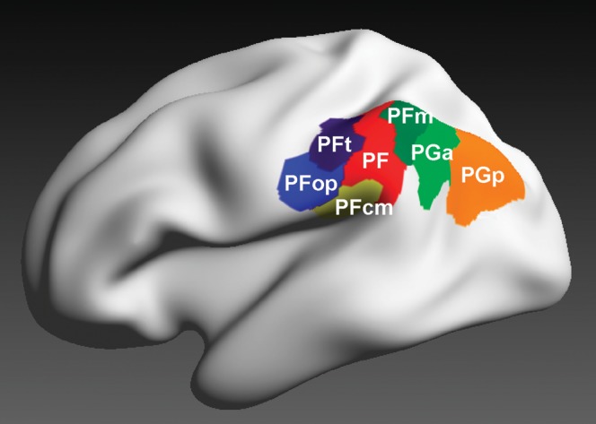
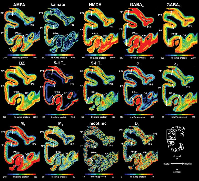
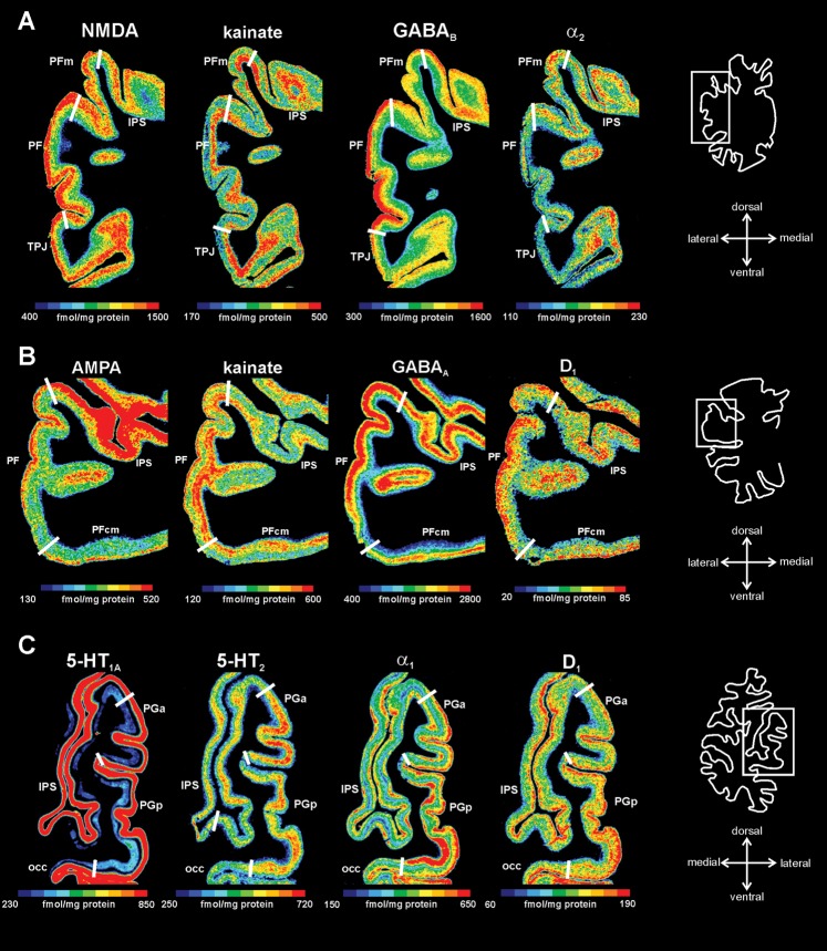
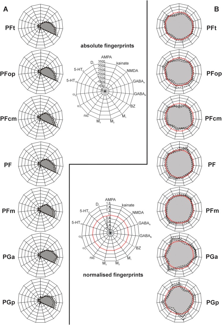
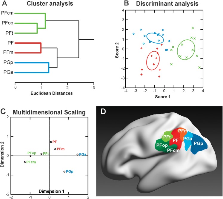
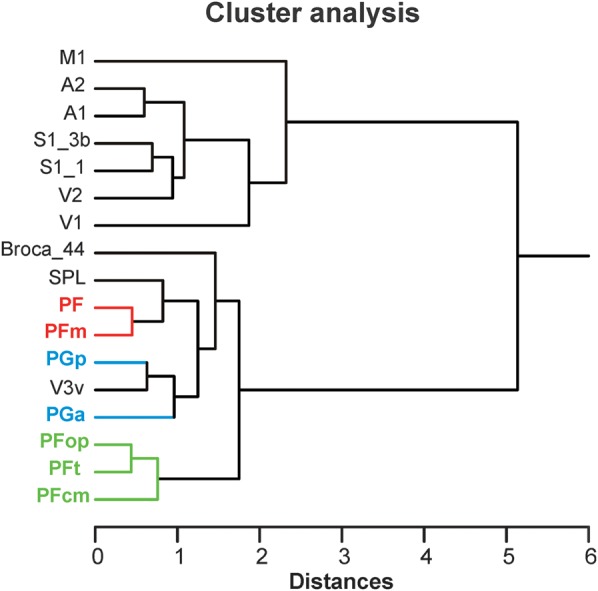
Human inferior parietal lobule (IPL) plays a key role in various cognitive functions. Its functional diversity, including attention, language, and action processing, is reflected by its structural segregation into 7 cytoarchitectonically distinct areas, each with characteristic connectivity patterns. We hypothesized that commonalities of the cytoarchitectonic, connectional, and functional diversity of the IPL should be reflected by a correlated transmitter receptor-based organization. Since the function of a cortical area requires a well-tuned receptor balance, the densities of 15 different receptors were measured in each IPL area. A hierarchical cluster analysis of the receptor balance revealed a tripartite segregation of the IPL into a rostral, middle, and caudal group. Comparison with other cortical areas showed strong similarities with Broca's region for all 3 groups, with the superior parietal cortex for the middle, and with extrastriate visual areas for the caudal group. Notably, caudal-most area PGp has a receptor fingerprint very similar to that of ventral extrastriate visual cortex. We therefore propose a new organizational model of the human IPL, consisting of 3 clusters, which corresponds to its known cytoarchitectonic, connectional, and functional diversity at the molecular level. This might reflect a general organizational principle of human IPL, beyond specific functional domains.
Overview
The claims and statistics of this paper haven’t been checked yet.
Run the check — free, no account needed. It reads the open-access text and checks the sources it cites.
Integrity signals
Registry
Cites
Statistics
Figures
Verification
Claims the paper makes
The claims and statistics of this paper haven’t been checked yet.
Run the check — free, no account needed. It reads the open-access text and checks the sources it cites.
Self-check
Percentages, sums and reported differences re-computed from the paper’s own numbers.
The self-check needs the paper’s full text.
Citations
Cites
Control of goal-directed and stimulus-driven attention in the brain
Corbetta, M., & Shulman, G. L. (2002) · Nature Reviews Neuroscience · doi:10.1038/nrn755 · Cited by 12,881
Separate visual pathways for perception and action
Goodale, M. A., & Milner, A. D. (1992) · Trends in Neurosciences · doi:10.1016/0166-2236(92)90344-8 · Cited by 6,519
Ungerleider, L. G. (1982) · Medical Entomology and Zoology · Cited by 4,531
The reorienting system of the human brain: from environment to theory of mind
Corbetta, M., Patel, G., & Shulman, G. L. (2008) · Neuron · doi:10.1016/j.neuron.2008.04.017 · Cited by 3,889
Milner, A. D., & Goodale, M. A. (2002) · The MIT Press eBooks · doi:10.7551/mitpress/7111.003.0023 · Cited by 3,345
A review of central 5-HT receptors and their function
Barnes, N. M., & Sharp, T. (1999) · Neuropharmacology · doi:10.1016/s0028-3908(99)00010-6 · Cited by 3,211
The Neural Bases of Cognitive Conflict and Control in Moral Judgment
Greene, J. D., Nystrom, L. E., Engell, A. D., Darley, J. M., & Cohen, J. D. (2004) · Neuron · doi:10.1016/j.neuron.2004.09.027 · Cited by 2,620
Hickok, G., & Poeppel, D. (2004) · Cognition · doi:10.1016/j.cognition.2003.10.011 · Cited by 2,212
Mountcastle, V. B., Lynch, J. C., Georgopoulos, A., Sakata, H., & Acuna, C. (1975) · Journal of Neurophysiology · doi:10.1152/jn.1975.38.4.871 · Cited by 2,061 · cited 2×
“…in sensorimotor integration and contains mirror neurons (Fogassi et al. 2005), whereas caudal IPL was found to participate in spatial attention, visuomotor, and auditory processes (Mountcastle et al. 1975; Hyvärinen 1982; Pandya and Seltzer 1982; Seltzer and Pandya 1984; Rozzi et al. 2008).”
Parietal Lobe: From Action Organization to Intention Understanding
Fogassi, L., Ferrari, P. F., Gesierich, B., Rozzi, S., Chersi, F., & Rizzolatti, G. (2005) · Science · doi:10.1126/science.1106138 · Cited by 1,990
Cited by
Sorted by citations
Natural speech reveals the semantic maps that tile human cerebral cortex
Huth, A. G., de Heer, W. A., Griffiths, T. L., Theunissen, F. E., & Gallant, J. L. (2016) · Nature · doi:10.1038/nature17637 · Cited by 1,504
Comprehensive cellular‐resolution atlas of the adult human brain
Ding, S., Royall, J. J., Sunkin, S. M., Ng, L., Facer, B. A. C., Lesnar, P., Guillozet‐Bongaarts, A., McMurray, B., Szafer, A., Dolbeare, T. A., et al. (2016) · Journal of Comparative Neurology · doi:10.1002/cne.24080 · Cited by 447
The inferior parietal lobule and temporoparietal junction: A network perspective
Igelström, K. M., & Graziano, M. S. A. (2017) · Neuropsychologia · doi:10.1016/j.neuropsychologia.2017.01.001 · Cited by 367
Boccia, M., Piccardi, L., Palermo, L., Nori, R., & Palmiero, M. (2015) · Frontiers in Psychology · doi:10.3389/fpsyg.2015.01195 · Cited by 310
Mapping the Human Cortical Surface by Combining Quantitative T1 with Retinotopy†
Sereno, M. I., Lutti, A., Weiskopf, N., & Dick, F. (2012) · Cerebral Cortex · doi:10.1093/cercor/bhs213 · Cited by 261
Cortical layers: Cyto-, myelo-, receptor- and synaptic architecture in human cortical areas
Palomero‐Gallagher, N., & Zilles, K. (2017) · NeuroImage · doi:10.1016/j.neuroimage.2017.08.035 · Cited by 249
Self-citationA unifying account of angular gyrus contributions to episodic and semantic cognition
Humphreys, G. F., Lambon Ralph, M. A., & Simons, J. S. (2021) · Trends in Neurosciences · doi:10.1016/j.tins.2021.01.006 · Cited by 220
Deschamps, I., Baum, S. R., & Gracco, V. L. (2013) · Neuropsychologia · doi:10.1016/j.neuropsychologia.2013.10.015 · Cited by 228
Schurz, M., Tholen, M. G., Perner, J., Mars, R. B., & Sallet, J. (2017) · Human Brain Mapping · doi:10.1002/hbm.23675 · Cited by 212
Four Functionally Distinct Regions in the Left Supramarginal Gyrus Support Word Processing
Oberhuber, M., Hope, T. M. H., Seghier, M. L., Jones, Ō. P., Prejawa, S., Green, D. W., & Price, C. J. (2016) · Cerebral Cortex · doi:10.1093/cercor/bhw251 · Cited by 191
Differential Diagnosis of Dysgraphia, Dyslexia, and OWL LD: Behavioral and Neuroimaging Evidence
Berninger, V. W., Richards, T. L., & Abbott, R. D. (2015) · Reading and Writing · doi:10.1007/s11145-015-9565-0 · Cited by 176
“Consistent with the highly connected human connectome (Caspers et al., 2012), brain systems involved in processing wordspecific spellings are not insular modules, but rather complex systems of multiple, interacting neural networks.”
Functional Heterogeneity in Posterior Parietal Cortex Across Attention and Episodic Memory Retrieval
Hutchinson, J. B., Uncapher, M. R., Weiner, K. S., Bressler, D. W., Silver, M. A., Preston, A. R., & Wagner, A. D. (2012) · Cerebral Cortex · doi:10.1093/cercor/bhs278 · Cited by 178
Differential connectivity within the Parahippocampal Place Area
Baldassano, C. A., Beck, D. M., & Fei-Fei, L. (2013) · NeuroImage · doi:10.1016/j.neuroimage.2013.02.073 · Cited by 170
Ventral lateral parietal cortex and episodic memory retrieval
Rugg, M. D., & King, D. R. (2017) · Cortex · doi:10.1016/j.cortex.2017.07.012 · Cited by 156
Martin, A., Kronbichler, M., & Richlan, F. (2016) · Human Brain Mapping · doi:10.1002/hbm.23202 · Cited by 167
The evolution of the temporoparietal junction and posterior superior temporal sulcus
Patel, G. H., Sestieri, C., & Corbetta, M. (2019) · Cortex · doi:10.1016/j.cortex.2019.01.026 · Cited by 168
Goldman, J. G., Stebbins, G. T., Dinh, V., Bernard, B. A., Merkitch, D., deToledo‐Morrell, L., & Goetz, C. G. (2014) · Brain · doi:10.1093/brain/awt360 · Cited by 155
Emotion regulation, attention to emotion, and the ventral attentional network
Viviani, R. (2013) · Frontiers in Human Neuroscience · doi:10.3389/fnhum.2013.00746 · Cited by 159
Multiple Transmitter Receptors in Regions and Layers of the Human Cerebral Cortex
Zilles, K., & Palomero‐Gallagher, N. (2017) · Frontiers in Neuroanatomy · doi:10.3389/fnana.2017.00078 · Cited by 150
Self-citationTwo distinct scene processing networks connecting vision and memory
Baldassano, C., Esteva, A., Fei-Fei, L., & Beck, D. M. (2016) · eneuro · doi:10.1523/eneuro.0178-16.2016 · Cited by 144
The macaque lateral grasping network: A neural substrate for generating purposeful hand actions
Borra, E., Gerbella, M., Rozzi, S., & Luppino, G. (2017) · Neuroscience & Biobehavioral Reviews · doi:10.1016/j.neubiorev.2017.01.017 · Cited by 149
Price, A. R., Peelle, J. E., Bonner, M., Grossman, M., & Hamilton, R. H. (2016) · Journal of Neuroscience · doi:10.1523/jneurosci.3120-15.2016 · Cited by 144
Naito, E., Morita, T., & Amemiya, K. (2015) · Neuroscience Research · doi:10.1016/j.neures.2015.10.013 · Cited by 131
Gong, J., Wang, J., Luo, X., Chen, G., Huang, H., Huang, R., Huang, L., & Wang, Y. (2019) · Journal of Psychiatry and Neuroscience · doi:10.1503/jpn.180245 · Cited by 131
Wig, G. S., Laumann, T. O., Cohen, A. L., Power, J. D., Nelson, S. M., Glasser, M. F., Miezin, F. M., Snyder, A. Z., Schlaggar, B. L., & Petersen, S. E. (2013) · Cerebral Cortex · doi:10.1093/cercor/bht056 · Cited by 126
Gryglewski, G., Seiger, R., James, G. M., Godbersen, G. M., Komorowski, A., Unterholzner, J., Michenthaler, P., Hahn, A., Wadsak, W., Mitterhauser, M., et al. (2018) · NeuroImage · doi:10.1016/j.neuroimage.2018.04.068 · Cited by 112
Wang, J., Xie, S., Guo, X., Becker, B., Fox, P. T., Eickhoff, S. B., & Jiang, T. (2017) · Human Brain Mapping · doi:10.1002/hbm.23488 · Cited by 106
Functional neuroanatomy of developmental dyslexia: the role of orthographic depth
Richlan, F. (2014) · Frontiers in Human Neuroscience · doi:10.3389/fnhum.2014.00347 · Cited by 106
Biological Brain Age Prediction Using Cortical Thickness Data: A Large Scale Cohort Study
Aycheh, H. M., Seong, J., Shin, J., Na, D. L., Kang, B., Seo, S. W., & Sohn, K.-A. (2018) · Frontiers in Aging Neuroscience · doi:10.3389/fnagi.2018.00252 · Cited by 102
On the Neuroanatomy and Functional Role of the Inferior Parietal Lobule and Intraparietal Sulcus
Binkofski, F., Klann, J., & Caspers, S. (2015) · Elsevier eBooks · doi:10.1016/b978-0-12-407794-2.00004-3 · Cited by 100
Self-citationOrganization and evolution of parieto-frontal processing streams in macaque monkeys and humans
Caminiti, R., Innocenti, G. M., & Battaglia‐Mayer, A. (2015) · Neuroscience & Biobehavioral Reviews · doi:10.1016/j.neubiorev.2015.06.014 · Cited by 97
Richards, T. L., Grabowski, T. J., Boord, P., Yagle, K. J., Askren, M. K., Mestre, Z., Robinson, P., Welker, O., Gulliford, D., Nagy, W. E., et al. (2015) · NeuroImage Clinical · doi:10.1016/j.nicl.2015.03.018 · Cited by 101
Ruschel, M., Knösche, T. R., Friederici, A. D., Turner, R., Geyer, S., & Anwander, A. (2013) · Cerebral Cortex · doi:10.1093/cercor/bht098 · Cited by 95
Pan, P., Zhan, H., Xia, M., Zhang, Y., Guan, D., & Xu, Y. (2016) · Neuroscience & Biobehavioral Reviews · doi:10.1016/j.neubiorev.2016.11.018 · Cited by 93
Favila, S. E., Samide, R., Sweigart, S. C., & Kuhl, B. A. (2018) · Journal of Neuroscience · doi:10.1523/jneurosci.0564-18.2018 · Cited by 99
Berryhill, M. E. (2012) · Frontiers in Integrative Neuroscience · doi:10.3389/fnint.2012.00031 · Cited by 91
Parcellation of left parietal tool representations by functional connectivity
Garcea, F. E., & Mahon, B. Z. (2014) · Neuropsychologia · doi:10.1016/j.neuropsychologia.2014.05.018 · Cited by 87
Zilles, K., Bacha‐Trams, M., Palomero‐Gallagher, N., Amunts, K., & Friederici, A. D. (2014) · Cortex · doi:10.1016/j.cortex.2014.07.007 · Cited by 88
Self-citationIdentifying Individuals with Antisocial Personality Disorder Using Resting-State fMRI
Tang, Y., Jiang, W., Liao, J., Wang, W., & Luo, A. (2013) · PLoS ONE · doi:10.1371/journal.pone.0060652 · Cited by 84
Gray matter atrophy and freezing of gait in Parkinson's disease: Is the evidence black‐on‐white?
Herman, T., Rosenberg‐Katz, K., Jacob, Y., Giladi, N., & Hausdorff, J. M. (2013) · Movement Disorders · doi:10.1002/mds.25697 · Cited by 77
Cyto- and receptor architectonic mapping of the human brain
Palomero‐Gallagher, N., & Zilles, K. (2018) · Handbook of clinical neurology · doi:10.1016/b978-0-444-63639-3.00024-4 · Cited by 74
Self-citationWang, Z., Xia, M., Dai, Z., Liang, X., Song, H., He, Y., & Li, K. (2015) · Brain Structure and Function · doi:10.1007/s00429-013-0681-9 · Cited by 73
Cytoarchitectonics, Receptorarchitectonics, and Network Topology of Language
Amunts, K., & Catani, M. (2015) · Elsevier eBooks · doi:10.1016/b978-0-12-397025-1.00211-6 · Cited by 87
Self-citationA Comparative Perspective on the Human Temporal Lobe
Bryant, K., & Preuss, T. M. (2017) · Springer eBooks · doi:10.1007/978-4-431-56582-6_16 · Cited by 72
The parietal lobe evolution and the emergence of material culture in the human genus
Bruner, E., Battaglia‐Mayer, A., & Caminiti, R. (2022) · Brain Structure and Function · doi:10.1007/s00429-022-02487-w · Cited by 70
Corticocortical Systems Underlying High-Order Motor Control
Battaglia‐Mayer, A., & Caminiti, R. (2019) · Journal of Neuroscience · doi:10.1523/jneurosci.2094-18.2019 · Cited by 75
Antonenko, D., Hayek, D., Netzband, J., Grittner, U., & Flöel, A. (2019) · Scientific Reports · doi:10.1038/s41598-019-38630-7 · Cited by 66
Parietal Functional Connectivity in Numerical Cognition
Park, J., Park, D. C., & Polk, T. A. (2012) · Cerebral Cortex · doi:10.1093/cercor/bhs193 · Cited by 61
Lin, J., Lv, X., Niu, M., Liu, L., Chen, J., Xie, F., Zhong, M., Qiu, S., Li, L., & Huang, R. (2017) · NeuroImage Clinical · doi:10.1016/j.nicl.2017.02.025 · Cited by 60
Obligatory and facultative brain regions for voice-identity recognition
Roswandowitz, C., Kappes, C., Obrig, H., & von Kriegstein, K. (2017) · Brain · doi:10.1093/brain/awx313 · Cited by 64
Sarıçiçek, A., Yalın, N., Hıdıroğlu, C., Çavuşoğlu, B., Taş, C., Ceylan, D., Zorlu, N., Ada, E., Tunca, Z., & Özerdem, A. (2015) · Journal of Affective Disorders · doi:10.1016/j.jad.2015.06.055 · Cited by 60
Miao, H., Chen, X., Yan, Y., He, X., Hu, S., Kong, J., Wu, M., Wei, Y., Zhou, Y., Wang, L., et al. (2016) · Neuroradiology · doi:10.1007/s00234-016-1708-8 · Cited by 62
Wang, J., Zhang, J., Rong, M., Wei, X., Zheng, D., Fox, P. T., Eickhoff, S. B., & Jiang, T. (2016) · Human Brain Mapping · doi:10.1002/hbm.23311 · Cited by 58
Functional anatomy of the macaque temporo-parieto-frontal connectivity.
Borra, E., & Luppino, G. (2017) · Cortex; a journal devoted to the study of the nervous system and behavior · doi:10.1016/j.cortex.2016.12.007 · Cited by 53
Pan, P., Zhang, Y., Liu, Y., Zhang, H., Guan, D., & Xu, Y. (2017) · Scientific Reports · doi:10.1038/srep40469 · Cited by 58
Cerebral perfusion changes in presymptomatic genetic frontotemporal dementia: a GENFI study
Mutsaerts, H. J. M. M., Mirza, S. S., Petr, J., Thomas, D. L., Cash, D. M., Bocchetta, M., Vita, E. D., Metcalfe, A. W. S., Shirzadi, Z., Robertson, A. D., et al. (2019) · Brain · doi:10.1093/brain/awz039 · Cited by 60
The extrinsic risk and its association with neural alterations in spasmodic dysphonia
Xavier, L. D. L., & Simonyan, K. (2019) · Parkinsonism & Related Disorders · doi:10.1016/j.parkreldis.2019.05.034 · Cited by 56
Microarchitecture and connectivity of the parietal lobe
Caspers, S., & Zilles, K. (2018) · Handbook of clinical neurology · doi:10.1016/b978-0-444-63622-5.00003-6 · Cited by 59
Self-citationCaspers, J., Palomero‐Gallagher, N., Caspers, S., Schleicher, A., Amunts, K., & Zilles, K. (2013) · Brain Structure and Function · doi:10.1007/s00429-013-0646-z · Cited by 58
Self-citationJockwitz, C., Caspers, S., Lux, S., Jütten, K., Schleicher, A., Eickhoff, S. B., Amunts, K., & Zilles, K. (2016) · Brain Structure and Function · doi:10.1007/s00429-016-1202-4 · Cited by 53
Self-citationDifferential Connectivity Within the Parahippocampal Place Area
Baldassano, C. A., Beck, D. M., & Fei-Fei, L. (2013) · Journal of Vision · doi:10.1167/13.9.1096 · Cited by 53
Sendi, M. S. E., Zendehrouh, E., Fu, Z., Liu, J., Du, Y., Mormino, E. C., Salat, D. H., Calhoun, V. D., & Miller, R. L. (2021) · Brain Connectivity · doi:10.1089/brain.2020.0847 · Cited by 52
Electric-field-based dosing for TMS
Numssen, O., Kuhnke, P., Weise, K., & Hartwigsen, G. (2024) · Imaging Neuroscience · doi:10.1162/imag_a_00106 · Cited by 52
Zhang, W., Sweeney, J. A., Li, Y., Li, S., Zeng, J., Xu, M., Tallman, M. J., Gong, Q., DelBello, M. P., Lui, S., et al. (2020) · Neuropsychopharmacology · doi:10.1038/s41386-020-0687-y · Cited by 52
Receptor-driven, multimodal mapping of the human amygdala
Kedo, O., Zilles, K., Palomero‐Gallagher, N., Schleicher, A., Mohlberg, H., Bludau, S., & Amunts, K. (2017) · Brain Structure and Function · doi:10.1007/s00429-017-1577-x · Cited by 52
Self-citationCytoarchitectonic and receptorarchitectonic organization in Broca's region and surrounding cortex
Zilles, K., & Amunts, K. (2018) · Current Opinion in Behavioral Sciences · doi:10.1016/j.cobeha.2018.02.011 · Cited by 51
Self-citationRamyead, A., Studerus, E., Kometer, M., Uttinger, M., Gschwandtner, U., Fuhr, P., & Riecher‐Rössler, A. (2015) · The World Journal of Biological Psychiatry · doi:10.3109/15622975.2015.1083614 · Cited by 52
Xia, X., Fan, L., Chen, C., Eickhoff, S. B., Chen, J., Li, H., & Jiang, T. (2017) · Human Brain Mapping · doi:10.1002/hbm.23636 · Cited by 48
A Connectomic Atlas of the Human Cerebrum—Chapter 7: The Lateral Parietal Lobe
Baker, C. M., Burks, J. D., Briggs, R. G., Conner, A. K., Glenn, C. A., Taylor, K. N., Sali, G., McCoy, T. M., Battiste, J. D., OʼDonoghue, D. L., et al. (2018) · Operative Neurosurgery · doi:10.1093/ons/opy261 · Cited by 48
Cai, J., Lee, S., Ba, F., Garg, S., Kim, L. J., Liu, A., Kim, D. J., Wang, Z., & McKeown, M. J. (2018) · Frontiers in Neuroscience · doi:10.3389/fnins.2018.00101 · Cited by 47
Borra, E., & Luppino, G. (2018) · Cortex · doi:10.1016/j.cortex.2018.09.024 · Cited by 49
Metzger, F. L., Auer, T., Helms, G., Paulus, W., Frahm, J., Sommer, M., & Neef, N. E. (2017) · Brain Structure and Function · doi:10.1007/s00429-017-1476-1 · Cited by 43
Morita, T., Saito, D. N., Ban, M., Shimada, K., Okamoto, Y., Kosaka, H., Okazawa, H., Asada, M., & Naito, E. (2017) · Neuroscience · doi:10.1016/j.neuroscience.2017.02.031 · Cited by 35
Embodied Semantics in a Second Language: Critical Review and Clinical Implications
Monaco, E. L., Jost, L. B., Gygax, P. M., & Annoni, J. (2019) · Frontiers in Human Neuroscience · doi:10.3389/fnhum.2019.00110 · Cited by 43
Multimodal 3D atlas of the macaque monkey motor and premotor cortex
Rapan, L., Froudist‐Walsh, S., Niu, M., Xu, T., Funck, T., Zilles, K., & Palomero‐Gallagher, N. (2020) · NeuroImage · doi:10.1016/j.neuroimage.2020.117574 · Cited by 40
Self-citationZhang, L. J., Wen, J., Liang, X., Qi, R., Schoepf, U. J., Wichmann, J. L., Milliken, C. M., Chen, H., Kong, X., & Lu, G. (2015) · Radiology · doi:10.1148/radiol.2015150004 · Cited by 41
van den Heuvel, M. P., Scholtens, L. H., Turk, E., Mantini, D., Vanduffel, W., & Barrett, L. F. (2016) · Human Brain Mapping · doi:10.1002/hbm.23229 · Cited by 40
Samara, Z., Evers, E. A. T., Peeters, F., Uylings, H. B. M., Rajkowska, G., Ramaekers, J. G., & Stiers, P. (2018) · Biological Psychiatry: Cognitive Neuroscience and Neuroimaging · doi:10.1016/j.bpsc.2018.01.004 · Cited by 40
Lee, J. S., Shin, H. Y., Kim, Y. J., Jang, Y. K., Jung, N., Lee, J., Kim, Y. J., Chun, P., Yang, J., Lee, J., et al. (2016) · Scientific Reports · doi:10.1038/srep24284 · Cited by 43
Kumar, V., Beckmann, C. F., Scheffler, K., & Grodd, W. (2022) · Communications Biology · doi:10.1038/s42003-022-04126-w · Cited by 38
Alhilali, L. M., Delic, J. A., & Fakhran, S. (2017) · American Journal of Neuroradiology · doi:10.3174/ajnr.a5073 · Cited by 36
Ten key reasons for continuing research on pharmacotherapy for post-stroke aphasia
Berthier, M. L. (2020) · Aphasiology · doi:10.1080/02687038.2020.1769987 · Cited by 37
Elman, J. A., Cohn‐Sheehy, B. I., & Shimamura, A. P. (2012) · Neuropsychologia · doi:10.1016/j.neuropsychologia.2012.12.013 · Cited by 37
Transmitter Receptor Distribution in the Human Brain
Palomero‐Gallagher, N., Amunts, K., & Zilles, K. (2015) · Elsevier eBooks · doi:10.1016/b978-0-12-397025-1.00221-9 · Cited by 34
Self-citationThe role of the inferior parietal lobule in writer’s cramp
Merchant, S. H. I., Frangos, E., Parker, J. A., Bradson, M. L., Wu, T., Felipe Vial, U., Leodori, G., Bushnell, M. C., Horovitz, S. G., Hallett, M., et al. (2020) · Brain · doi:10.1093/brain/awaa138 · Cited by 35
Myeloarchitecture and Maps of the Cerebral Cortex
Zilles, K., Palomero‐Gallagher, N., & Amunts, K. (2015) · Elsevier eBooks · doi:10.1016/b978-0-12-397025-1.00209-8 · Cited by 34
Self-citationWho gets afraid in the MRI-scanner? Neurogenetics of state-anxiety changes during an fMRI experiment
Mutschler, I., Wieckhorst, B., Meyer, A. H., Schweizer, T., Klarhöfer, M., Wilhelm, F. H., Seifritz, E., & Tonio Ball, A. (2014) · Neuroscience Letters · doi:10.1016/j.neulet.2014.09.021 · Cited by 33
Li, J., Yuan, Y., Wang, M., Zhang, J., Zhang, L., Jiang, S., Wang, X., Ding, J., & Zhang, K. (2018) · Parkinsonism & Related Disorders · doi:10.1016/j.parkreldis.2018.03.015 · Cited by 33
Structural brain aging and speech production: a surface-based brain morphometry study
Tremblay, P., & Deschamps, I. (2015) · Brain Structure and Function · doi:10.1007/s00429-015-1100-1 · Cited by 35
Naito, E., Morita, T., Saito, D. N., Ban, M., Shimada, K., Okamoto, Y., Kosaka, H., Okazawa, H., & Asada, M. (2017) · Cerebral Cortex · doi:10.1093/cercor/bhx223 · Cited by 33
Abstract categories of functions in anterior parietal lobe
Leshinskaya, A., & Caramazza, A. (2015) · Neuropsychologia · doi:10.1016/j.neuropsychologia.2015.01.014 · Cited by 35
Receptor-driven, multimodal mapping of cortical areas in the macaque monkey intraparietal sulcus
Niu, M., Impieri, D., Rapan, L., Funck, T., Palomero‐Gallagher, N., & Zilles, K. (2020) · eLife · doi:10.7554/elife.55979 · Cited by 32
Self-citationLambert, C., Zrinzo, L. U., Nagy, Z., Lutti, A., Hariz, M. I., Foltynie, T., Draganski, B., Ashburner, J., & Frackowiak, R. S. J. (2015) · NeuroImage · doi:10.1016/j.neuroimage.2015.01.038 · Cited by 30
Daselaar, S. M., Huijbers, W., Eklund, K., Moscovitch, M., & Cabeza, R. E. L. (2013) · Frontiers in Human Neuroscience · doi:10.3389/fnhum.2013.00038 · Cited by 30
Chaim, T. M., Zhang, T., Zanetti, M. V., da Silva, M. A., Louzã, M. R., Doshi, J. J., Serpa, M. H., Duran, F., Caetano, S. C., Davatzikos, C. A., et al. (2014) · PLoS ONE · doi:10.1371/journal.pone.0110199 · Cited by 29
Niu, M., Rapan, L., Funck, T., Froudist‐Walsh, S., Zhao, L., Zilles, K., & Palomero‐Gallagher, N. (2021) · NeuroImage · doi:10.1016/j.neuroimage.2021.117843 · Cited by 29
Self-citationEvidence of cortical thickness reduction and disconnection in high myopia
Wu, Y., Wu, N., Huang, X., Rao, J., Li, Y., Shi, L., Huang, H., Li, S., Zhou, F., & Wu, X. (2020) · Scientific Reports · doi:10.1038/s41598-020-73415-3 · Cited by 28
Niu, M., & Palomero‐Gallagher, N. (2022) · Brain Structure and Function · doi:10.1007/s00429-022-02509-7 · Cited by 27
Self-citationNeural Substrates of Muscle Co-contraction during Dynamic Motor Adaptation
Babadi, S., Vahdat, S., & Milner, T. E. (2021) · Journal of Neuroscience · doi:10.1523/jneurosci.2924-19.2021 · Cited by 26
Neural Correlates of Explicit Social Judgments on Vocal Stimuli
Hensel, L., Bzdok, D., Müller, V. I., Zilles, K., & Eickhoff, S. B. (2013) · Cerebral Cortex · doi:10.1093/cercor/bht307 · Cited by 26
Self-citation
Metadata
- Authors
- Caspers, S., Schleicher, A., Bacha-Trams, M., Palomero-Gallagher, N., Amunts, K., & Zilles, K.
- Affiliations
- Forschungszentrum Jülich (DE)
- Heinrich Heine University Düsseldorf (DE)
- Corresponding author
- Caspers, Svenja
- Venue
- Cerebral Cortex
- Publisher
- Oxford University Press (OUP)
- Published
- 2012-02-28
- Volume
- 23
- Issue
- 3
- Pages
- 615-628
- ISSN
- 1047-3211 (ISSN-L)
- 1460-2199
- PMC
- PMC3563340
- OpenAlex
- W2099052030
- Cited by
- 236
- References
- 84
- Citations by year
- 2026: 9 · 2025: 16 · 2024: 13 · 2023: 16 · 2022: 24 · 2021: 13 · 2020: 13 · 2019: 13 · 2018: 21 · 2017: 26 · 2016: 21 · 2015: 21 · 2014: 10 · 2013: 13 · 2012: 6
- FWCI
- 6.59
- Type
- journal article
- Language
- en
- Open access
- hybrid
- doi.org
- Licence
- cc-by-nc
- Locations
- Cerebral Cortex (publishedVersion, cc-by-nc)
- Europe PMC (PubMed Central) (publishedVersion, cc-by-nc)
- PubMed Central (publishedVersion, cc-by-nc)
- PubMed (publishedVersion)
- MPG.PuRe (Max Planck Society) (submittedVersion, other-oa)
- Max Planck Digital Library (submittedVersion, other-oa)
- JuSER (Forschungszentrum Jülich) (submittedVersion, other-oa)
- RWTH Publications (RWTH Aachen) (submittedVersion)
- Topics
- Life Sciences › Neuroscience › Cellular and Molecular Neuroscience › Neuroscience and Neuropharmacology Research
- Life Sciences › Neuroscience › Cognitive Neuroscience › Neural dynamics and brain function
- Life Sciences › Neuroscience › Cognitive Neuroscience › Functional Brain Connectivity Studies
- Keywords
- inferior parietal lobule, receptor structure, neurotransmitter receptors, cytoarchitecture, cortical organization, Broca's area, superior parietal cortex, extrastriate cortex, posterior parietal cortex, functional segregation
- MeSH terms
- Aged, Autoradiography, Brain Mapping, Female, Humans, Male, Parietal Lobe, Cluster Analysis, Receptors, Neurotransmitter
About these checks
Registry, retraction, Cites and statistics results are computed, not judged, with one exception: a printed reference that no index lists is read once by a model before it counts as a citation error. Claims are extracted by a model and kept only when their quote appears verbatim in the text; each cited source is then checked, with a model’s help, for whether it says what the claim relies on. Those verdicts can be wrong — read the claim’s quote and the cited source before relying on one.
Statistics and arithmetic are recomputed, not judged.
Confidence bands are shown only for fields calibrated on our annotated test set. None is calibrated yet, so no band is shown.
What counts as a claim ›How we measure confidence ›How we check this ›
Are you an author, or do you see a problem with this page? Request a review
