

The DOI matches its Crossref record; 63 of the 63 references it cites were found in a registry or an index.
N1-methyl-pseudouridine in mRNA enhances translation through eIF2α-dependent and independent mechanisms by increasing ribosome density
Open accessAbstract
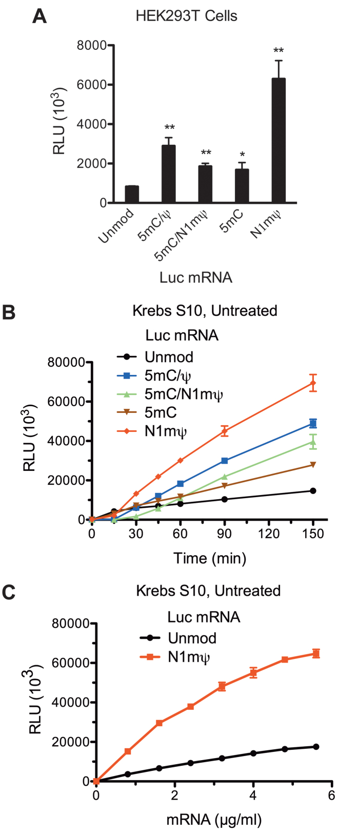
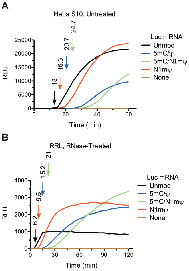
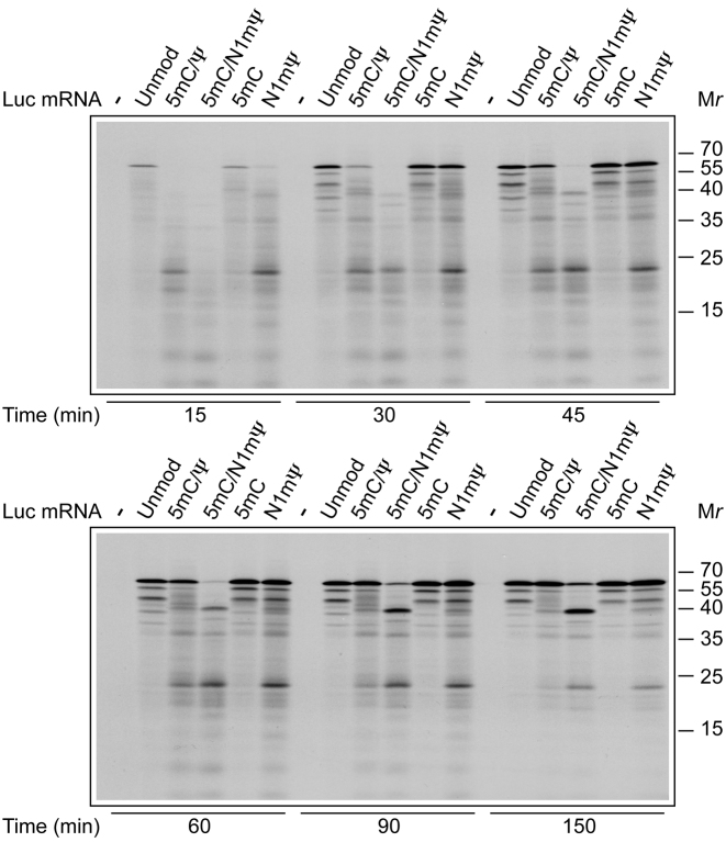
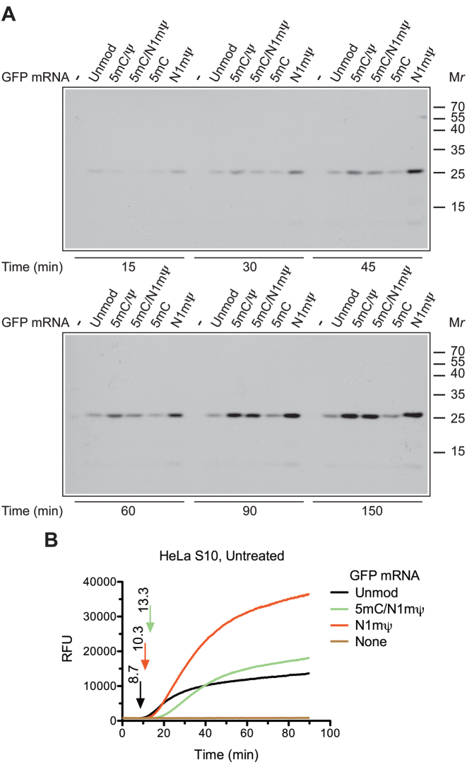
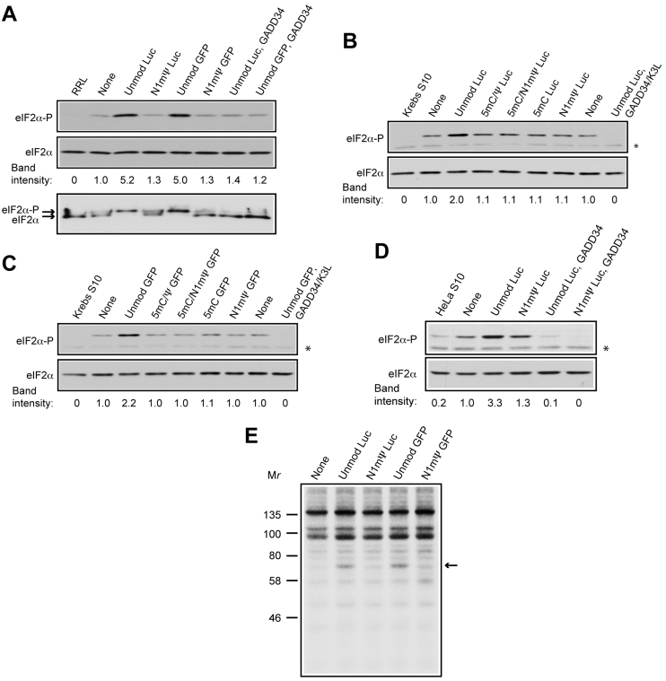
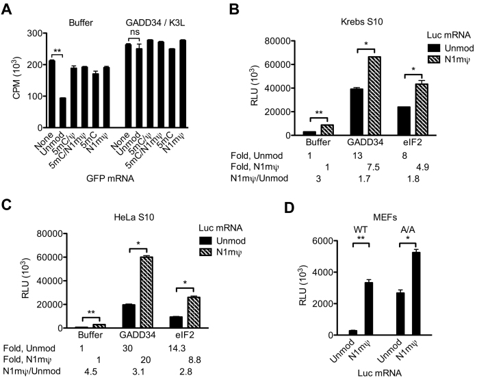
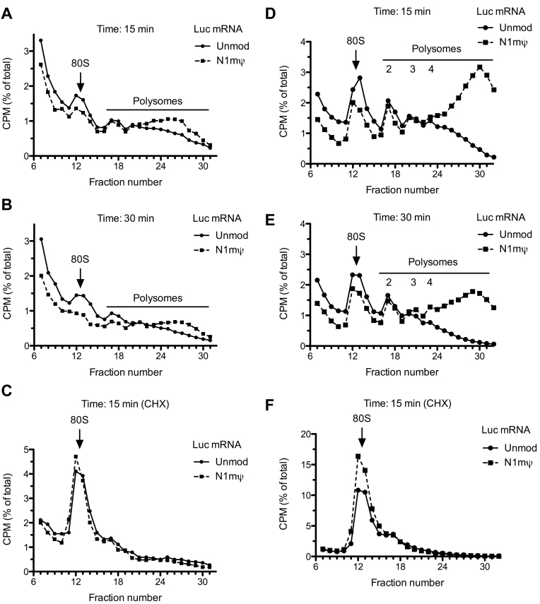
Certain chemical modifications confer increased stability and low immunogenicity to in vitro transcribed mRNAs, thereby facilitating expression of therapeutically important proteins. Here, we demonstrate that N1-methyl-pseudouridine (N1mΨ) outperforms several other nucleoside modifications and their combinations in terms of translation capacity. Through extensive analysis of various modified transcripts in cell-free translation systems, we deconvolute the different components of the effect on protein expression independent of mRNA stability mechanisms. We show that in addition to turning off the immune/eIF2α phosphorylation-dependent inhibition of translation, the incorporated N1mΨ nucleotides dramatically alter the dynamics of the translation process by increasing ribosome pausing and density on the mRNA. Our results indicate that the increased ribosome loading of modified mRNAs renders them more permissive for initiation by favoring either ribosome recycling on the same mRNA or de novo ribosome recruitment.
Overview
The claims and statistics of this paper haven’t been checked yet.
Run the check — free, no account needed. It reads the open-access text and checks the sources it cites.
Integrity signals
Registry
Cites
Statistics
Figures
Verification
Claims the paper makes
The claims and statistics of this paper haven’t been checked yet.
Run the check — free, no account needed. It reads the open-access text and checks the sources it cites.
Claims other papers make about this one
Each is checked against this paper’s own text: does it say what the citing paper says it does?
The cited source says what the claim attributes to it.
In the citing paper
“Nucleoside-modified mRNA can reduce the activation of TLRs, retinoic acid-inducible gene I (RIG-I), protein kinase R (PKR), and 2′-5′-oligoadenylate synthetase (OAS). Additionally, nucleoside modification increases translation activity and resistance against RNase L-mediated degradation.54,81,173–176”
From Advances in COVID-19 mRNA vaccine development (2022) · cites this paper as [176]
Self-check
Percentages, sums and reported differences re-computed from the paper’s own numbers.
The self-check needs the paper’s full text.
Citations
Cites
Topology of the human and mouse m6A RNA methylomes revealed by m6A-seq
Dominissini, D., Moshitch-Moshkovitz, S., Schwartz, S., Salmon-Divon, M., Ungar, L., Osenberg, S., Cesarkas, K., Jacob-Hirsch, J., Amariglio, N., Kupiec, M., et al. (2012) · Nature · doi:10.1038/nature11112 · Cited by 4,884
Comprehensive Analysis of mRNA Methylation Reveals Enrichment in 3′ UTRs and near Stop Codons
Meyer, K. D., Saletore, Y., Zumbo, P., Elemento, O., Mason, C. E., & Jaffrey, S. R. (2012) · Cell · doi:10.1016/j.cell.2012.05.003 · Cited by 4,223
Regulation of Translation Initiation in Eukaryotes: Mechanisms and Biological Targets
Sonenberg, N., & Hinnebusch, A. G. (2009) · Cell · doi:10.1016/j.cell.2009.01.042 · Cited by 3,304
Self-citationKarikó, K., Buckstein, M., Ni, H., & Weissman, D. (2005) · Immunity · doi:10.1016/j.immuni.2005.06.008 · Cited by 2,728
Warren, L., Manos, P. D., Ahfeldt, T., Loh, Y.-H., Li, H., Lau, F., Ebina, W., Mandal, P. K., Smith, Z. D., Meissner, A., et al. (2010) · Cell Stem Cell · doi:10.1016/j.stem.2010.08.012 · Cited by 2,662
mRNA-based therapeutics--developing a new class of drugs
Sahin, U., Karikó, K., & Türeci, Ö. (2014) · Nature Reviews Drug Discovery · doi:10.1038/nrd4278 · Cited by 2,474
Ingolia, N. T., Lareau, L. F., & Weissman, J. S. (2011) · Cell · doi:10.1016/j.cell.2011.10.002 · Cited by 2,187
Insights into RNA Biology from an Atlas of Mammalian mRNA-Binding Proteins
Castello, A., Fischer, B., Eichelbaum, K., Horos, R., Beckmann, B. M., Strein, C., Davey, N. E., Humphreys, D. T., Preiss, T., Steinmetz, L. M., et al. (2012) · Cell · doi:10.1016/j.cell.2012.04.031 · Cited by 2,132
5′ UTR m6A Promotes Cap-Independent Translation
Meyer, K. D., Patil, D. P., Zhou, J. L., Zinoviev, A., Skabkin, M. A., Elemento, O., Pestova, T. V., Qian, S., & Jaffrey, S. R. (2015) · Cell · doi:10.1016/j.cell.2015.10.012 · Cited by 1,812
Feedback Inhibition of the Unfolded Protein Response by GADD34-Mediated Dephosphorylation of eIF2α
Novoa, I., Zeng, H., Harding, H. P., & Ron, D. (2001) · The Journal of Cell Biology · doi:10.1083/jcb.153.5.1011 · Cited by 1,434
Cited by
Sorted by citations
Items 201–278 of 278
Critical applications and prospects of RNA modification in mRNA vaccines
Liu, C., Peng, J., Zhang, M., Xiang, H., & Yi, C. (2024) · Chinese Science Bulletin (Chinese Version) · doi:10.1360/tb-2023-0800 · Cited by 2
ADME: Assessing Pharmacokinetic–Pharmacodynamic Parameters of Oligonucleotides
Jansson‐Löfmark, R., Ahlström, C., & Gennemark, P. (2019) · Methods in molecular biology · doi:10.1007/978-1-4939-9670-4_19 · Cited by 3
Structure Characterization of Escherichia coli Pseudouridine Kinase PsuK
Li, X., Li, K., Guo, W., Wen, Y., Meng, C., & Wu, B. (2022) · Frontiers in Microbiology · doi:10.3389/fmicb.2022.926099 · Cited by 3
Deconvolution of synthetic mRNA expression: Nucleoside chemistry alters translatability
Moradian, H., Schwestka, M., Roch, T., & Gossen, M. (2023) · Bioengineering & Translational Medicine · doi:10.1002/btm2.10622 · Cited by 3
Nakanishi, H., & Saito, H. (2021) · Methods in molecular biology · doi:10.1007/978-1-0716-1441-9_5 · Cited by 3
Localized 2′-OH Acylation at Poly(A) Extends RNA Translation
Fang, L., Zhong, W., Xiao, L., Shioi, R., Lee, A., Bravo, C. H., & Kool, E. T. (2025) · Journal of the American Chemical Society · doi:10.1021/jacs.5c11900 · Cited by 3
Pioneering next-generation mRNA therapeutics through molecular engineering and delivery optimization
Qi, L., Li, Z., Feng, Z., Liu, J., & Chen, X. S. (2025) · Materials Today · doi:10.1016/j.mattod.2025.08.023 · Cited by 3
Parikh, C. N., DeMarco, K. D., Bhalerao, N., Giwa, H. K., Kane, G. I., Dinnell, R. W., Ma, B., Mori, H., Brassil, M. L., Murphy, K. C., et al. (2026) · Nature Communications · doi:10.1038/s41467-026-74574-z · Cited by 3
Yamada, K., Sung, Y., Lee, T., & Siegwart, D. J. (2025) · Molecular Therapy — Nucleic Acids · doi:10.1016/j.omtn.2025.102749 · Cited by 5
Rational design of therapeutic RNA molecules
Fang, F., & Wei, B. (2025) · Trends in Chemistry · doi:10.1016/j.trechm.2025.09.016 · Cited by 2
A novel plasmid-based co-tethered transcription platform for high yield, high purity mRNA synthesis
Mala, P., Banerjee, R., Abek, A., Forster, J., Pinjari, A., Kulkarni, A. A., & Martin, C. T. (2025) · Nucleic Acids Research · doi:10.1093/nar/gkaf1355 · Cited by 2
Fitzgerald, N. J., Huang, W., & Clark, K. D. (2026) · Analytica Chimica Acta · doi:10.1016/j.aca.2026.345154 · Cited by 2
Antigen anisotropy: The overlooked geometric determinant of vaccine immunogenicity and fidelity
Santiago, D. (2026) · Human Vaccines & Immunotherapeutics · doi:10.1080/21645515.2026.2638640 · Cited by 2
Seo, H. Y., Jung, H., Lee, S., Jung, H.-G., Cho, H., Son, Y.-M., Bak, Y., Hwang, S.-Y., Kim, J.-H., Park, I. H., et al. (2026) · Molecular Therapy — Nucleic Acids · doi:10.1016/j.omtn.2026.102886 · Cited by 3
An, J. S., Lim, S. B., Lee, K. H., Kim, S., Kim, S., Lee, J., Kim, D., Han, S. R., Naito, M., Miyata, K., et al. (2026) · ACS Biomaterials Science & Engineering · doi:10.1021/acsbiomaterials.5c02147 · Cited by 2
A comprehensive analysis of the use of nucleoside analogues in RNA therapeutics
Kukushkina-Zvezdova, A. A., Zherebtsova, A. A., Telegina, D. V., Skvortsov, I. A., Ivanov, R. A., & Reshetnikov, V. V. (2026) · Journal of Translational Medicine · doi:10.1186/s12967-026-08083-4 · Cited by 3
Liu, Y. H., Liu, R., Meng, C., Li, J., Yang, K., Zhang, F., Xia, X., Ye, G., & Yuan, Y. (2026) · Frontiers in Immunology · doi:10.3389/fimmu.2026.1873905 · Cited by 2
The current landscape of mRNA therapy and the strategies for mRNA purification and dsRNA removal
Liu, J., Zang, J., Jing-Ru, X., Ran, X., Su, M., Zhang, W.-M., Ge, Z.-W., Sun, Q.-Y., & Ding, L. (2026) · Journal of Biomedical Science · doi:10.1186/s12929-026-01277-4 · Cited by 2
Brown, L. R. (2022) · Springer eBooks · doi:10.1007/978-3-030-73317-9_25-1 · Cited by 2
Enzymatic Synthesis of Base-Modified Nucleic Acids
Hollenstein, M. (2023) · Springer eBooks · doi:10.1007/978-981-16-1313-5_23-1 · Cited by 2
Guo, Y., Sun, T., Li, M., Chen, Z., Liu, Y., Luo, X., Chen, Y., Li, Y., Kuai, L., Xue, Y., et al. (2025) · Pharmaceutical Research · doi:10.1007/s11095-024-03807-x · Cited by 2
Asa, T. A., Singh, C. D., Singh, T. S., Salahi, S., Alom, K. M., & Seo, Y. J. (2025) · Bioorganic Chemistry · doi:10.1016/j.bioorg.2025.108328 · Cited by 2
The Possible Mechanistic Basis of Individual Susceptibility to Spike Protein Injury
Halma, M. T. J., Vottero, P., Thorp, J. A., Peers, T., Tuszyński, J. A., & Marik, P. E. (2025) · Advances in Virology · doi:10.1155/av/7990876 · Cited by 2
Preparation of Synthetic mRNAs—Overview and Considerations
Chan, S., & Roy, B. (2022) · RNA technologies · doi:10.1007/978-3-031-08415-7_9 · Cited by 2
Karalė, K., Bollmark, M., Stulz, R., Honcharenko, D., Tedebark, U., & Strömberg, R. (2021) · Molecules · doi:10.3390/molecules26226927 · Cited by 2
Wang, Y., Pajdzik, K., Zhao, Y., Ju, C., Stone, T. B., Zhang, L., Zhang, W., Assari, M., Zhao, W. R., Sepich‐Poore, C., et al. (2026) · Nature Communications · doi:10.1038/s41467-026-70597-8 · Cited by 2
Regulation of RNA methylation by therapy treatment, promotes tumor survivalPreprint
Bukhari, S. I. A., Truesdell, S. S., Datta, C., Choudhury, P., Wu, K. Q., Shrestha, J., Maharjan, R., Plotsker, E., Elased, R. K., Laisa, S., et al. (2023) · bioRxiv (Cold Spring Harbor Laboratory) · doi:10.1101/2023.05.19.540602 · Cited by 2
Disarming the Sword of Damocles: Biomarkers and Susceptibilities to Spike Protein DiseasesPreprint
Halma, M. T. J. (2023) · Preprints.org · doi:10.20944/preprints202307.0840.v2 · Cited by 1
“Goldilocks Modifications” for mRNA Therapeutics Won the Nobel Prize
Li, L. (2024) · Nucleic Acid Therapeutics · doi:10.1089/nat.2023.0062 · Cited by 1
Kimura-Yoshida, C., Mochida, K., Matsuno, C., Kano, K., Mishiro‐Sato, E., & Matsuo, I. (2025) · Development · doi:10.1242/dev.204700 · Cited by 1
Effect of Modified Nucleotides on the Immunogenicity of the H1N1 Influenza mRNA Vaccine
Krasnikova, S. I., Sharabrin, S. V., Kisakov, D. N., Borgoyakova, M. B., Starostina, E. V., Tigeeva, E. V., Makarova, K. P., Yakovlev, V. A., Ivanova, K. I., Рудометов, А. П., et al. (2025) · Bulletin of Experimental Biology and Medicine · doi:10.1007/s10517-026-06688-9 · Cited by 1
mRNA-based immunotherapy platform targeting endometrial cancer
Freyberg, M., Dierks, M., Naß, N., George, C., Geffken, M., Ignatov, A., Kalinski, T., & Ignatova, Z. (2025) · Journal of Translational Medicine · doi:10.1186/s12967-025-07497-w · Cited by 2
mRNA based in vivo MG1113 antibody production as a potential therapeutic approach for hemophilia
Oh, M., Jeong, Y., Jung, H., Byeon, Y., Kwak, H., Kim, S., Seo, W., Jung, J., & Lee, H. (2025) · Journal of Controlled Release · doi:10.1016/j.jconrel.2025.114520 · Cited by 1
Ligase-mediated programmable genomic integration (L-PGI)
Nan, A. X., Chickering, M., Bartolome, C. L., Shadija, N., Li, D., Estes, B., Stetina, J. R. V., Li, W., Andresen, J. L., Molugu, K., et al. (2025) · Nature Communications · doi:10.1038/s41467-025-67255-w · Cited by 5
He, W., Xi, J., Wu, X. P. P., Geng, Q., Zhang, X., Ye, J., He, Y.-C., & Jin, Q. (2026) · International Journal of Biological Macromolecules · doi:10.1016/j.ijbiomac.2026.151789 · Cited by 2
Weaning Date for Spring Calving Cows Grazing Sandhills Range
Ciminski, L. A., Adams, D. C., Klopfenstein, T. J., Clark, D., Applegarth, A., Musgrave, J. A., & Sandberg, R. (2002) · Lincoln (University of Nebraska) · Cited by 1
Optimization of Synthesis of Modified mRNA
Yoo, J., & Zangi, L. (2022) · Methods in molecular biology · doi:10.1007/978-1-0716-2707-5_7 · Cited by 1
Codon Optimality Modulates Cellular Stress and Innate Immune Responses Triggered by Exogenous RNAs
Seephetdee, C., Bejar, N., Castillo, K., Chau, E. P., Nguyen, T. Y., Godin, B., & Kiss, D. (2026) · Journal of Molecular Biology · doi:10.1016/j.jmb.2026.169995 · Cited by 1
Use and dual use of synthetic biology
Danchin, A. (2025) · Comptes Rendus Biologies · doi:10.5802/crbiol.173 · Cited by 1
Strzelecka, D., Śmietański, M., Sikorski, P. J., Warmiński, M., Kowalska, J., & Jemielity, J. (2020) · bioRxiv (Cold Spring Harbor Laboratory) · doi:10.1101/2020.07.02.184598 · Cited by 1
Editorial: Post COVID-19: the nucleoside-modified messenger RNA (modRNA) platform
Roncati, L., & Huo, Q. T. (2024) · Frontiers in Medicine · doi:10.3389/fmed.2023.1324610 · Cited by 1
Modified or Unmodified mRNA Vaccines? – The Biochemistry of Pseudouridine and mRNA Pseudouridylation
Morais, P., & Yu, Y. (2024) · Wiley eBooks · doi:10.1002/9783527838394.ch3 · Cited by 2
A módosított messenger RNS-terápia lehetőségei a kardiális regenerációban
Szabó, G. T. (2020) · Cardiologia Hungarica · doi:10.26430/chungarica.2020.50.2.111
Vaccines against Covid-19, Different Strategies towards the same Goal
Charles, R. (2021) · CHIMIA International Journal for Chemistry · doi:10.2533/chimia.2021.982
Improving the fidelity of uridine analog incorporation during in vitro transcriptionPreprint
Chen, T.-H., Potapov, V., Dai, N., Ong, J. L., & Roy, B. (2022) · bioRxiv (Cold Spring Harbor Laboratory) · doi:10.1101/2022.04.12.488100
Lipid Nanoparticles to Harness the Therapeutic Potential of mRNA for Cancer Treatment
Guevara, M. L., Persano, F., & Persano, S. (2022) · RNA technologies · doi:10.1007/978-3-031-08415-7_14
Computational prediction shows spike protein translation fragments are aggregation-prone (Preprint)Preprint
Halma, M. T. J. (2022) · doi:10.2196/preprints.43261
The pivotal role of uridine modifications in the development of mRNA technology
Rzymski, P. (2023) · Journal of Medical Science · doi:10.20883/medical.e938
Eggert, T. W., & Kleiner, R. E. (2024) · Methods and principles in medicinal chemistry · doi:10.1002/9783527840458.ch12
COVID-19 Vaccine Manufacturing Processes: Making the Molecules to Solve the Pandemic
Romine, J. A. S., Kolodziej, S. A., Vetter, T., & Dux, M. P. (2024) · Springer eBooks · doi:10.1007/978-3-031-62007-2_8
Bene, A. D., D’Aniello, A., Mottola, S., Mazzarella, V., Cutolo, R., Campagna, E., Benedetti, R., Altucci, L., Cosconati, S., Maro, S. D., et al. (2025) · RSC Medicinal Chemistry · doi:10.1039/d5md00032g
Optimization of mRNA synthesis and its cell expression for vaccine developmentPreprint
Das, S. K., Dutt, G., Goswami, V., Faiz, A., & Joshi, A. B. (2025) · bioRxiv (Cold Spring Harbor Laboratory) · doi:10.1101/2025.05.13.653856
Rubtsova, M., Mokrushina, Y., Andreev, D., Poteshnova, M., Shepelev, N., Koryagina, M., Moiseeva, E., Malabuiok, D., Prokopenko, Y., Terekhov, S., et al. (2025) · Vaccines · doi:10.3390/vaccines13050530
Assessing the Immunogenicity of Synthetic RNA Using Blood Cells
Sioud, M. (2025) · Methods in molecular biology · doi:10.1007/978-1-0716-4742-4_6
Yu, H., Yang, Y., Lin, P., Liu, C., Wen, Y., Fang, Z., Hu, Z., & Huang, S. (2025) · bioRxiv (Cold Spring Harbor Laboratory) · doi:10.1101/2025.10.27.684966
Re, A., Politano, G. M. M., Reinmets, K., Benso, A., Girbal, L., Cocaign‐Bousquet, M., & Valgepea, K. (2025) · bioRxiv (Cold Spring Harbor Laboratory) · doi:10.64898/2025.12.18.695208
Introduction to RNAi therapeutics
Bunekar, N. (2026) · Elsevier eBooks · doi:10.1016/b978-0-443-45006-8.00013-6
Li, K., Li, X., Zhang, C., Dai, S., Li, L., Wang, J., Zhang, H., & Zhao, L. (2026) · Frontiers in Immunology · doi:10.3389/fimmu.2026.1737558
Platform-dependent performance of P2A and IRES linkers in multicistronic gene expression
Ju, M., & Han, J. (2026) · Genes & Genomics · doi:10.1007/s13258-026-01771-x
Xu, Y., Zhang, X.-B., & Zhang, J.-P. (2026) · Human Gene Therapy · doi:10.1177/10430342261454725
Silencing the alarm: nucleoside modifications suppress innate immune sensing of RNA
Sweileh, R. B. A., & Pardi, N. (2026) · The Journal of Immunology · doi:10.1093/jimmun/vkaf345
D’Amico, C., Miska, M., Saarinen, S., Säkkinen, V., & Kostiainen, M. A. (2026) · bioRxiv (Cold Spring Harbor Laboratory) · doi:10.64898/2026.06.25.734245
Li, K., Ma, Z., Li, X., Chi, X., Zhang, Y., Zhang, C., Li, W., Cui, M., Qi, W., Zhao, Y., et al. (2026) · Drug Resistance Updates · doi:10.1016/j.drup.2026.101439
Niazi, S. K. (2026) · Pharmaceutics · doi:10.3390/pharmaceutics18080941
Targeted delivery of mRNA to immune cells for in vivo cell therapy
Wang, D., Li, Y., Deng, J., Li, M., & Wu, L. (2026) · Drug Delivery · doi:10.1080/10717544.2026.2716510
Next-generation CAR therapies enabled by mRNA engineering and delivery
Zhao, Q., Wen, Q., Pu, P., Xu, C., Hu, Q., Zhou, Z., Hua, X., & Zhang, X. (2026) · Acta Pharmaceutica Sinica B · doi:10.1016/j.apsb.2026.09.001
Small Nucleic Acid Therapeutics for Ocular Diseases: Progress, Challenges, and Future Perspectives
Guo, Q., Chen, L., Wu, Z., Zhang, Q., Xue, J., & Zhang, H. (2026) · Pharmaceutics · doi:10.3390/pharmaceutics18091189
Enkmann, V., & Rajicic, N. (2026) · AI Chemistry · doi:10.3390/aichem1030015 · Cited by 1
Recoding by N1-methylpseudouridine guides rational mRNA vaccine design
Poulis, P., Robecchi, G., Kurochkina, A., Felletti, M., & Rodnina, M. V. (2026) · Nature Communications · doi:10.1038/s41467-026-77796-3
RNA vaccines in motion: A multidisciplinary exploration of design, delivery, and deployment
Abuhassan, Q., Aldulaimi, A., Waleed, O. S., Ganesan, S., Kavitha, V., Maharana, L., Sharma, R., Latipova, M., Madatova, N., Jumanazarov, D., et al. (2026) · Bioimpacts · doi:10.34172/bi.33056
Lipid-based mRNA chimeric antigen receptor T cells for enhanced in vitro antitumor activity
Gao, H., Fang, Y., Tian, Z., Guo, C., Wang, P., Liu, T., Sun, Y., Zhang, P., & Qian, Q. (2026) · iScience · doi:10.1016/j.isci.2026.117390
Bioengineering strategies for improving the immunogenicity of mRNA vaccines
Wang, H., Zhuo, R., Wu, J., & Ouyang, S. (2026) · Signal Transduction and Targeted Therapy · doi:10.1038/s41392-026-02991-z
AI-integrated RNA therapeutics in precision oncology
Symmans, W. S. O., Li, Z., Calin, G. A., & Samuel, A. (2026) · Cell Reports Medicine · doi:10.1016/j.xcrm.2026.103100
Retrotransposons: A New Frontier in In Vivo Genome Engineering
Kieffer-Kwon, P., Harris, M. T., Getts, D. R., & Hofmeister, R. (2026) · Pharmaceutics · doi:10.3390/pharmaceutics18101316
Adaptation of a Fluorogenic RNA to N1‐Methylpseudouridine Nucleoside Chemistry
Bertrand, P. P. A., Cubi, R., Husser, C., Frugier, M., & Ryckelynck, M. (2026) · Angewandte Chemie · doi:10.1002/ange.8507021
Adaptation of a Fluorogenic RNA to N1‐Methylpseudouridine Nucleoside Chemistry
Bertrand, P. P. A., Cubi, R., Husser, C., Frugier, M., & Ryckelynck, M. (2026) · Angewandte Chemie International Edition · doi:10.1002/anie.8507021
Metadata
- Authors
- Svitkin, Y. V., Cheng, Y. M., Chakraborty, T., Presnyak, V., John, M., & Sonenberg, N.
- Affiliations
- Institut du cancer Rosalind & Morris Goodman (CA)
- Moderna Therapeutics (United States) (US)
- Corresponding author
- Svitkin, Yuri V.
- Sonenberg, Nahum
- Venue
- Nucleic Acids Research
- Publisher
- Oxford University Press (OUP)
- Published
- 2017-02-23
- Volume
- 45
- Issue
- 10
- Pages
- 6023-6036
- ISSN
- 0305-1048 (ISSN-L)
- 1362-4962
- PMC
- PMC5449617
- OpenAlex
- W2589945527
- Cited by
- 278
- References
- 63
- Citations by year
- 2026: 38 · 2025: 49 · 2024: 31 · 2023: 32 · 2022: 48 · 2021: 29 · 2020: 22 · 2019: 13 · 2018: 13 · 2017: 2
- FWCI
- 5.58
- Type
- journal article
- Language
- en
- Open access
- gold
- doi.org
- Licence
- Locations
- Nucleic Acids Research (publishedVersion, cc-by-nc)
- Europe PMC (PubMed Central) (publishedVersion, cc-by-nc)
- PubMed Central (publishedVersion, cc-by-nc)
- PubMed (publishedVersion)
- eScholarship@McGill (McGill) (submittedVersion)
- Funders
- Canadian Institutes of Health Research
- Topics
- Life Sciences › Biochemistry, Genetics and Molecular Biology › Molecular Biology › RNA and protein synthesis mechanisms
- Life Sciences › Biochemistry, Genetics and Molecular Biology › Molecular Biology › RNA modifications and cancer
- Life Sciences › Biochemistry, Genetics and Molecular Biology › Molecular Biology › RNA Interference and Gene Delivery
- Keywords
- nucleoside-modified mRNA, translation efficiency, eIF2α phosphorylation, ribosome density, ribosome pausing, ribosome recycling, in vitro translation, in vitro transcribed mRNA
- MeSH terms
- Animals, Cell Line, Cell-Free System, Enzyme Activation, Fibroblasts, HeLa Cells, Humans, Phosphorylation, Polyribosomes, Protein Processing, Post-Translational, Pseudouridine, RNA, RNA, Messenger, Transfection, Protein Biosynthesis, Eukaryotic Initiation Factor-2, eIF-2 Kinase, RNA Stability, Mice, HEK293 Cells
About these checks
Registry, retraction, Cites and statistics results are computed, not judged, with one exception: a printed reference that no index lists is read once by a model before it counts as a citation error. Claims are extracted by a model and kept only when their quote appears verbatim in the text; each cited source is then checked, with a model’s help, for whether it says what the claim relies on. Those verdicts can be wrong — read the claim’s quote and the cited source before relying on one.
Statistics and arithmetic are recomputed, not judged.
Confidence bands are shown only for fields calibrated on our annotated test set. None is calibrated yet, so no band is shown.
What counts as a claim ›How we measure confidence ›How we check this ›
Are you an author, or do you see a problem with this page? Request a review
