

The DOI matches its Crossref record; 123 of the 124 references it cites were found in a registry or an index, and 1 are not checked yet.
Structural and functional ramifications of antigenic drift in recent SARS-CoV-2 variants
Open accessAbstract
The protective efficacy of neutralizing antibodies (nAbs) elicited during natural infection with SARS-CoV-2 and by vaccination based on its spike protein has been compromised with emergence of the recent SARS-CoV-2 variants. Residues E484 and K417 in the receptor-binding site (RBS) are both mutated in lineages first described in South Africa (B.1.351) and Brazil (B.1.1.28.1). The nAbs isolated from SARS-CoV-2 patients are preferentially encoded by certain heavy-chain germline genes and the two most frequently elicited antibody families (IGHV3-53/3-66 and IGHV1-2) can each bind the RBS in two different binding modes. However, their binding and neutralization are abrogated by either the E484K or K417N mutation, whereas nAbs to the cross-reactive CR3022 and S309 sites are largely unaffected. This structural and functional analysis illustrates why mutations at E484 and K417 adversely affect major classes of nAbs to SARS-CoV-2 with consequences for next-generation COVID-19 vaccines.
Overview
The claims and statistics of this paper haven’t been checked yet.
Run the check — free, no account needed. It reads the open-access text and checks the sources it cites.
Integrity signals
Registry
Cites
Statistics
Figures
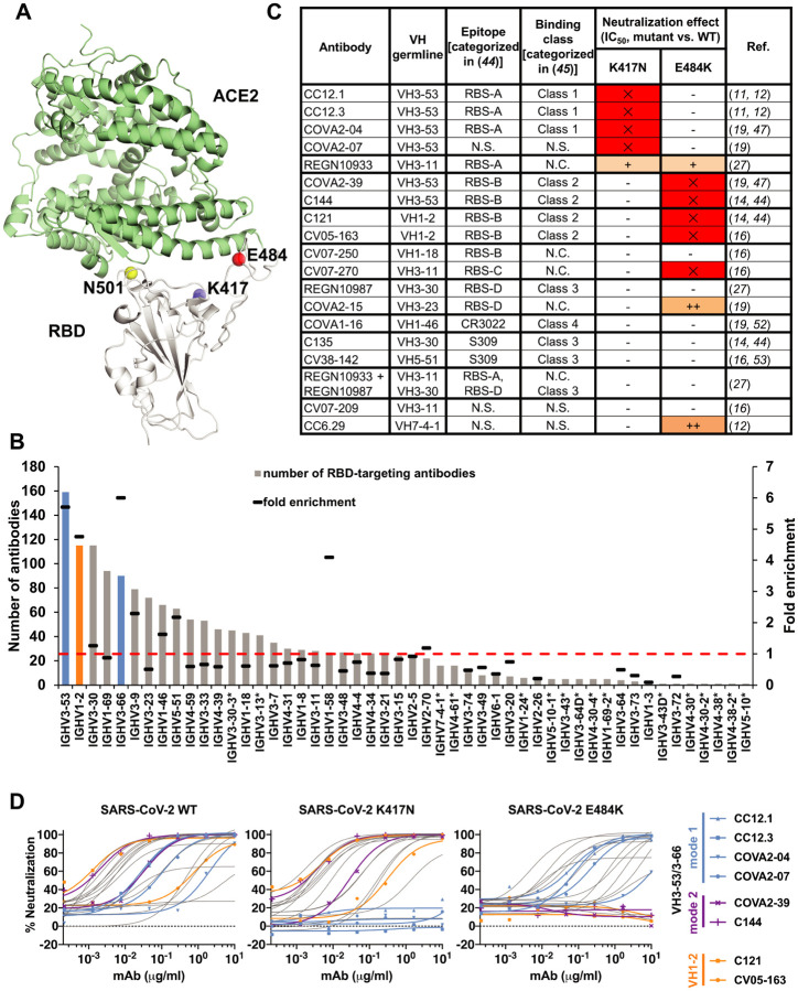
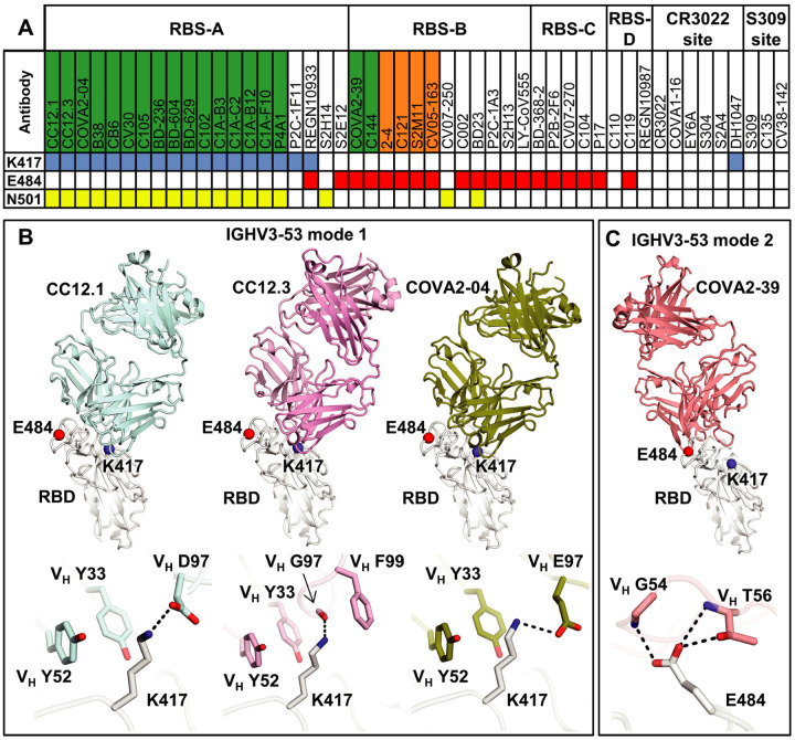
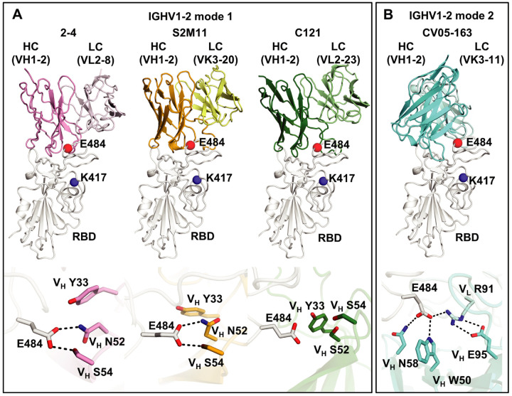
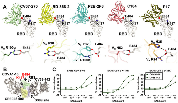
Verification
Claims the paper makes
The claims and statistics of this paper haven’t been checked yet.
Run the check — free, no account needed. It reads the open-access text and checks the sources it cites.
Claims other papers make about this one
Each is checked against this paper’s own text: does it say what the citing paper says it does?
In the citing paper
“The B.1.351 and P.1 variants have a glutamate (E) to lysine (K) mutation at position 484 (E484K) in the receptor-binding domain of the spike protein, which promotes immune evasion207.”
From mRNA vaccines for infectious diseases: principles, delivery and clinical translation (2021) · cites this paper as [207]
Self-check
Percentages, sums and reported differences re-computed from the paper’s own numbers.
The self-check needs the paper’s full text.
Citations
Cites
UCSF Chimera—A visualization system for exploratory research and analysis
Pettersen, E. F., Goddard, T. D., Huang, C. C., Couch, G. S., Greenblatt, D. M., Meng, E. C., & Ferrin, T. E. (2004) · Journal of Computational Chemistry · doi:10.1002/jcc.20084 · Cited by 45,882
[20] Processing of X-ray diffraction data collected in oscillation mode
Otwinowski, Z., & Minor, W. (1997) · Methods in Enzymology · doi:10.1016/s0076-6879(97)76066-x · Cited by 33,475
Features and development of Coot
Emsley, P., Lohkamp, B., Scott, W. G., & Cowtan, K. (2010) · Acta Crystallographica Section D Biological Crystallography · doi:10.1107/s0907444910007493 · Cited by 29,915
PHENIX : a comprehensive Python-based system for macromolecular structure solution
Adams, P. D., Afonine, P. V., Bunkóczi, G., Chen, V. B., Davis, I. W., Echols, N., Headd, J. J., Hung, L.-W., Kapral, G. J., Grosse-Kunstleve, R. W., et al. (2010) · Acta Crystallographica Section D Biological Crystallography · doi:10.1107/s0907444909052925 · Cited by 23,716
Phaser crystallographic software
McCoy, A. J., Grosse-Kunstleve, R. W., Adams, P. D., Winn, M. D., Storoni, L. C., & Read, R. J. (2007) · Journal of Applied Crystallography · doi:10.1107/s0021889807021206 · Cited by 20,612
NMRPipe: A multidimensional spectral processing system based on UNIX pipes
Delaglio, F., Grzesiek, S., Vuister, G. W., Zhu, G., Pfeifer, J., & Bax, A. (1995) · Journal of Biomolecular NMR · doi:10.1007/bf00197809 · Cited by 15,961
Inference of Macromolecular Assemblies from Crystalline State
Krissinel, E., & Henrick, K. (2007) · Journal of Molecular Biology · doi:10.1016/j.jmb.2007.05.022 · Cited by 10,007
Structure, function and antigenicity of the SARS-CoV-2 spike glycoprotein
Walls, A. C., Park, Y.-J., Tortorici, M. A., Wall, A., McGuire, A. T., & Veesler, D. (2020) · doi:10.1101/2020.02.19.956581 · Cited by 8,586
PHENIX: a comprehensive Python-based system for macromolecular structure solution
Adams, P. D., Afonine, P. V., Bunkòczi, G., Chen, V. B., Davis, I., Echols, N., Headd, J. J., Hung, L., Kapral, G. J., Grosse‐Kunstleve, R. W., et al. (2012) · International Tables for Crystallography · doi:10.1107/97809553602060000865 · Cited by 7,334
Structure of the SARS-CoV-2 spike receptor-binding domain bound to the ACE2 receptor
Lan, J., Ge, J., Yu, J., Shan, S., Zhou, H., Fan, S., Zhang, Q., Shi, X., Wang, Q., Zhang, L., et al. (2020) · Nature · doi:10.1038/s41586-020-2180-5 · Cited by 5,983
Research works printed but not linked: 20
- 10.1101/2020.08.19.253369 · DOI
- 10.1101/2020.10.06.323634 · DOI
- 10.1101/2020.12.31.424987 · DOI
- 10.1126/science.abb7269 · DOI
- 10.1126/science.abc7520 · DOI · Paper page
- 10.1016/j.bbrc.2020.10.012 · DOI
- 10.1126/science.abd2321 · DOI · Paper page
- 10.1126/science.abc2241 · DOI
- 2020, A human neutralizing antibody targets the receptor-binding site of SARS-CoV-2, Nature, 584, 120, 10.1038/s41586-020-2381-y · DOI · Paper page
- 10.1038/s41467-020-19231-9 · DOI
- 10.1016/j.cell.2020.06.025 · DOI · Paper page
- 2020, Structurally resolved SARS-CoV-2 antibody shows high efficacy in severely infected hamsters and provides a potent cocktail pairing strategy, Cell, 183, 1013, 10.1016/j.cell.2020.09.035 · DOI
- 2020, SARS-CoV-2 neutralizing antibody structures inform therapeutic strategies, Nature, 588, 682, 10.1038/s41586-020-2852-1 · DOI · Paper page
- 10.1101/2020.11.13.381533 · DOI
- 10.21203/rs.3.rs-78945/v1 · DOI
- 10.1038/s41586-020-2571-7 · DOI · Paper page
- 10.1126/science.abe3354 · DOI · Paper page
- 2020, Structure of the SARS-CoV-2 spike receptor-binding domain bound to the ACE2 receptor, Nature, 581, 215, 10.1038/s41586-020-2180-5 · DOI · Paper page
- 2020, Convergent antibody responses to SARS-CoV-2 in convalescent individuals, Nature, 584, 437, 10.1038/s41586-020-2456-9 · DOI · Paper page
- 10.1126/science.abd0827 · DOI · Paper page
Cited by
Sorted by citations
Items 201–300 of 365
SARS-CoV-2 Portrayed against HIV: Contrary Viral Strategies in Similar Disguise
Duerr, R., Crosse, K. M., Valero-Jimenez, A. M., & Dittmann, M. (2021) · Microorganisms · doi:10.3390/microorganisms9071389 · Cited by 12
Multiviral Quartet Nanocages Elicit Broad Anti-Coronavirus Responses for Proactive VaccinologyPreprint
Hills, R. A., Tan, T. K., Cohen, A. A., Keeffe, J. R., Keeble, A. H., Gnanapragasam, P. N. P., Storm, K. N., Hill, M. L., Liu, S., Gilbert‐Jaramillo, J., et al. (2023) · bioRxiv (Cold Spring Harbor Laboratory) · doi:10.1101/2023.02.24.529520 · Cited by 12
Liu, B., Niu, X., Deng, Y., Zhang, Z., Wang, Y., Gao, X., Liang, H., Li, Z., Wang, Q., Cheng, Y., et al. (2024) · Cell Reports · doi:10.1016/j.celrep.2024.114265 · Cited by 10
Hou, X.-D., Zhang, Z., Gao, J., & Wang, Y. (2021) · Molecular Systems Design & Engineering · doi:10.1039/d1me00086a · Cited by 10
New Frontiers for Machine Learning in Protein Science
Morgunov, A. S., Saar, K. L., Vendruscolo, M., & Knowles, T. P. J. (2021) · Journal of Molecular Biology · doi:10.1016/j.jmb.2021.167232 · Cited by 11
An encodable multiplex microsphere-phase amplification sensing platform detects SARS-CoV-2 mutations
Zhong, Z., Wang, J., He, S., Su, X., Huang, W., Chen, M., Zhuo, Z., Zhu, X., Fang, M., Li, T., et al. (2022) · Biosensors and Bioelectronics · doi:10.1016/j.bios.2022.114032 · Cited by 11
Self-citationEvolving spike-protein N -glycosylation in SARS-CoV-2 variantsPreprint
Baboo, S., Diedrich, J. K., Torres, J. L., Copps, J., Singh, B., Garrett, P. T., Ward, A. B., Paulson, J. C., & Yates, J. R. (2023) · bioRxiv (Cold Spring Harbor Laboratory) · doi:10.1101/2023.05.08.539897 · Cited by 10
Self-citationPublic antibodies: convergent signatures in human humoral immunity against pathogens
Rao, V. N., & Coelho, C. H. (2025) · mBio · doi:10.1128/mbio.02247-24 · Cited by 10
Verkhivker, G. M. (2022) · International Journal of Molecular Sciences · doi:10.3390/ijms23042172 · Cited by 9
Agudelo, M., Muecksch, F., Schaefer-Babajew, D. J., Cho, A., DaSilva, J., Bednarski, E., Ramos, V., Oliveira, T. Y., Cipolla, M., Gazumyan, A., et al. (2022) · The Journal of Experimental Medicine · doi:10.1084/jem.20220367 · Cited by 9
Tian, X., Duan, W., Zhang, X., Wu, X., Zhang, C., Wang, Z., Cao, G., Gu, Y., Shao, F., & Yan, T. (2022) · Frontiers in Public Health · doi:10.3389/fpubh.2022.888064 · Cited by 11
Mehra, R., & Kepp, K. P. (2022) · European Biophysics Journal · doi:10.1007/s00249-022-01619-8 · Cited by 10
An RBD bispecific antibody effectively neutralizes a SARS-CoV-2 Omicron variant
Yuan, M., Zhu, Y., Liu, G., Wang, Y., Wang, G., Zhang, G., Ye, L., Qian, Z., & Liu, P. (2023) · One Health Advances · doi:10.1186/s44280-023-00012-0 · Cited by 10
Self-citationLiu, J., Chen, X., Liu, Y., Lin, J., Shen, J., Zhang, H., Yin, J., Pu, R., Ding, Y., & Cao, G. (2021) · Infectious Diseases of Poverty · doi:10.1186/s40249-021-00895-4 · Cited by 10
Sulbarán, G., Maisonnasse, P., Amen, A., Effantin, G., Guilligay, D., Dereuddre‐Bosquet, N., Burger, J. A., Poniman, M., Grobben, M., Buisson, M., et al. (2022) · Cell Reports Medicine · doi:10.1016/j.xcrm.2022.100528 · Cited by 11
Self-citationModelling SARS-CoV-2 spike-protein mutation effects on ACE2 binding
Thakur, S., Verma, R. K., Kepp, K. P., & Mehra, R. (2022) · Journal of Molecular Graphics and Modelling · doi:10.1016/j.jmgm.2022.108379 · Cited by 13
Zhang, G. F., Meng, W., Chen, L., Ding, L., Sun, S., Wang, X., Huang, Y., Guo, H., & Gao, S. (2022) · Journal of Medical Virology · doi:10.1002/jmv.28437 · Cited by 9
Tachibana, K., Nakamura, Y., Ly, T. K., Kihara, T., Kawada, H., Yamamoto, N., & Ando, K. (2024) · Biochemical and Biophysical Research Communications · doi:10.1016/j.bbrc.2024.150120 · Cited by 9
Fung, K.-M., Lai, S.-J., Lin, T.-L., & Tseng, T. (2022) · Frontiers in Molecular Biosciences · doi:10.3389/fmolb.2022.797132 · Cited by 9
Giancotti, R., Lomoio, U., Puccio, B., Tradigo, G., Vizza, P., Torti, C., Veltri, P., & Guzzi, P. H. (2024) · Biology · doi:10.3390/biology13020090 · Cited by 9
Bruun, T. U. J., Do, J., Weidenbacher, P. A. B., Utz, A., & Kim, P. S. (2024) · ACS Central Science · doi:10.1021/acscentsci.4c00722 · Cited by 9
Alturaiki, W., Mubarak, A., Jurayyan, A. A., & Hemida, M. G. (2021) · Human Vaccines & Immunotherapeutics · doi:10.1080/21645515.2021.1935172 · Cited by 10
Geromichalou, E. G., & Geromichalos, G. D. (2022) · Molecules · doi:10.3390/molecules27217572 · Cited by 8
Synergism of interferon-beta with antiviral drugs against SARS-CoV-2 variants
Bojková, D., Stack, R. S., Rothenburger, T., Kandler, J. D., Ciesek, S., Wass, M. N., Michaelis, M., & Cinatl, J. J. (2022) · Journal of Infection · doi:10.1016/j.jinf.2022.07.023 · Cited by 9
Bakshi, R., Kaur, S., Kaur, K., Kaur, R., Boparai, J. K., Ghai, R., Kaur, T., Kaur, A., Kaur, J., Verma, K., et al. (2021) · Journal of Pure and Applied Microbiology · doi:10.22207/jpam.15.4.07 · Cited by 8
Viral entry mechanisms: the role of molecular simulation in unlocking a key step in viral infections
Valério, M., Buga, C. C., Melo, M. N., Soares, C. M., & Lousa, D. (2024) · FEBS Open Bio · doi:10.1002/2211-5463.13908 · Cited by 8
Wang, J., Ni, S., Chen, Q., Wang, C., Liu, H., Huang, L., Nasir, M. W., Wang, W. J., Zhang, X., Wu, J., et al. (2024) · Journal of Medical Virology · doi:10.1002/jmv.70073 · Cited by 8
Self-citationNeutralizing Antibodies vs. Viruses: Interacting Mechanisms and Escape Tactics
Lu, H., Liu, Y., Song, Y., Chen, L., Zhang, L., Li, R., Nie, X., Zhu, G., Ding, X., & Wang, L. (2025) · Microorganisms · doi:10.3390/microorganisms13092199 · Cited by 9
Li, L., Gao, M., Li, J., Xie, X., Zhao, H., Wang, Y., Xu, X., Zu, S., Chen, C., Wan, D., et al. (2022) · EBioMedicine · doi:10.1016/j.ebiom.2022.104401 · Cited by 8
Structural basis of spike RBM-specific human antibodies counteracting broad SARS-CoV-2 variants
Shitaoka, K., Higashiura, A., Kawano, Y., Yamamoto, A., Mizoguchi, Y., Hashiguchi, T., Nishimichi, N., Huang, S., Ito, A., Ohki, S., et al. (2023) · Communications Biology · doi:10.1038/s42003-023-04782-6 · Cited by 9
Vinzón, S. E., Lopez, M. V., Cafferata, E. G. A., Soto, A. S., Berguer, P. M., Vázquez, L., Nusblat, L., Pontoriero, A., Belotti, E. M., Salvetti, N. R., et al. (2023) · npj Vaccines · doi:10.1038/s41541-023-00737-4 · Cited by 8
Antibody engineering improves neutralization activity against K417 spike mutant SARS-CoV-2 variants
Li, L., Gao, M., Jiao, P., Zu, S., Deng, Y., Wan, D., Cao, Y., Duan, J., Aliyari, S. R., Li, J., et al. (2022) · Cell & Bioscience · doi:10.1186/s13578-022-00794-7 · Cited by 8
Li, Y., Fan, Q., Zhou, B., Shen, Y., Zhang, Y., Cheng, L., Qi, F., Song, S., Guo, Y., Yan, R., et al. (2022) · iScience · doi:10.1016/j.isci.2022.104431 · Cited by 8
An Engineered IgG–VHH Bispecific Antibody against SARS‐CoV‐2 and Its Variants
Chi, H., Wang, L., Liu, C., Cheng, X., Zheng, H., Lv, L., Tan, Y., Zhang, N., Zhao, S.-Q., Wu, M., et al. (2022) · Small Methods · doi:10.1002/smtd.202200932 · Cited by 8
Fiedler, S., Devenish, S. R. A., Morgunov, A. S., Ilsley, A., Ricci, F., Emmenegger, M., Kosmoliaptsis, V., Theel, E. S., Mills, J. R., Sholukh, A. M., et al. (2022) · Scientific Reports · doi:10.1038/s41598-022-22214-z · Cited by 8
Rationally designed immunogens enable immune focusing to the SARS-CoV-2 receptor binding motifPreprint
Hauser, B. M., Sangesland, M., Denis, K. S., Windsor, I. W., Feldman, J. E., Lam, E. C., Kannegieter, T., Balazs, A. B., Lingwood, D., & Schmidt, A. G. (2021) · bioRxiv (Cold Spring Harbor Laboratory) · doi:10.1101/2021.03.15.435440 · Cited by 8
Reduced antigenicity of Omicron lowers host serologic responsePreprint
Tubiana, J., Xiang, Y., Fan, L., Wolfson, H. J., Chen, K., Schneidman‐Duhovny, D., & Shi, Y. (2022) · bioRxiv (Cold Spring Harbor Laboratory) · doi:10.1101/2022.02.15.480546 · Cited by 8
Recall of pre-existing cross-reactive B cell memory following Omicron breakthrough infectionPreprint
Kaku, C., Bergeron, A. J., Ahlm, C., Normark, J., Sakharkar, M., Forsell, M., & Walker, L. (2022) · bioRxiv · doi:10.1101/2022.04.01.486726 · Cited by 12
Ruiz, F., Foreman, W. B., Lilly, M., Baharani, V. A., Depierreux, D. M., Chohan, V., Taylor, A. L., Guenthoer, J., Ralph, D., Matsen IV, F. A., et al. (2024) · PLOS Pathogens · doi:10.1371/journal.ppat.1012650 · Cited by 7
Ovchynnykova, O., Капуста, К., Sizochenko, N., Sukhyy, K. M., Kołodziejczyk, W., Hill, G., & Saloni, J. (2022) · Molecules · doi:10.3390/molecules27217336 · Cited by 7
Predicting the immune escape of SARS-CoV-2 neutralizing antibodies upon mutation
Sharma, D., Rawat, P., Greiff, V., Vani, J., & Gromiha, M. M. (2023) · Biochimica et Biophysica Acta (BBA) - Molecular Basis of Disease · doi:10.1016/j.bbadis.2023.166959 · Cited by 7
Balasco, N., Damaggio, G., Esposito, L., Villani, F., Berisio, R., Colonna, V., & Vitagliano, L. (2021) · Scientific Reports · doi:10.1038/s41598-021-04147-1 · Cited by 7
Advances in COVID-19 Vaccines and New Coronavirus Variants
Liu, M., & Li, Y. (2022) · Frontiers in Medicine · doi:10.3389/fmed.2022.888631 · Cited by 7
Azuma, H., Kawano, Y., Shitaoka, K., Kawahara, T., Ito, A., Higashiura, A., Kitajima, Y., Ohki, S., & Yasuda, T. (2022) · International Immunology · doi:10.1093/intimm/dxac055 · Cited by 6
Xu, H., Wang, T., Sun, P., Hou, X., Gong, X., Zhang, B., Wu, J., & Liu, B. (2023) · Frontiers in Microbiology · doi:10.3389/fmicb.2022.1093080 · Cited by 7
Fiori, F., Cossu, F. L., Salis, F., Carboni, D., Stagi, L., Forni, D. D., Poddesu, B., Malfatti, L., Khalel, A., Salis, A., et al. (2023) · Nanomaterials · doi:10.3390/nano13243090 · Cited by 7
Hassan, S. S., Basu, P., Redwan, E. M. M. M., Lundstrom, K. H., Choudhury, P. P., Serrano‐Aroca, Á., Azad, G. K., Aljabali, A. A. A., Palù, G., El‐Aziz, T. M. A., et al. (2021) · Environmental Research · doi:10.1016/j.envres.2021.112092 · Cited by 7
COVID-19 knowledge deconstruction and retrieval: an intelligent bibliometric solution
Wu, M., Zhang, Y., Markley, M., Cassidy, C., Newman, N. C., & Porter, A. L. (2023) · Scientometrics · doi:10.1007/s11192-023-04747-w · Cited by 7
Sokal, A., Broketa, M., Meola, A., Spaeth, G. B., Fernández, I., Fourati, S., Azzaoui, I., Selle, A. D. L., Vandenberghe, A., Roeser, A., et al. (2021) · bioRxiv (Cold Spring Harbor Laboratory) · doi:10.1101/2021.12.21.473528 · Cited by 7
Ju, B., Zheng, Q., Guo, H., Fan, Q., Li, T., Song, S., Sun, H., Shen, S., Zhou, X., Cheng, L., et al. (2022) · bioRxiv (Cold Spring Harbor Laboratory) · doi:10.1101/2022.01.19.476892 · Cited by 7
Functional and structural basis of Omicron BA.3.2.1 spike
Wang, Y., Hu, Y., Chen, Z., Zou, J., Zhang, K., Ren, P., Shi, P., Liang, B., & Xie, X. (2026) · Cell Reports · doi:10.1016/j.celrep.2026.117812 · Cited by 6
Teruel, N., Crown, M. R., Rajsbaum, R., Bashton, M., & Najmanovich, R. (2025) · Virus Evolution · doi:10.1093/ve/veaf027 · Cited by 7
Almanza, G., Clark, A. E., Kouznetsova, V. L., Olmedillas, E., Castro, A., Tsigelny, I. F., Wu, Y., Gao, G. F., Leibel, S. L., Bray, W. A., et al. (2022) · PLoS Pathogens · doi:10.1371/journal.ppat.1010686 · Cited by 5
Rey, F. A. (2021) · Comptes Rendus Biologies · doi:10.5802/crbiol.53 · Cited by 11
Development of an In Vivo Probe to Track SARS-CoV-2 Infection in Rhesus Macaques
Madden, P. J., Arif, M. S., Becker, M. E., McRaven, M. D., Carias, A. M., Lorenzo-Redondo, R., Xiao, S., Midkiff, C. C., Blair, R. V., Potter, E. L., et al. (2021) · Frontiers in Immunology · doi:10.3389/fimmu.2021.810047 · Cited by 7
Peissert, F., Pedotti, M., Corbellari, R., Simonelli, L., Gasparo, R. D., Tamagnini, E., Plüss, L., Elsayed, A., Matasci, M., Luca, R. D., et al. (2023) · Global Challenges · doi:10.1002/gch2.202300088 · Cited by 8
Liu, H., Kaku, C. I., Song, G., Yuan, M., Andrabi, R., Burton, D. R., Walker, L. M., & Wilson, I. A. (2021) · bioRxiv (Cold Spring Harbor Laboratory) · doi:10.1101/2021.12.15.472864 · Cited by 6
Self-citationGontu, A., Misra, S., Chothe, S. K., Ramasamy, S., Jakka, P., Byukusenge, M., LaBella, L. C., Nair, M. S., Jayarao, B. M., Archetti, M., et al. (2025) · npj Vaccines · doi:10.1038/s41541-025-01166-1 · Cited by 6
Feng, Y., Zhang, Z., & Zhang, J. (2024) · Journal of the Knowledge Economy · doi:10.1007/s13132-024-01751-0 · Cited by 7
RetractedPlant‐produced SARS ‐ CoV ‐2 antibody engineered towards enhanced potency and in vivo efficacy
de Taeye, S. W., Faye, L. J.-Y., Morel, B., Schriek, A. I., Umotoy, J. C., Yuan, M., Kuzmina, N. A., Turner, H. L., Zhu, X., Grünwald‐Gruber, C., et al. (2024) · Plant Biotechnology Journal · doi:10.1111/pbi.14458 · Cited by 5
Self-citationNature of Acquired Immune Responses, Epitope Specificity and Resultant Protection from SARS-CoV-2
Gorczynski, R. M., Lindley, R. A., Steele, E. J., & Wickramasinghe, N. C. (2021) · Journal of Personalized Medicine · doi:10.3390/jpm11121253 · Cited by 7
Verkhivker, G. M. (2022) · International Journal of Molecular Sciences · doi:10.3390/ijms23031646 · Cited by 5
Gong, H., Hu, Y., Li, X., Yau, T. C. C., Zhang, B., & Huang, J.-D. (2022) · ACS Infectious Diseases · doi:10.1021/acsinfecdis.2c00488 · Cited by 6
Ji, C., Han, N., Cheng, Y., Shang, J., Weng, S., Yang, R., Zhou, H., & Wu, A. (2022) · Viruses · doi:10.3390/v14051087 · Cited by 5
Sokal, A., Spaeth, G. B., Fernández, I., Broketa, M., Azzaoui, I., Selle, A. D. L., Vandenberghe, A., Fourati, S., Roeser, A., Meola, A., et al. (2021) · bioRxiv (Cold Spring Harbor Laboratory) · doi:10.1101/2021.06.17.448459 · Cited by 5
An elite broadly neutralizing antibody protects SARS-CoV-2 Omicron variant challengePreprint
Zhou, B., Zhou, R., Chan, J. F., Luo, M., Peng, Q., Yuan, S., Mok, B. W.-Y., Chen, B., Wang, P., Poon, V. K., et al. (2022) · bioRxiv (Cold Spring Harbor Laboratory) · doi:10.1101/2022.01.05.475037 · Cited by 5
Yuan, M., West, B. R., Foreman, W. B., Powers, C., Feng, Z., Taylor, A. L., Yu, X., Wang, G., Walker, L. M., Starr, T. N., et al. (2025) · bioRxiv (Cold Spring Harbor Laboratory) · doi:10.1101/2025.08.28.672883 · Cited by 5
Self-citationMane, V. P., Mehta, R., Álvarez, N., Sharma, V., Park, S., Fox, A., DeCarlo, C., Yang, X., Perlin, D. S., & Powell, R. L. R. (2024) · Human Vaccines & Immunotherapeutics · doi:10.1080/21645515.2024.2303226 · Cited by 4
Structural Insights into the SARS-CoV-2 Spike Protein and Its Implications for Antibody Resistance
Yamamoto, Y., & Noguchi, K. (2025) · Biomolecules · doi:10.3390/biom15111489 · Cited by 5
O’Reilly, S. R., Kenny, G., Alrawahneh, T., Francois, N., Gu, L., Angeliadis, M., d’Autume, V. D. M., Leon, A. G., Feeney, E. R., Yousif, O., et al. (2023) · PLoS ONE · doi:10.1371/journal.pone.0294262 · Cited by 4
Milon, T. I., Sarkar, T., Chen, Y., Grider, J. M., Chen, F., Ji, J., Jois, S. D. S., Kousoulas, K. G., Raghavan, V. V., & Xu, W. (2025) · Frontiers in Chemistry · doi:10.3389/fchem.2025.1395374 · Cited by 5
Hua, R.-H., Zhang, S., Niu, B., Ge, J., Lan, T., & Bu, Z. (2023) · Microbiology Spectrum · doi:10.1128/spectrum.01190-23 · Cited by 4
Li, K., Huntwork, R. H. C., Horn, G. Q., Abraha, M., Hastie, K. M., Li, H., Rayaprolu, V., Olmedillas, E., Feeney, E., Cronin, K. D., et al. (2023) · Journal of Virology · doi:10.1128/jvi.01070-23 · Cited by 4
Role of the complement system in Long COVIDPreprint
Farztdinov, V. M., Zühlke, B. M., Sotzny, F., Steinbeis, F., Seifert, M., Kedor, C., Wittke, K., Tober‐Lau, P., Zöller, T., Textoris‐Taube, K., et al. (2024) · medRxiv · doi:10.1101/2024.03.14.24304224 · Cited by 4
Optimizing the breadth of SARS-CoV-2-neutralizing antibodies in vivo and in silico
Kuroda, D., Moriyama, S., Sasaki, H., & Takahashi, Y. (2025) · Human Vaccines & Immunotherapeutics · doi:10.1080/21645515.2025.2526873 · Cited by 3
Yaghoobizadeh, F., Ardakani, M. R., Ranjbar, M. M., Khosravi, M., & Galehdari, H. (2024) · Computers in Biology and Medicine · doi:10.1016/j.compbiomed.2024.108091 · Cited by 5
Alshahrani, M., Parikh, V., Foley, B., Raisinghani, N., & Verkhivker, G. M. (2025) · International Journal of Molecular Sciences · doi:10.3390/ijms26041507 · Cited by 3
Li, S., Wu, J., Jiang, W., He, H., Zhou, Y., Wu, W., Gao, Y., Xie, M., Xia, A., He, J., et al. (2022) · Journal of Medical Virology · doi:10.1002/jmv.28440 · Cited by 3
Sandhya, K. S., Kishore, A. A., Unni, A., Sunitha, P., V, Sajithra, C. V., & Nair, A. S. (2024) · Journal of Molecular Graphics and Modelling · doi:10.1016/j.jmgm.2024.108719 · Cited by 3
Zhu, Y., Lü, Y., Zhou, C., Tong, G., Gao, M., Zhan, Y., Wang, Y., Liang, R., Li, Y., Gao, T., et al. (2022) · Med · doi:10.1016/j.medj.2022.05.003 · Cited by 3
A Pre-Vaccination Baseline of SARS-CoV-2 Genetic Surveillance and Diversity in the United States
Capoferri, A. A., Shao, W., Spindler, J., Coffin, J. M., Rausch, J. W., & Kearney, M. F. (2022) · Viruses · doi:10.3390/v14010104 · Cited by 3
In silico design of miniprotein to inhibit SARS-CoV-2 variant Omicron spike protein
Wu, J., Zhang, H., & Zhang, J. (2023) · Physical Chemistry Chemical Physics · doi:10.1039/d3cp01167d · Cited by 3
Structural Ramifications of Spike Protein D614G Mutation in SARS-CoV-2Preprint
Dokainish, H. M., & Sugita, Y. (2022) · bioRxiv (Cold Spring Harbor Laboratory) · doi:10.1101/2022.01.24.477651 · Cited by 3
Curtis, N. C., Shin, S., Hederman, A. P., Connor, R. I., Wieland‐Alter, W., Ionov, S., Boylston, J. A., Rose, J., Sakharkar, M., Dorman, D. B., et al. (2023) · bioRxiv (Cold Spring Harbor Laboratory) · doi:10.1101/2023.09.08.556349 · Cited by 4
Yan, Q., Huang, X., Liu, B., Yao, Y., Zheng, H., Song, Y., Wu, F., He, Z., Li, S., Chen, F., et al. (2025) · bioRxiv (Cold Spring Harbor Laboratory) · doi:10.64898/2025.12.10.693372 · Cited by 3
Tang, C., Lupala, C. S., Wang, D., Li, X., Tang, L., & Li, X. (2025) · International Journal of Molecular Sciences · doi:10.3390/ijms26083776 · Cited by 2
Enhanced Sampling Reveals Metastable Conformations Driving K417N-Mediated Class I Antibody Escape
Pan, X., Tadokoro, T., Onodera, T., Kita, S., Takahashi, Y., Maenaka, K., & Dokainish, H. M. (2025) · Journal of Chemical Information and Modeling · doi:10.1021/acs.jcim.5c01396 · Cited by 2
Kwon, H. J., Zhang, J., Kosikova, M., Tang, W., Ortega‐Rodriguez, U., Peng, H., Meseda, C. A., Pedro, C. L., Schmeisser, F., Lu, J., et al. (2023) · Journal of Medical Virology · doi:10.1002/jmv.28673 · Cited by 2
Modeling of adaptive immunity uncovers disease tolerance mechanisms
Jonas, D., Kirby, M. J. L., Schenkel, A. R., & Dangelmayr, G. (2023) · Journal of Theoretical Biology · doi:10.1016/j.jtbi.2023.111498 · Cited by 2
Matveev, A. L., Pyankov, O. V., Khlusevich, Y. A., Tyazhelkova, O. V., Emelyanova, L. A., Timofeeva, A. M., Shipovalov, A. V., Владимирович, Ч. А., Zaitseva, N. S., Kudrov, G. A., et al. (2023) · Biochemistry (Moscow) · doi:10.1134/s000629792309002x · Cited by 2
Prathiviraj, R., Prinsha, K., Hassan, S., Kumar, S., Kiran, G. S., & Selvin, J. (2023) · Medicine in Omics · doi:10.1016/j.meomic.2023.100024 · Cited by 2
Samudio, A., Sotelo, P. H., González-Maldonado, P., Alfonso, L., Valiente‐Echeverría, F., Soto‐Rifo, R., & Langjahr, P. (2023) · The Journal of Infection in Developing Countries · doi:10.3855/jidc.16955 · Cited by 2
Coronaviruses: Pandemic SARS-CoV-2
Cowling, B. J., & Wong, S. (2024) · Springer eBooks · doi:10.1007/978-1-4939-9544-8_68-1 · Cited by 2
Fan, Q., Liu, C., Guo, H., Tang, S., Wang, H., Zhou, B., Sun, Y., Wang, M., Ge, X., Liu, L., et al. (2025) · Structure · doi:10.1016/j.str.2025.04.005 · Cited by 2
Efficacy and effectiveness of COVID-19 vaccines
Li, Y., & Lu, J. (2021) · One Health Bulletin · doi:10.4103/2773-0344.329027 · Cited by 2
Hou, X.-D., Gao, J., & Wang, Y. (2022) · The Journal of Physical Chemistry Letters · doi:10.1021/acs.jpclett.2c02209 · Cited by 2
Bormotova, E., Gupalova, T. V., & Suvorov, A. N. (2023) · Russian Journal for Personalized Medicine · doi:10.18705/2782-3806-2023-3-1-64-71 · Cited by 2
Gutlapalli, S. D., Ganipineni, V. D. P., Danda, S., Fabián, D., Okorie, I. J., Paramsothy, J., Kailayanathan, T., Umyarova, R., Aviles, C., Garlapati, S. K. P., et al. (2023) · Cureus · doi:10.7759/cureus.36809 · Cited by 2
Mechanistic study of the transmission pattern of the SARS‐CoV ‐2 omicron variant
An, K., Yang, X., Luo, M., Yan, J., Xu, P., Zhang, H., Li, Y., Wu, S., Warshel, A., & Bai, C. (2024) · Proteins Structure Function and Bioinformatics · doi:10.1002/prot.26663 · Cited by 2
Chen, C., Boorla, V. S., Chowdhury, R., Nissly, R. H., Gontu, A., Chothe, S. K., LaBella, L. C., Jakka, P., Ramasamy, S., Vandegrift, K. J., et al. (2022) · bioRxiv (Cold Spring Harbor Laboratory) · doi:10.1101/2022.03.22.485413 · Cited by 2
Metadata
- Authors
- Yuan, M., Huang, D., Lee, C.-C. D., Wu, N. C., Jackson, A. M., Zhu, X., Liu, H., Peng, L., van Gils, M. J., Sanders, R. W., Burton, D. R., Reincke, S. M., Prüss, H., Kreye, J., Nemazee, D., Ward, A. B., & Wilson, I. A.
- Affiliations
- Scripps Research Institute (US)
- University of Illinois Urbana-Champaign (US)
- Cornell University (US)
- German Center for Neurodegenerative Diseases (DE)
- Corresponding author
- Wilson, Ian A.
- ORCID
- Venue
- Science
- Publisher
- openRxiv
- Published
- 2021-02-17
- Volume
- 373
- Issue
- 6556
- Pages
- 818-823
- ISSN
- 0036-8075 (ISSN-L)
- PMC
- PMC7899451
- OpenAlex
- W3160471157
- Cited by
- 364
- References
- 112
- Citations by year
- 2026: 11 · 2025: 27 · 2024: 35 · 2023: 60 · 2022: 138 · 2021: 92
- FWCI
- 25.73
- Type
- journal article
- Language
- en
- Open access
- hybrid
- doi.org
- Licence
- cc-by
- Locations
- Science (publishedVersion, cc-by)
- bioRxiv (Cold Spring Harbor Laboratory) (acceptedVersion, cc-by-nc)
- PubMed Central (publishedVersion, cc-by)
- PubMed (publishedVersion)
- Pure Amsterdam UMC (submittedVersion, cc-by)
- PubMed Central (submittedVersion, cc-by-nc)
- Data Archiving and Networked Services (DANS) (submittedVersion, other-oa)
- OSTI OAI (U.S. Department of Energy Office of Scientific and Technical Information) (submittedVersion)
- DZNE Pub (submittedVersion)
- Funders
- U.S. Department of Energy (DE‐ AC02‐06CH11357, COVID-19, Contract No. DE-AC02-06CH11357., AC02‐06CH11357, 06CH11357, No. DE-AC02-06CH11357, DE-AC02-)
- Bill and Melinda Gates Foundation (INV-004923, OPP1170236, OPP1170236 and INV-004923)
- Helmholtz-Gemeinschaft (HIL-A03 and SO-097)
- Deutsche Forschungsgemeinschaft (PR1274/5–1, and PR 1274/5-1, DE-AC02-06CH11357, PR1274/3–1, PR 1274/3-1, and PR 1274/5-1)
- Bundesministerium für Bildung und Forschung (01GM1908D)
- Nederlandse Organisatie voor Wetenschappelijk Onderzoek
- National Institutes of Health (COVID-19, DE-AC02–06CH11357, AI110657)
- Office of Science (Contract No. DE-AC02-06CH11357, AC02‐06CH11357, DE-AC02, No. DE-AC02-06CH11357., 06CH11357, DE-AC02-06CH11357)
- National Institute of Allergy and Infectious Diseases (R01AI142945, R01AI132317, P01AI110657, R00AI139445)
- Argonne National Laboratory (DE- AC02-06-CH11357, DE-AC02, AC02–06CH11357, No. DE-AC02-06CH11357, 06CH11357, Contract No. DE-AC02-06CH11357.)
- Topics
- Health Sciences › Medicine › Infectious Diseases › SARS-CoV-2 and COVID-19 Research
- Life Sciences › Biochemistry, Genetics and Molecular Biology › Molecular Biology › vaccines and immunoinformatics approaches
- Life Sciences › Agricultural and Biological Sciences › Animal Science and Zoology › Animal Virus Infections Studies
- Keywords
- SARS-CoV-2, antigenic drift, Beta variant, P.1, spike protein mutations, K417N, E484K, receptor binding sites, ACE2 binding, neutralizing antibodies, CR3022, vaccine design, antibody therapy
About these checks
Registry, retraction, Cites and statistics results are computed, not judged, with one exception: a printed reference that no index lists is read once by a model before it counts as a citation error. Claims are extracted by a model and kept only when their quote appears verbatim in the text; each cited source is then checked, with a model’s help, for whether it says what the claim relies on. Those verdicts can be wrong — read the claim’s quote and the cited source before relying on one.
Statistics and arithmetic are recomputed, not judged.
Confidence bands are shown only for fields calibrated on our annotated test set. None is calibrated yet, so no band is shown.
What counts as a claim ›How we measure confidence ›How we check this ›
Are you an author, or do you see a problem with this page? Request a review
