

The DOI matches its Crossref record; 1 of the 1 references it cites were found in a registry or an index.
CDK1 and PLK1 cooperate to regulate BICD2, dynein activation and recruitment to the nucleus as well as centrosome separation in G2/M
Open accessPreprint — published as 10.1038/s41467-023-38116-1 · Paper page
Abstract
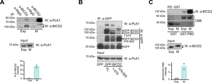
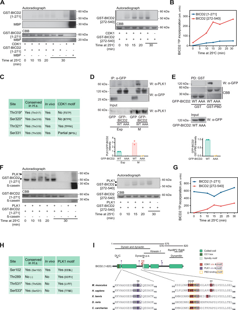
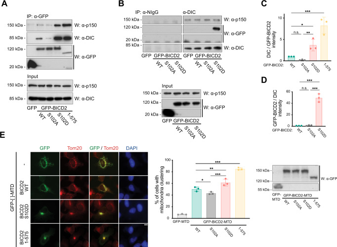
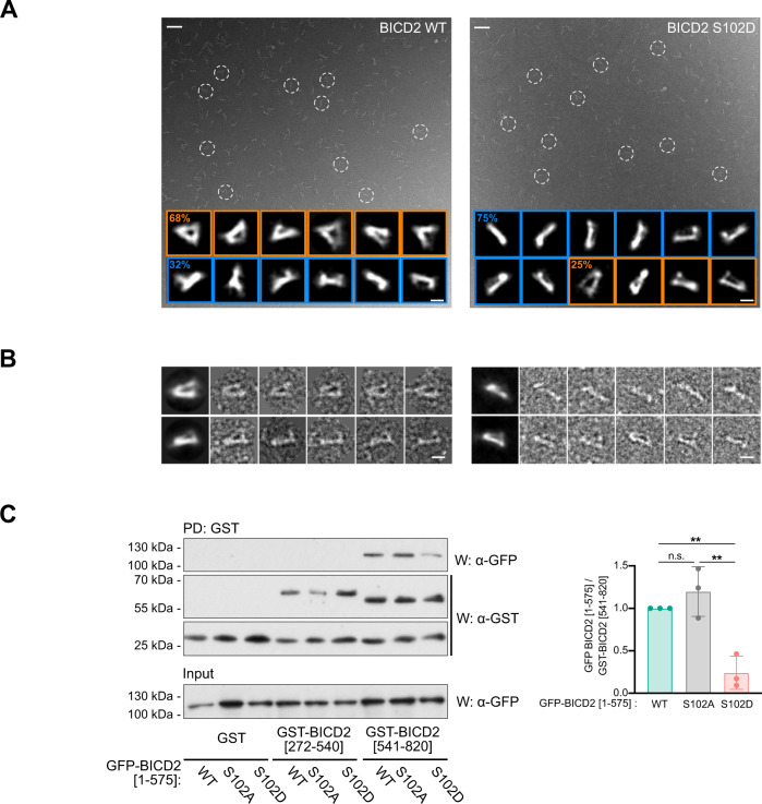
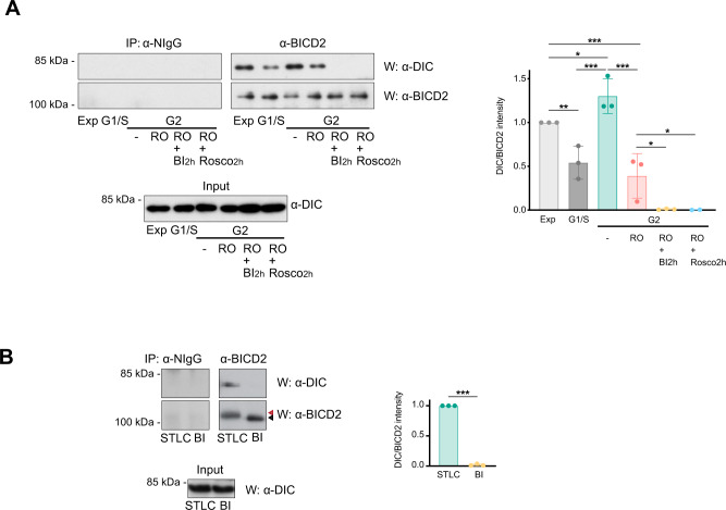
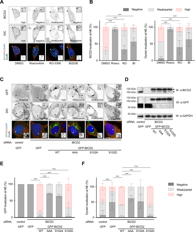
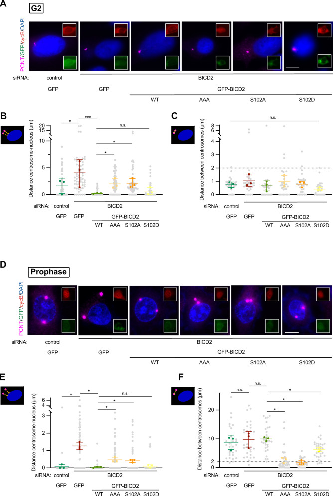
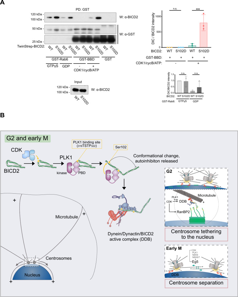
Summary: The activity of dynein is regulated by a number of adaptors that mediate its interaction with dynactin, effectively activating the motor complex while also connecting it to different cargos. The regulation of adaptors is consequently central to dynein physiology, but remains largely unexplored. We now describe that one of the best-known dynein adaptors, BICD2, is effectively activated through phosphorylation. In G2 phosphorylation of BICD2 by CDK1 promotes its interaction with PLK1. In turn, PLK1 phosphorylation of a single residue in the N-terminus of BICD2 results in a conformational change that facilitates interaction with dynein and dynactin, allowing the formation of active motor complexes. BICD2 phosphorylation is central for dynein recruitment to the nuclear envelope, centrosome tethering to the nucleus and centrosome separation in G2/M. This work reveals adaptor activation through phosphorylation as crucial for the spatiotemporal regulation of dynein activity.
Overview
The claims and statistics of this paper haven’t been checked yet.
Run the check — free, no account needed. It reads the open-access text and checks the sources it cites.
Integrity signals
Registry
Cites
Statistics
Figures
Verification
Claims the paper makes
The claims and statistics of this paper haven’t been checked yet.
Run the check — free, no account needed. It reads the open-access text and checks the sources it cites.
Self-check
Percentages, sums and reported differences re-computed from the paper’s own numbers.
The self-check needs the paper’s full text.
Citations
Cites
Highly accurate protein structure prediction with AlphaFold
Jumper, J., Evans, R., Pritzel, A., Green, T., Figurnov, M., Ronneberger, O., Tunyasuvunakool, K., Bates, R., Žídek, A., Potapenko, A., et al. (2021) · Nature · doi:10.1038/s41586-021-03819-2 · Cited by 43,764
Research works printed but not linked: 73
- Agircan, F.G. , Schiebel, E. , and Mardin, B.R . (2014). Separate to operate: control of centrosome positioning and separation. Philos Trans R Soc Lond B Biol Sci 369, pii: 20130461. · DOI
- 10.1126/scisignal.2001796 · DOI
- 10.1074/jbc.M111.226605 · DOI
- 10.1016/j.devcel.2015.04.022 · DOI
- 10.1038/emboj.2011.179 · DOI
- 10.1016/0092-8674(95)90142-6 · DOI · Paper page
- 10.1083/jcb.201007118 · DOI
- 10.1016/j.tibs.2012.09.005 · DOI
- 2021, Structure and Mechanics of Dynein Motors, Annu. Rev. Biophys, 50, 549, 10.1146/annurev-biophys-111020-101511 · DOI
- 10.1016/j.sbi.2015.12.003 · DOI
- 2017, A Splice Variant of Centrosomin Converts Mitochondria to Microtubule-Organizing Centers, Curr. Biol. CB, 27, 1928, 10.1016/j.cub.2017.05.090 · DOI
- 2005, BicD-dependent localization processes: from Drosophilia development to human cell biology, Ann. Anat. Anat. Anz. Off. Organ Anat. Ges, 187, 539
- 10.1083/jcb.127.6.1671 · DOI
- 10.1016/j.cub.2017.11.046 · DOI
- 10.1126/science.1079079 · DOI
- 10.1016/S0092-8674(03)00725-6 · DOI
- 10.1038/sj.emboj.7601459 · DOI
- 10.1083/jcb.201610108 · DOI
- 10.1083/jcb.147.1.135 · DOI
- 10.1016/j.devcel.2007.06.010 · DOI · Paper page
- 10.1016/j.tcb.2016.01.001 · DOI
- 10.1093/emboj/20.15.4041 · DOI
- 10.1093/emboj/cdg592 · DOI
- 10.1016/j.cell.2013.08.024 · DOI
- 10.1126/science.1181348 · DOI
- 10.1083/jcb.201703201 · DOI
- 10.1016/S0171--9335(99)80020--X · DOI
- 10.1038/s41467-018-03412-8 · DOI
- 10.1073/pnas.1006615107 · DOI
- 10.1016/j.cub.2010.05.050 · DOI
- 10.1038/ncb891 · DOI · Paper page
- 2006, Paircoil2: improved prediction of coiled coils from sequence, Bioinforma. Oxf. Engl, 22, 356, 10.1093/bioinformatics/bti797 · DOI · Paper page
- 10.1038/nchembio825 · DOI
- 10.1126/science.1254198 · DOI
- 10.1111/j.1432-1033.1997.t01-2-00527.x · DOI · Paper page
- 2001, Centrosome cohesion is regulated by a balance of kinase and phosphatase activities, J Cell Sci, 114, 3749, 10.1242/jcs.114.20.3749 · DOI
- 10.1242/dev.02565 · DOI
- 10.1074/jbc.C300126200 · DOI
- Noell, C.R. , Loftus, K.M. , Cui, H. , Grewer, C.T. , Kizer, M. , Debler, E.W. , and Solmaz, S.R . (2018). A quantitative model for BicD2/cargo interactions. Biochemistry. · DOI
- Noell, C.R. , Loh, J.Y. , Debler, E.W. , Loftus, K.M. , Cui, H. , Russ, B.B. , Zhang, K. , Goyal, P. , and Solmaz, S.R . (2019). Role of Coiled-coil Registry Shifts in Activation of Human Bicaudal D2 for Dynein Recruitment Upon Cargo-Binding. J. Phys. Chem. Lett. · DOI
- 10.1242/jcs.227132 · DOI
- 10.1074/jbc.M116.738211 · DOI
- 10.1038/nmeth.4169 · DOI · Paper page
- 10.1007/s00412-014-0468-7 · DOI
- 10.1038/emboj.2012.272 · DOI
- 10.1038/s41580-018-0004-3 · DOI
- 10.7554/eLife.28257 · DOI
- 10.1083/jcb.146.3.597 · DOI
- 10.1101/gad.972202 · DOI
- 10.1016/S0092-8674(01)00628-6 · DOI
- 10.1038/emboj.2010.51 · DOI · Paper page
- 10.15252/embj.201488792 · DOI
- 10.1186/s12896-017-0400-3 · DOI
- 10.1091/mbc.E11-02-0095 · DOI
- 10.1074/jbc.M511735200 · DOI
- 10.7554/eLife.36306 · DOI
- 1994, Use of an oriented peptide library to determine the optimal substrates of protein kinases, Curr. Biol. CB, 4, 973, 10.1016/S0960-9822(00)00221-9 · DOI
- 10.1371/journal.pbio.1000350 · DOI
- 10.1091/mbc.E12-03-0210 · DOI
- 10.1016/j.cub.2006.12.037 · DOI
- 10.1016/j.celrep.2020.107681 · DOI
- 10.1016/S0171-9335(99)80061-2 · DOI
- 10.1016/0092-8674(91)90365-6 · DOI
- 10.1038/emboj.2008.242 · DOI
- 10.4161/cib.13780 · DOI
- 10.1016/j.bbrc.2015.03.054 · DOI
- 10.1126/science.aaa4080 · DOI · Paper page
- 10.1038/nature25462 · DOI
- 10.1073/pnas.0600447103 · DOI
- 2018, Expressing Multi-subunit Complexes Using biGBac, Methods Mol. Biol. Clifton NJ, 1764, 329, 10.1007/978-1-4939-7759-8_21 · DOI
- 10.1083/jcb.201707081 · DOI
- 10.1101/cshperspect.a005322 · DOI
- 10.1038/nrm3819 · DOI
Metadata
- Authors
- Gallisà-Suñé, N., Sànchez-Fernàndez-de-Landa, P., Zimmermann, F., Serna, M., Paz, J., Regué, L., Llorca, O., Lüders, J., & Roig, J.
- Affiliations
- Consejo Superior de Investigaciones Científicas (ES)
- Institute for Research in Biomedicine (ES)
- Spanish National Cancer Research Centre (ES)
- Corresponding author
- Roig, Joan
- Venue
- Nature Communications
- Publisher
- openRxiv
- Published
- 2021-11-04
- Volume
- 14
- Issue
- 1
- Pages
- 2434-2434
- ISSN
- 2041-1723 (ISSN-L)
- PMC
- PMC10140047
- OpenAlex
- W4367176084
- Cited by
- 26
- References
- 1
- Citations by year
- 2026: 9 · 2025: 10 · 2024: 4 · 2023: 3
- FWCI
- 3.76
- Type
- journal article
- Language
- en
- Open access
- gold
- doi.org
- Licence
- cc-by
- Locations
- Nature Communications (publishedVersion, cc-by)
- bioRxiv (Cold Spring Harbor Laboratory) (acceptedVersion, cc-by-nc-nd)
- PubMed Central (publishedVersion, cc-by)
- PubMed (publishedVersion)
- DIGITAL.CSIC (Spanish National Research Council (CSIC)) (submittedVersion, cc-by)
- RePEc: Research Papers in Economics (submittedVersion)
- Dipòsit Digital de la Universitat de Barcelona (Universitat de Barcelona) (submittedVersion, cc-by)
- DOAJ (DOAJ: Directory of Open Access Journals) (submittedVersion, cc-by-sa)
- Funders
- Institute for Research in Biomedicine
- Ministerio de Ciencia, Innovación y Universidades (MCIN/ AEI /10.13039/501100011033/FEDER, 10.13039.501100011033, 10.13039/501100011033/FEDER, RED2018-102723-T, 2017 SGR 1089, 713673, / AEI10.13039/501100011033, 100010434, SAF2017-82632-P, 13039, MCIN/AEI/ 10.13039/501100011033, PGC 2018, 10.13039, .13039/501100011033, PGC2018-099562-B-I00, 501100011033, BFU2015-69275-P)
- Centres de Recerca de Catalunya (501100011033)
- European Commission (100010434, RED2018-102723, MCIN /AEI/ 10.13039/501100011033, 10.13039/501100011033, 13039/501100011033, 501100011033, HORIZON2020, /AEI/10.13039/501100011033, 713673, ID 100010434, RED2018-102723-T)
- Universiteit Utrecht
- Generalitat de Catalunya (10.13039/501100011033, 501100011033, 713673, ID100010434, 2017-SGR, 100010434, MCIN /AEI/10.13039/501100011033, SGR 1089)
- Ministerio de Economía y Competitividad (PID2021-127045NB-I00), BFU2014-58422-P, PGC2018-096307-B-I00)
- Ministerio de Ciencia e Innovación (713673, B-I00, 2017 SGR 1089, .13039/501100011033, PGC2018-099562-B-I00, ID100010434, 10.13039, RED2018-102723-T, 13039/501100011033/FEDER, / AEI/10, MCIN/AEI/10.13039/501100011033,, SAF2017-82632-P, 13039, 10.13039/501100011033/, PGC2018, 100010434, 501100011033, PID2020, PID2020-114429RB-I00, 10.13039/501100011033/FEDER, MCIN /AEI /10.13039, /AEI/10.13039/ 501100011033)
- Fundación Bancaria Caixa d'Estalvis i Pensions de Barcelona (ID 100010434, 100010434)
- Agència de Gestió d'Ajuts Universitaris i de Recerca (2017 SGR 1089, MCIN/AEI/10.13039/501100011033/, 10.13039/501100011033, 2017SGR, 13039/501100011033, RED2018-102723-T)
- Instituto de Salud Carlos III (ID 100010434, MCIN/ AEI /10.13039/501100011033, /10.13039/501100011033, 100010434, Y2018/BIO4747, P2018/NMT4443, 713673, RED2018-102723-T)
- European Regional Development Fund (MCIN/AEI/10.13039/ 501100011033/FEDER, RED2018-102723-T, SAF2017, PID2020-114429RB-I00, 10.13039/501100011033, 713673, 13039/501100011033, AEI/10.13039/501100011033, 100010434, 501100011033, SAF2017-82632-P, P2018/NMT4443, PGC2018, MCIN/ AEI/ 10.13039/501100011033, Y2018/BIO4747)
- Agencia Estatal de Investigación (AEI//10.13039/501100011033/, BFU2014-58422-P, 501100011033, 10.13039/501100011033, MCIN/AEI/10.13039/501 100 011 033, 13039, .13039/501100011033, PGC2018-096307-B-I00, 10.13039, 100010434, 501100011033/FEDER, AEI/10, PGC2018, PID2021-127045NB-I00, RED2018-102723-T)
- European Social Fund (501100011033)
- Topics
- Life Sciences › Biochemistry, Genetics and Molecular Biology › Cell Biology › Microtubule and mitosis dynamics
- Life Sciences › Biochemistry, Genetics and Molecular Biology › Molecular Biology › Nuclear Structure and Function
- Life Sciences › Biochemistry, Genetics and Molecular Biology › Molecular Biology › Genomics and Chromatin Dynamics
- Keywords
- BICD2, dynein, dynactin, phosphorylation, CDK1, PLK1, RanBP2, centrosome separation, nuclear envelope, G2/M phase
- MeSH terms
- Dynactin Complex, Cell Cycle, Dyneins, Microtubule-Associated Proteins, Phosphorylation, Centrosome
About these checks
Registry, retraction, Cites and statistics results are computed, not judged, with one exception: a printed reference that no index lists is read once by a model before it counts as a citation error. Claims are extracted by a model and kept only when their quote appears verbatim in the text; each cited source is then checked, with a model’s help, for whether it says what the claim relies on. Those verdicts can be wrong — read the claim’s quote and the cited source before relying on one.
Statistics and arithmetic are recomputed, not judged.
Confidence bands are shown only for fields calibrated on our annotated test set. None is calibrated yet, so no band is shown.
What counts as a claim ›How we measure confidence ›How we check this ›
Are you an author, or do you see a problem with this page? Request a review
