PreprintCited by 11Parkinson's Disease Mechanisms and TreatmentsCellular transport and secretionProtein Kinase Regulation and GTPase Signaling

The DOI matches its Crossref record; 1 of the 1 references it cites were found in a registry or an index.

Endogenous Rab38 regulates LRRK2’s membrane recruitment and substrate Rab phosphorylation in melanocytes

Open access
Unapanta, Alexandra, Shavarebi, Farbod, Porath, Jacob, Balen, Carson, Nguyen, Albert, Tseng, Josh, Shen, Yiyi, Liu, Michelle, Lis, Pawel, Di Pietro, Santiago M., Hiniker, Annie·2022·Journal of Biological Chemistry

Preprint — published as 10.1016/j.jbc.2023.105192 · Paper page

Abstract

Point mutations in LRRK2 cause Parkinson’s Disease and augment LRRK2’s kinase activity. However, cellular pathways that enhance LRRK2 kinase function have not been identified. While overexpressed Rab29 draws LRRK2 to Golgi membranes to increase LRRK2 kinase activity, there is little evidence that endogenous Rab29 performs this function under physiological conditions. Here we identify Rab38 as a novel physiological regulator of LRRK2. In mouse melanocytes, which express high levels of Rab38, Rab32, and Rab29, knockdown of Rab38 but not Rab32 or Rab29 decreases phosphorylation of multiple LRRK2 substrates, including Rab10 and Rab12, by both exogenous and endogenous LRRK2. In B16-F10 mouse melanoma cells, Rab38 drives LRRK2 membrane association, and overexpressed kinase-active but not kinase-inactive LRRK2 shows striking pericentriolar recruitment, which is dependent on the presence of endogenous Rab38 but not Rab32 or Rab29. Deletion or mutation of LRRK2 at the Rab38 binding site in the N-terminal armadillo domain decreases LRRK2 membrane association, pericentriolar recruitment, and ability to phosphorylate Rab10. Consistently, overexpression of LRRK2 350-550 , a fragment that encompasses the Rab38 binding site, blocks endogenous LRRK2’s phosphorylation of Thr73-Rab10. Finally, disruption of BLOC-3, the guanine nucleotide exchange factor for Rab38 and 32, inhibits Rab38’s regulation of LRRK2. In sum, our data identify Rab38 as a physiologic regulator of LRRK2 function and lend support to a model in which LRRK2 plays a central role in Rab GTPase coordination of vesicular trafficking.

Overview

The claims and statistics of this paper haven’t been checked yet.

Run the check — free, no account needed. It reads the open-access text and checks the sources it cites.

A few free checks a day · papers already verified open instantly · results are public

Integrity signals

Registry

Crossref · 10.1101/2022.06.20.496629

Cites

1 of 1 references verified0 citation errors · 0 not checkable

Statistics

Figures

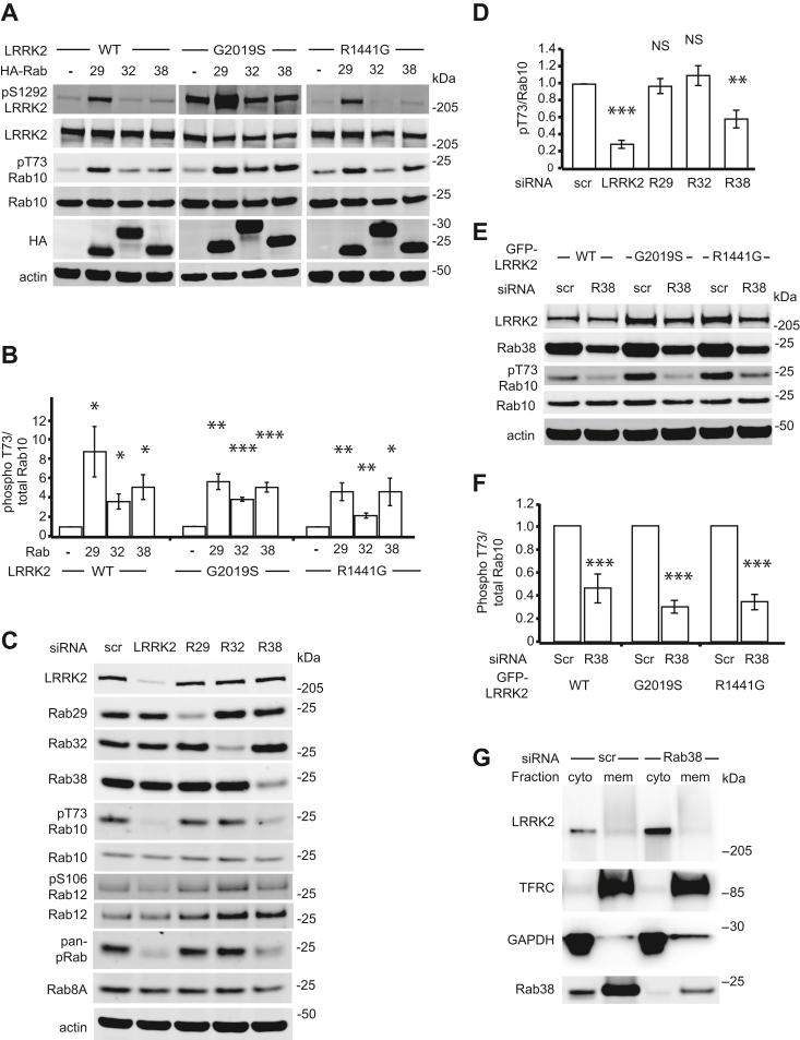
Figure 1 Rab38 regulates LRRK2’s substrate Rab phosphorylation and drives LRRK2 membrane association.A, immunoblot of phosphorylated LRRK2 Ser1292 and Rab10 Thr73 in the presence of transiently transfected HA-tagged Rab29, Rab32, and Rab38 in stably expressing WT GFP-LRRK2 (left), GFP-LRRK2 PD-mutant G2019S (middle), and GFP-LRRK2 PD-mutant R1441G (right) in HEK-293T cells. B, quantification of Rab10 phosphorylation in (A) from four independent experiments. C, representative immunoblot of B16 melanocytes following knockdown of LRRK2, Rab29, Rab32, and Rab38. D, quantification of endogenous phosphorylated Rab10 levels in (C) from six independent experiments. pThr73-Rab10/total Rab10 = 29% ± 5% in LRRK2 knockdown, 97% ± 9% in Rab29 knockdown, 110% ± 12% in Rab32 knockdown, and 59% ± 11% in Rab38 knockdown (mean ± SEM). Knockdown was quantified in Fig. S1C. E, representative immunoblot of B16 melanocytes with Rab38 versus control siRNA knockdown in the presence of transiently transfected GFP-LRRK2 WT (left), G2019S (middle), and R1441G (right). F, quantification of endogenous phosphorylated Rab10 levels in (E) from seven independent experiments. With Rab38 knockdown, pThr73-Rab10/total Rab10 = 46% ± 5% in WT GFP-LRRK2, 30% ± 2% in G2019S GFP-LRRK2, 34% ± 2% in R1441G GFP-LRRK2 (mean ± SEM). Knockdown was quantified in Fig. S1G. G, chemiluminescent immunoblot of endogenous LRRK2 in cytoplasmic and membrane fractions of B16 melanocytes with Rab38 versus control siRNA knockdown. TFRC and GAPDH are used as membrane and cytoplasmic markers, respectively. Result is representative of four replicates. Knockdown was quantified in Fig. S1H. Significance testing for panel B was performed using Kruskal–Wallis test with post hoc Dunn correction when applicable. Significance testing for panels D and F was performed using a two-tailed Student’s t test and panel D also used Bonferroni correction for multiple comparisons. Asterisks represent significant p-values in the following manner: ∗ = p < 0.05; ∗∗ = p < 0.01; ∗∗∗ = p < 0.001. LRRK2, leucine-rich repeat kinase 2; PD, Parkinson's disease.
Figure 1 Rab38 regulates LRRK2’s substrate Rab phosphorylation and drives LRRK2 membrane association.A, immunoblot of phosphorylated LRRK2 Ser1292 and Rab10 Thr73 in the presence of transiently transfected HA-tagged Rab29, Rab32, and Rab38 in stably expressing WT GFP-LRRK2 (left), GFP-LRRK2 PD-mutant G2019S (middle), and GFP-LRRK2 PD-mutant R1441G (right) in HEK-293T cells. B, quantification of Rab10 phosphorylation in (A) from four independent experiments. C, representative immunoblot of B16 melanocytes following knockdown of LRRK2, Rab29, Rab32, and Rab38. D, quantification of endogenous phosphorylated Rab10 levels in (C) from six independent experiments. pThr73-Rab10/total Rab10 = 29% ± 5% in LRRK2 knockdown, 97% ± 9% in Rab29 knockdown, 110% ± 12% in Rab32 knockdown, and 59% ± 11% in Rab38 knockdown (mean ± SEM). Knockdown was quantified in Fig. S1C. E, representative immunoblot of B16 melanocytes with Rab38 versus control siRNA knockdown in the presence of transiently transfected GFP-LRRK2 WT (left), G2019S (middle), and R1441G (right). F, quantification of endogenous phosphorylated Rab10 levels in (E) from seven independent experiments. With Rab38 knockdown, pThr73-Rab10/total Rab10 = 46% ± 5% in WT GFP-LRRK2, 30% ± 2% in G2019S GFP-LRRK2, 34% ± 2% in R1441G GFP-LRRK2 (mean ± SEM). Knockdown was quantified in Fig. S1G. G, chemiluminescent immunoblot of endogenous LRRK2 in cytoplasmic and membrane fractions of B16 melanocytes with Rab38 versus control siRNA knockdown. TFRC and GAPDH are used as membrane and cytoplasmic markers, respectively. Result is representative of four replicates. Knockdown was quantified in Fig. S1H. Significance testing for panel B was performed using Kruskal–Wallis test with post hoc Dunn correction when applicable. Significance testing for panels D and F was performed using a two-tailed Student’s t test and panel D also used Bonferroni correction for multiple comparisons. Asterisks represent significant p-values in the following manner: ∗ = p < 0.05; ∗∗ = p < 0.01; ∗∗∗ = p < 0.001. LRRK2, leucine-rich repeat kinase 2; PD, Parkinson's disease.CC BY · PubMed Central
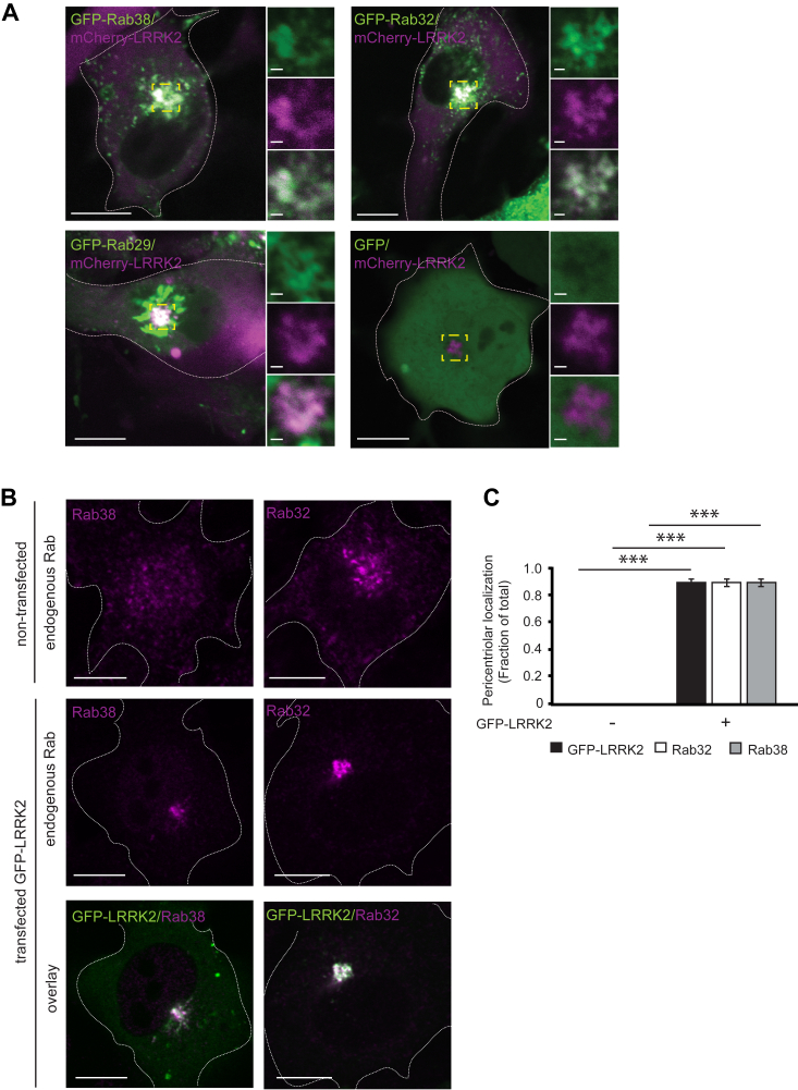
Figure 2 Overexpressed LRRK2 accumulates in a pericentriolar location with endogenous Rab38 and Rab32.A, live-cell confocal microscopy of mCherry-LRRK2 (magenta) and GFP-tagged Rab proteins (green) in B16 cells (overlay is white). Top panels show GFP-Rab38 (left) and GFP-Rab32 (right) accumulating at the pericentriolar region with mCherry-LRRK2. Bottom panels show GFP-Rab29 (left) is Golgi-localized and GFP alone (right) is cytoplasmic in the presence of mCherry-LRRK2. Insets show higher magnification of region in yellow box. Individual channels are shown in Fig. S4A. B, immunofluorescence confocal microscopy of endogenous Rab38 and Rab32 with and without GFP-LRRK2 in B16 cells. Top row: cellular distribution of Rab38 and Rab32 in the absence of GFP-LRRK2. Middle row: isolated channel for endogenous Rab protein from image in bottom row. Bottom row: overlay of GFP-LRRK2 (green) and each Rab (magenta) at pericentriolar region. C, quantification of GFP-LRRK2, Rab38, and Rab32 localization in (B). Quantification includes three replicates of ≥50 cells per transfection condition. Percent of cells with pericentriolar GFP-LRRK2, Rab38, and Rab32 was 0% ± 0% in nontransfected cells and 88.9% ± 3% in GFP-LRRK2–transfected cells. All quantifications show mean with error bars showing SEM. Significance testing for panel C was performed using a two-tailed Student’s t test and Bonferroni correction for multiple comparisons. Asterisks represent significant p-values in the following manner: ∗ = p < 0.05; ∗∗ = p < 0.01; ∗∗∗ = p < 0.001. Scale bars represent 10 μm in main panel, 1 μm in magnified region. LRRK2, leucine-rich repeat kinase 2.
Figure 2 Overexpressed LRRK2 accumulates in a pericentriolar location with endogenous Rab38 and Rab32.A, live-cell confocal microscopy of mCherry-LRRK2 (magenta) and GFP-tagged Rab proteins (green) in B16 cells (overlay is white). Top panels show GFP-Rab38 (left) and GFP-Rab32 (right) accumulating at the pericentriolar region with mCherry-LRRK2. Bottom panels show GFP-Rab29 (left) is Golgi-localized and GFP alone (right) is cytoplasmic in the presence of mCherry-LRRK2. Insets show higher magnification of region in yellow box. Individual channels are shown in Fig. S4A. B, immunofluorescence confocal microscopy of endogenous Rab38 and Rab32 with and without GFP-LRRK2 in B16 cells. Top row: cellular distribution of Rab38 and Rab32 in the absence of GFP-LRRK2. Middle row: isolated channel for endogenous Rab protein from image in bottom row. Bottom row: overlay of GFP-LRRK2 (green) and each Rab (magenta) at pericentriolar region. C, quantification of GFP-LRRK2, Rab38, and Rab32 localization in (B). Quantification includes three replicates of ≥50 cells per transfection condition. Percent of cells with pericentriolar GFP-LRRK2, Rab38, and Rab32 was 0% ± 0% in nontransfected cells and 88.9% ± 3% in GFP-LRRK2–transfected cells. All quantifications show mean with error bars showing SEM. Significance testing for panel C was performed using a two-tailed Student’s t test and Bonferroni correction for multiple comparisons. Asterisks represent significant p-values in the following manner: ∗ = p < 0.05; ∗∗ = p < 0.01; ∗∗∗ = p < 0.001. Scale bars represent 10 μm in main panel, 1 μm in magnified region. LRRK2, leucine-rich repeat kinase 2.CC BY · PubMed Central
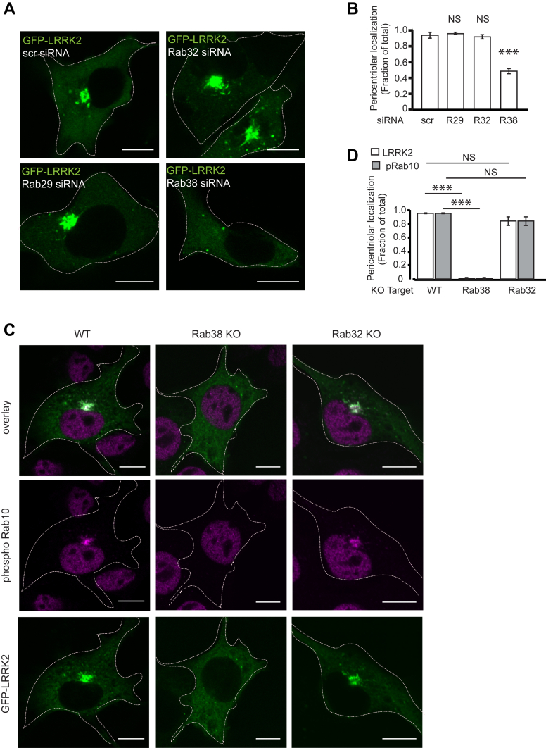
Figure 3 Endogenous Rab38 drives pericentriolar recruitment of overexpressed LRRK2.A, immunofluorescence confocal microscopy of GFP-LRRK2 in B16 cells with scrambled control siRNA (top left), knockdown of Rab29 (bottom left), knockdown of Rab32 (top right), or knockdown of Rab38 (bottom right). B, quantification of GFP-LRRK2 pericentriolar recruitment in (A). Quantification includes three replicates of ≥50 cells per knockdown condition. Percent of cells with pericentriolar GFP-LRRK2 was 94% ± 4% with control siRNA, 96% ± 1% with Rab29 knockdown, 92% ± 2% with Rab32 knockdown, and 49% ± 2% with Rab38 knockdown. Knockdown was quantified in Fig. S7A. C, immunofluorescence confocal microscopy of GFP-LRRK2 and phosphoT73-Rab10 in B16-F10 WT (left column), Rab38 KO (middle column), and Rab32 KO (right column) cells. GFP-LRRK2 and phosphorylated Rab10 colocalize at the pericentriolar region in WT and Rab32 KO cells but do not accumulate at the pericentriolar region in Rab38 KO cells. D, quantification of GFP-LRRK2 and phosphoT73-Rab10 pericentriolar localization in (C). Quantification includes three replicates of ≥50 cells per knockdown condition. Percent of cells with pericentriolar GFP-LRRK2 and phosphorylated Rab10 was 94.8% ± 0.7% in WT cells, 0.6% ± 0.6% in Rab38 KO cells, and 83.6% ± 6% in the Rab32 KO cells. All quantifications show mean with error bars showing SEM. Significance testing for panels B and D was performed using a two-tailed Student’s t test and panel D also used Bonferroni correction for multiple comparisons. Asterisks represent significant p-values in the following manner: ∗ = p < 0.05; ∗∗ = p < 0.01; ∗∗∗ = p < 0.001. Scale bars represent 10 μm. LRRK2, leucine-rich repeat kinase 2.
Figure 3 Endogenous Rab38 drives pericentriolar recruitment of overexpressed LRRK2.A, immunofluorescence confocal microscopy of GFP-LRRK2 in B16 cells with scrambled control siRNA (top left), knockdown of Rab29 (bottom left), knockdown of Rab32 (top right), or knockdown of Rab38 (bottom right). B, quantification of GFP-LRRK2 pericentriolar recruitment in (A). Quantification includes three replicates of ≥50 cells per knockdown condition. Percent of cells with pericentriolar GFP-LRRK2 was 94% ± 4% with control siRNA, 96% ± 1% with Rab29 knockdown, 92% ± 2% with Rab32 knockdown, and 49% ± 2% with Rab38 knockdown. Knockdown was quantified in Fig. S7A. C, immunofluorescence confocal microscopy of GFP-LRRK2 and phosphoT73-Rab10 in B16-F10 WT (left column), Rab38 KO (middle column), and Rab32 KO (right column) cells. GFP-LRRK2 and phosphorylated Rab10 colocalize at the pericentriolar region in WT and Rab32 KO cells but do not accumulate at the pericentriolar region in Rab38 KO cells. D, quantification of GFP-LRRK2 and phosphoT73-Rab10 pericentriolar localization in (C). Quantification includes three replicates of ≥50 cells per knockdown condition. Percent of cells with pericentriolar GFP-LRRK2 and phosphorylated Rab10 was 94.8% ± 0.7% in WT cells, 0.6% ± 0.6% in Rab38 KO cells, and 83.6% ± 6% in the Rab32 KO cells. All quantifications show mean with error bars showing SEM. Significance testing for panels B and D was performed using a two-tailed Student’s t test and panel D also used Bonferroni correction for multiple comparisons. Asterisks represent significant p-values in the following manner: ∗ = p < 0.05; ∗∗ = p < 0.01; ∗∗∗ = p < 0.001. Scale bars represent 10 μm. LRRK2, leucine-rich repeat kinase 2.CC BY · PubMed Central
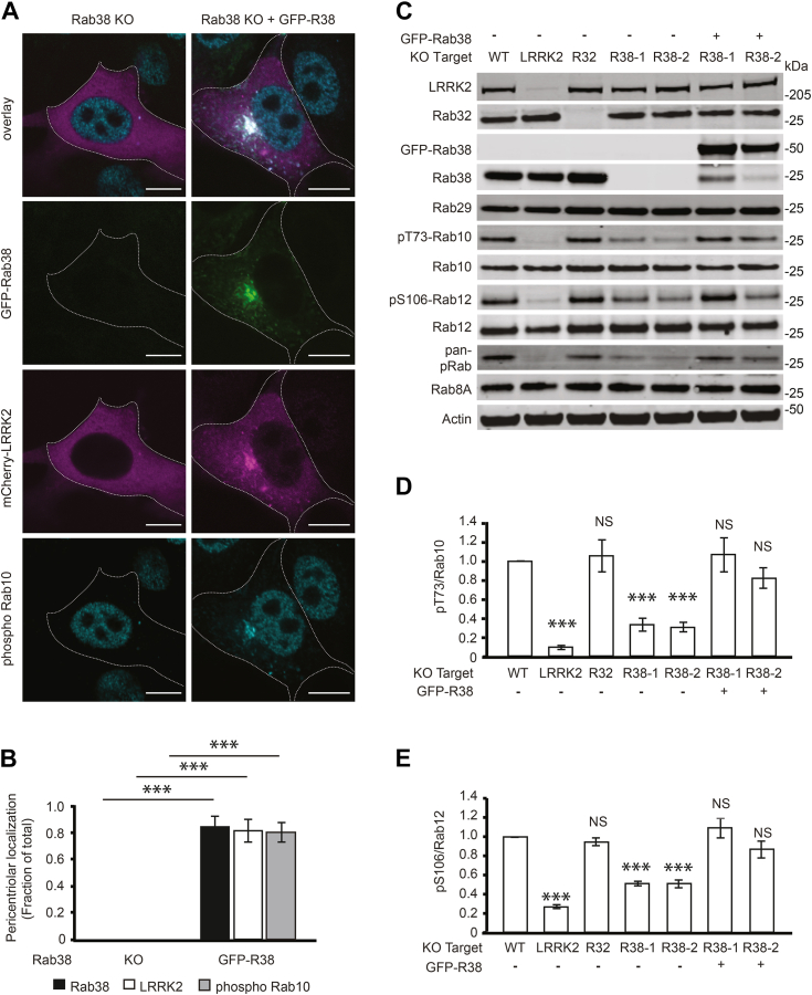
Figure 4 Overexpression of Rab38 in Rab38 KO cells rescues LRRK2 pericentriolar localization and substrate Rab phosphorylation.A, immunofluorescence confocal microscopy of GFP-Rab38 (green), mcherry-LRRK2 (magenta), and phosphorylated Rab10 (cyan) localization in B16 Rab38 KO cells or Rab38 KO cells with overexpression of GFP-Rab38. B, quantification of pericentriolar localization in (A). Quantification includes three replicates of ≥50 cells per condition. Percent of cells with pericentriolar GFP-Rab38 = 0% ± 0% in Rab38 knockout and 83.4% ± 8% in Rab38 knockout with overexpression of GFP-Rab38. Percent of cells with pericentriolar mCherry-LRRK2 = 0% ± 0% in Rab38 knockout and 80.2% ± 8% in Rab38 knockout with overexpression of GFP-Rab38. Percent of cells with pericentriolar phosphoT73-Rab10 = 0% ± 0% in Rab38 knockout and 79.0% ± 7% in Rab38 knockout with overexpression of GFP-Rab38. C, representative immunoblot of B16 melanocytes following CRISPR knockout of endogenous LRRK2, Rab32, and Rab38, as well as GFP-Rab38 overexpression in Rab38 KO lines to rescue the KO phenotype. Two independent Rab38 KO monoclonal lines arising from two different guide RNAs are shown. D, quantification of endogenous phospho T73-Rab10 from six independent experiments. pThr73-Rab10/total Rab10 = 8.8% ± 2% in LRRK2 knockout, 106.1% ± 17% in Rab32 knockout, 32.9% ± 7% in Rab38-1 knockout, 30.3% ± 5% in Rab38-2 knockout, 106.7% ± 18% in Rab38-1 knockout + GFP-Rab38, and 82.1% ± 11% in Rab38-2 knockout + GFP-Rab38 (mean ± SEM). E, quantification of endogenous phosphorylated Rab12 from six independent experiments. pSer106-Rab12/total Rab12 = 26.7% ± 2% in LRRK2 knockout, 94.9% ± 4% in Rab32 knockout, 51.2% ± 2% in Rab38-1 knockout, 51.0% ± 4% in Rab38-2 knockout, 109.3% ± 10% in Rab38-1 knockout + GFP-Rab38, and 86.8% ± 9% in Rab38-2 knockout + GFP-Rab38 (mean ± SEM). All quantifications show mean with error bars showing SEM. Significance testing for panels B, D, and E was performed using a two-tailed Student’s t test and Bonferroni correction for multiple comparisons. Asterisks represent significant p-values in the following manner: ∗ = p < 0.05; ∗∗ = p < 0.01; ∗∗∗ = p < 0.001. Scale bars represent 10 μm. LRRK2, leucine-rich repeat kinase 2.
Figure 4 Overexpression of Rab38 in Rab38 KO cells rescues LRRK2 pericentriolar localization and substrate Rab phosphorylation.A, immunofluorescence confocal microscopy of GFP-Rab38 (green), mcherry-LRRK2 (magenta), and phosphorylated Rab10 (cyan) localization in B16 Rab38 KO cells or Rab38 KO cells with overexpression of GFP-Rab38. B, quantification of pericentriolar localization in (A). Quantification includes three replicates of ≥50 cells per condition. Percent of cells with pericentriolar GFP-Rab38 = 0% ± 0% in Rab38 knockout and 83.4% ± 8% in Rab38 knockout with overexpression of GFP-Rab38. Percent of cells with pericentriolar mCherry-LRRK2 = 0% ± 0% in Rab38 knockout and 80.2% ± 8% in Rab38 knockout with overexpression of GFP-Rab38. Percent of cells with pericentriolar phosphoT73-Rab10 = 0% ± 0% in Rab38 knockout and 79.0% ± 7% in Rab38 knockout with overexpression of GFP-Rab38. C, representative immunoblot of B16 melanocytes following CRISPR knockout of endogenous LRRK2, Rab32, and Rab38, as well as GFP-Rab38 overexpression in Rab38 KO lines to rescue the KO phenotype. Two independent Rab38 KO monoclonal lines arising from two different guide RNAs are shown. D, quantification of endogenous phospho T73-Rab10 from six independent experiments. pThr73-Rab10/total Rab10 = 8.8% ± 2% in LRRK2 knockout, 106.1% ± 17% in Rab32 knockout, 32.9% ± 7% in Rab38-1 knockout, 30.3% ± 5% in Rab38-2 knockout, 106.7% ± 18% in Rab38-1 knockout + GFP-Rab38, and 82.1% ± 11% in Rab38-2 knockout + GFP-Rab38 (mean ± SEM). E, quantification of endogenous phosphorylated Rab12 from six independent experiments. pSer106-Rab12/total Rab12 = 26.7% ± 2% in LRRK2 knockout, 94.9% ± 4% in Rab32 knockout, 51.2% ± 2% in Rab38-1 knockout, 51.0% ± 4% in Rab38-2 knockout, 109.3% ± 10% in Rab38-1 knockout + GFP-Rab38, and 86.8% ± 9% in Rab38-2 knockout + GFP-Rab38 (mean ± SEM). All quantifications show mean with error bars showing SEM. Significance testing for panels B, D, and E was performed using a two-tailed Student’s t test and Bonferroni correction for multiple comparisons. Asterisks represent significant p-values in the following manner: ∗ = p < 0.05; ∗∗ = p < 0.01; ∗∗∗ = p < 0.001. Scale bars represent 10 μm. LRRK2, leucine-rich repeat kinase 2.CC BY · PubMed Central
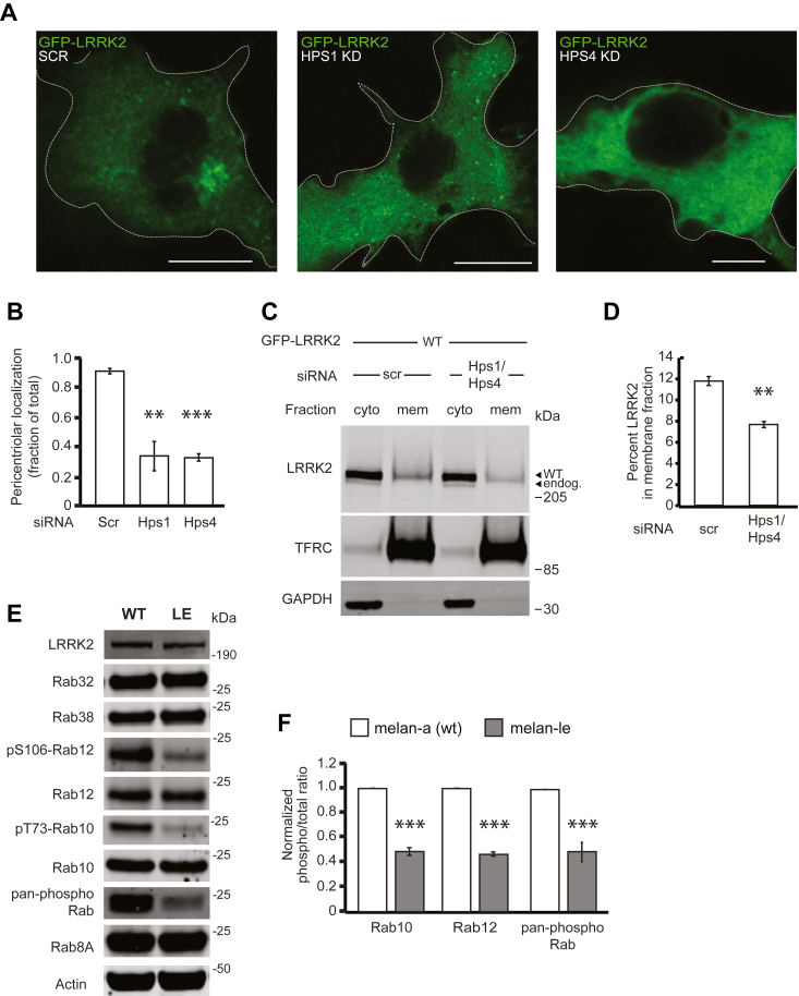
Figure 5 The guanine nucleotide exchange factor BLOC-3 regulates LRRK2 activity.A, immunofluorescence confocal microscopy of GFP-LRRK2 in B16 cells with scrambled control siRNA (left), knockdown of Hps1 (middle), or knockdown of Hps4 (right). B, quantification of GFP-LRRK2 in (A). Quantification includes three replicates of ≥50 cells per LRRK2 variant. Percent of cells with pericentriolar LRRK2 with control siRNA = 91% ± 2%, with Hps1 knockdown = 34% ± 10%, and with Hps4 knockdown = 33% ± 3%. Knockdown was quantified in Fig. S7B. C, immunoblot of cytoplasmic and membrane fractions of B16 cells expressing GFP-LRRK2 WT after knockdown with scrambled control siRNA or siRNAs targeting both Hps1 and Hps4. TFRC and GAPDH are used as membrane and cytoplasmic markers, respectively. D, quantification of membrane-associated GFP-LRRK2 in (C) from three independent experiments. After control siRNA, 11.8% ± 0.4% of GFP-LRRK2 WT was membrane-associated and after Hps1/Hps4 knockdown, 7.7% ± 0.3% of GFP-LRRK2 was membrane associated (mean ± SEM). Knockdown was quantified in Fig. S7C. E, representative immunoblot of WT melan-a and melan-le cells, which lack functional BLOC-3. F, quantification of endogenous phosphorylated Rab10, Rab12, and pan-phospho Rab levels from six independent experiments. Relative phosphorylation of Rabs in melan-le versus melan-a: pThr73-Rab10/total Rab10 = 42% ± 6%, pSer106-Rab12/total Rab12 = 47% ± 2% (mean ± SEM), pan-phospho Rab normalized to WT = 49% ± 8%. All quantifications show mean with error bars showing SEM. Significance testing for panels B, D, and F was performed using a two-tailed Student’s t test. Asterisks represent significant p-values in the following manner: ∗ = p < 0.05; ∗∗ = p < 0.01; ∗∗∗ = p < 0.001. Scale bars represent 10 μm.LRRK2, leucine-rich repeat kinase 2.
Figure 5 The guanine nucleotide exchange factor BLOC-3 regulates LRRK2 activity.A, immunofluorescence confocal microscopy of GFP-LRRK2 in B16 cells with scrambled control siRNA (left), knockdown of Hps1 (middle), or knockdown of Hps4 (right). B, quantification of GFP-LRRK2 in (A). Quantification includes three replicates of ≥50 cells per LRRK2 variant. Percent of cells with pericentriolar LRRK2 with control siRNA = 91% ± 2%, with Hps1 knockdown = 34% ± 10%, and with Hps4 knockdown = 33% ± 3%. Knockdown was quantified in Fig. S7B. C, immunoblot of cytoplasmic and membrane fractions of B16 cells expressing GFP-LRRK2 WT after knockdown with scrambled control siRNA or siRNAs targeting both Hps1 and Hps4. TFRC and GAPDH are used as membrane and cytoplasmic markers, respectively. D, quantification of membrane-associated GFP-LRRK2 in (C) from three independent experiments. After control siRNA, 11.8% ± 0.4% of GFP-LRRK2 WT was membrane-associated and after Hps1/Hps4 knockdown, 7.7% ± 0.3% of GFP-LRRK2 was membrane associated (mean ± SEM). Knockdown was quantified in Fig. S7C. E, representative immunoblot of WT melan-a and melan-le cells, which lack functional BLOC-3. F, quantification of endogenous phosphorylated Rab10, Rab12, and pan-phospho Rab levels from six independent experiments. Relative phosphorylation of Rabs in melan-le versus melan-a: pThr73-Rab10/total Rab10 = 42% ± 6%, pSer106-Rab12/total Rab12 = 47% ± 2% (mean ± SEM), pan-phospho Rab normalized to WT = 49% ± 8%. All quantifications show mean with error bars showing SEM. Significance testing for panels B, D, and F was performed using a two-tailed Student’s t test. Asterisks represent significant p-values in the following manner: ∗ = p < 0.05; ∗∗ = p < 0.01; ∗∗∗ = p < 0.001. Scale bars represent 10 μm.LRRK2, leucine-rich repeat kinase 2.CC BY · PubMed Central
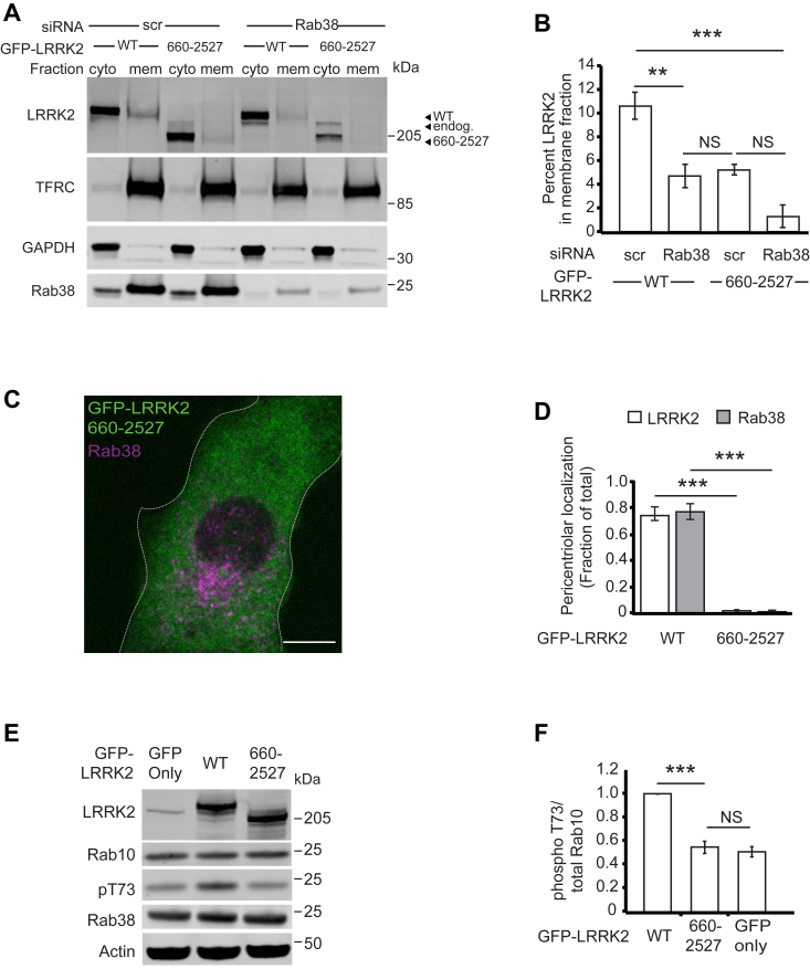
Figure 6 Disruption of Rab38 binding to LRRK2’s armadillo domain decreases LRRK2’s membrane association, pericentriolar recruitment, and kinase activity.A, immunoblot of cytoplasmic and membrane fractions of B16 cells expressing GFP-LRRK2 WT (full-length) or GFP-LRRK2660-2527 after Rab38 knockdown or scrambled control siRNA. TFRC and GAPDH are used as membrane and cytoplasmic markers, respectively. B, quantification of membrane-associated GFP-LRRK2 versus GFP-LRRK2660-2527 from four independent experiments. After control siRNA, 11% ± 1% of GFP-LRRK2 WT was membrane-associated, while after Rab38 knockdown, 5% ± 1% was membrane-associated (mean ± SEM). After control siRNA, 4% ± 1% of GFP-LRRK2660-2527 was membrane associated and after Rab38 knockdown, 2% ± 1% was membrane associated (mean ± SEM), which was not statistically significant. Knockdowns were quantified in Fig. S7E. C, immunofluorescence microscopy of GFP-LRRK2660-2527 (green) and endogenous Rab38 (magenta) in B16 cells. D, quantification of LRRK2 (white) and Rab38 (gray) pericentriolar recruitment in GFP-LRRK2 WT compared to GFP-LRRK2660-2527. Quantification includes three replicates of ≥50 cells per LRRK2 variant. The proportion of cells with pericentriolar GFP-LRRK2660-2527 was 2% ± 1% versus 74% ± 5% for GFP-LRRK2 WT (mean ± SEM). E, immunoblot of B16 cells expressing transiently transfected GFP alone, GFP-LRRK2 WT (full-length), and GFP-LRRK2660-2527. F, quantification of endogenous phosphorylated Rab10 levels (pThr73-Rab10/total Rab10) from four independent experiments. pThr73-Rab10 levels with GFP-LRRK2660-2527 = 54% ± 5% versus GFP alone = 51% ± 5%, with pThr73-Rab10 levels in the presence of GFP-LRRK2 WT set to 100%. All quantifications show mean with error bars showing SEM. Significance testing for panels B, D, and F was performed using a two-tailed Student’s t test and panel B also used Bonferroni correction for multiple comparisons. Asterisks represent significant p-values in the following manner: ∗ = p < 0.05; ∗∗ = p < 0.01; ∗∗∗ = p < 0.001. Scale bars represent 10 μm. LRRK2, leucine-rich repeat kinase 2.
Figure 6 Disruption of Rab38 binding to LRRK2’s armadillo domain decreases LRRK2’s membrane association, pericentriolar recruitment, and kinase activity.A, immunoblot of cytoplasmic and membrane fractions of B16 cells expressing GFP-LRRK2 WT (full-length) or GFP-LRRK2660-2527 after Rab38 knockdown or scrambled control siRNA. TFRC and GAPDH are used as membrane and cytoplasmic markers, respectively. B, quantification of membrane-associated GFP-LRRK2 versus GFP-LRRK2660-2527 from four independent experiments. After control siRNA, 11% ± 1% of GFP-LRRK2 WT was membrane-associated, while after Rab38 knockdown, 5% ± 1% was membrane-associated (mean ± SEM). After control siRNA, 4% ± 1% of GFP-LRRK2660-2527 was membrane associated and after Rab38 knockdown, 2% ± 1% was membrane associated (mean ± SEM), which was not statistically significant. Knockdowns were quantified in Fig. S7E. C, immunofluorescence microscopy of GFP-LRRK2660-2527 (green) and endogenous Rab38 (magenta) in B16 cells. D, quantification of LRRK2 (white) and Rab38 (gray) pericentriolar recruitment in GFP-LRRK2 WT compared to GFP-LRRK2660-2527. Quantification includes three replicates of ≥50 cells per LRRK2 variant. The proportion of cells with pericentriolar GFP-LRRK2660-2527 was 2% ± 1% versus 74% ± 5% for GFP-LRRK2 WT (mean ± SEM). E, immunoblot of B16 cells expressing transiently transfected GFP alone, GFP-LRRK2 WT (full-length), and GFP-LRRK2660-2527. F, quantification of endogenous phosphorylated Rab10 levels (pThr73-Rab10/total Rab10) from four independent experiments. pThr73-Rab10 levels with GFP-LRRK2660-2527 = 54% ± 5% versus GFP alone = 51% ± 5%, with pThr73-Rab10 levels in the presence of GFP-LRRK2 WT set to 100%. All quantifications show mean with error bars showing SEM. Significance testing for panels B, D, and F was performed using a two-tailed Student’s t test and panel B also used Bonferroni correction for multiple comparisons. Asterisks represent significant p-values in the following manner: ∗ = p < 0.05; ∗∗ = p < 0.01; ∗∗∗ = p < 0.001. Scale bars represent 10 μm. LRRK2, leucine-rich repeat kinase 2.CC BY · PubMed Central
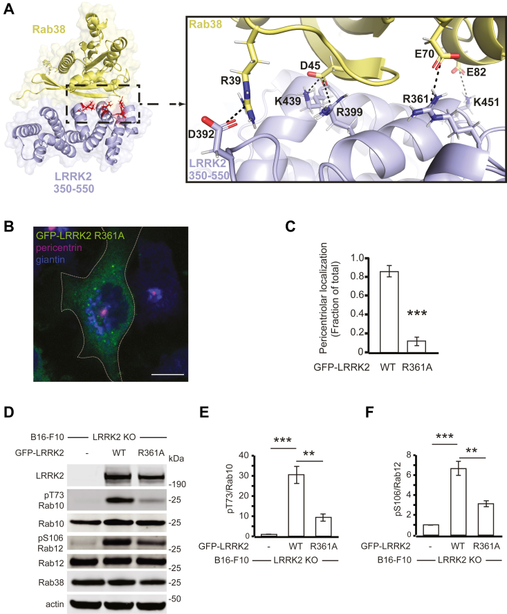
Figure 7 Disruption of the predicted Rab38-interacting LRRK2 residue R361 decreases LRRK2 pericentriolar recruitment and substrate Rab phosphorylation.A, model of LRRK2-Rab38–interacting domain (LRRK2350-550 in purple, Rab38 in yellow, predicted binding interface in red) with box showing details of key intermolecular electrostatic interactions; modeling of LRRK2350-550 and Rab38 complex binding was performed employing ColabFold using MMseqs2 for sequence alignment and AlphaFold2-multimer-v2 model for complex prediction; structure visualized using PyMOL. B, immunofluorescence confocal microscopy of GFP-LRRK2R361A (green), pericentrin (purple), and giantin (blue) in B16 cells. C, quantification of LRRK2 pericentriolar recruitment in GFP-LRRK2 WT versus GFP-LRRK2R361A. Quantification includes three replicates of ≥50 cells per LRRK2 variant. Percent of cells with pericentriolar GFP-LRRK2 R361A = 12% ± 4% versus 85% ± 6% for GFP-LRRK2 WT (mean ± SEM). D, immunoblot of B16 LRRK2 KO cells expressing transiently transfected GFP-LRRK2 WT (full-length) or GFP-LRRK2R361A mutant. E, quantification of endogenous Rab10 phosphorylation in (D) from five independent experiments. Relative to the nontransfected control, pThr73-Rab10/total Rab10 was increased 30.4 ± 4.2 -fold in cells expressing GFP-LRRK2 WT versus 9.3 ± 1.8-fold in cells expressing GFP-LRRK2 R361A. F, quantification of endogenous Rab12 phosphorylation in (D) from five independent experiments. Relative to the nontransfected control, pSer106-Rab12/total Rab12 was increased 6.7 ± 1.5 -fold in cells expressing GFP-LRRK2 WT versus 3.1 ± 0.7-fold in cells expressing GFP-LRRK2 R361A. All quantifications show mean with error bars showing SEM. Significance testing for panels C, E, and F was performed using a two-tailed Student’s t test. Asterisks represent significant p-values in the following manner: ∗ = p < 0.05; ∗∗ = p < 0.01; ∗∗∗ = p < 0.001. Scale bars represent 10 μm. LRRK2, leucine-rich repeat kinase 2.
Figure 7 Disruption of the predicted Rab38-interacting LRRK2 residue R361 decreases LRRK2 pericentriolar recruitment and substrate Rab phosphorylation.A, model of LRRK2-Rab38–interacting domain (LRRK2350-550 in purple, Rab38 in yellow, predicted binding interface in red) with box showing details of key intermolecular electrostatic interactions; modeling of LRRK2350-550 and Rab38 complex binding was performed employing ColabFold using MMseqs2 for sequence alignment and AlphaFold2-multimer-v2 model for complex prediction; structure visualized using PyMOL. B, immunofluorescence confocal microscopy of GFP-LRRK2R361A (green), pericentrin (purple), and giantin (blue) in B16 cells. C, quantification of LRRK2 pericentriolar recruitment in GFP-LRRK2 WT versus GFP-LRRK2R361A. Quantification includes three replicates of ≥50 cells per LRRK2 variant. Percent of cells with pericentriolar GFP-LRRK2 R361A = 12% ± 4% versus 85% ± 6% for GFP-LRRK2 WT (mean ± SEM). D, immunoblot of B16 LRRK2 KO cells expressing transiently transfected GFP-LRRK2 WT (full-length) or GFP-LRRK2R361A mutant. E, quantification of endogenous Rab10 phosphorylation in (D) from five independent experiments. Relative to the nontransfected control, pThr73-Rab10/total Rab10 was increased 30.4 ± 4.2 -fold in cells expressing GFP-LRRK2 WT versus 9.3 ± 1.8-fold in cells expressing GFP-LRRK2 R361A. F, quantification of endogenous Rab12 phosphorylation in (D) from five independent experiments. Relative to the nontransfected control, pSer106-Rab12/total Rab12 was increased 6.7 ± 1.5 -fold in cells expressing GFP-LRRK2 WT versus 3.1 ± 0.7-fold in cells expressing GFP-LRRK2 R361A. All quantifications show mean with error bars showing SEM. Significance testing for panels C, E, and F was performed using a two-tailed Student’s t test. Asterisks represent significant p-values in the following manner: ∗ = p < 0.05; ∗∗ = p < 0.01; ∗∗∗ = p < 0.001. Scale bars represent 10 μm. LRRK2, leucine-rich repeat kinase 2.CC BY · PubMed Central
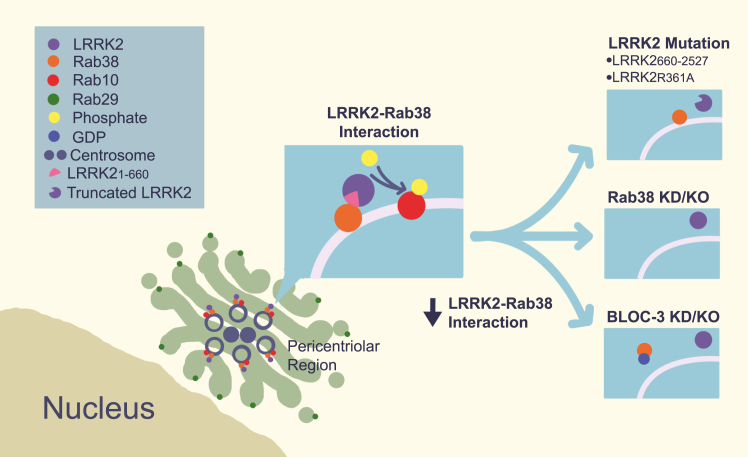
Figure 8 Schematic of LRRK2–Rab38 interactions and functional effects. LRRK2 colocalizes with Rab38 and phosphorylated Rab10 in the pericentriolar region (adjacent to Golgi-localized Rab29) in a Rab38-dependent manner. Disruption of (1) the LRRK2-Rab38 binding interface (top right), (2) Rab38 protein expression (middle right), or (3) Rab38’s active GTP-bound state (bottom right) blocks LRRK2 recruitment to the pericentriolar region and LRRK2’s phosphorylation of substrate Rabs. LRRK2, leucine-rich repeat kinase 2.
Figure 8 Schematic of LRRK2–Rab38 interactions and functional effects. LRRK2 colocalizes with Rab38 and phosphorylated Rab10 in the pericentriolar region (adjacent to Golgi-localized Rab29) in a Rab38-dependent manner. Disruption of (1) the LRRK2-Rab38 binding interface (top right), (2) Rab38 protein expression (middle right), or (3) Rab38’s active GTP-bound state (bottom right) blocks LRRK2 recruitment to the pericentriolar region and LRRK2’s phosphorylation of substrate Rabs. LRRK2, leucine-rich repeat kinase 2.CC BY · PubMed Central

Verification

Claims the paper makes

The claims and statistics of this paper haven’t been checked yet.

Run the check — free, no account needed. It reads the open-access text and checks the sources it cites.

A few free checks a day · papers already verified open instantly · results are public

Self-check

Percentages, sums and reported differences re-computed from the paper’s own numbers.

The self-check needs the paper’s full text.

Citations

Cites

Show all 1 references

Research works printed but not linked: 48

  1. 10.1038/ng.487 · DOI · Paper page
  2. Usmani, A. , Shavarebi, F. & Hiniker, A . The Cell Biology of LRRK2 in Parkinson’s Disease. Mol Cell Biol 41 (2021). · DOI
  3. Stormo, A.E.D. et al. The E3 ligase TRIM1 ubiquitinates LRRK2 and controls its localization, degradation, and toxicity. J Cell Biol 221 (2022). · DOI
  4. Steger, M. et al. Systematic proteomic analysis of LRRK2-mediated Rab GTPase phosphorylation establishes a connection to ciliogenesis. eLife 6 (2017). · DOI · Paper page
  5. Steger, M. et al. Phosphoproteomics reveals that Parkinson’s disease kinase LRRK2 regulates a subset of Rab GTPases. eLife 5 (2016). · DOI · Paper page
  6. 10.1093/hmg/ddz201 · DOI · Paper page
  7. Dhekne, H.S. et al. A pathway for Parkinson’s Disease LRRK2 kinase to block primary cilia and Sonic hedgehog signaling in the brain. eLife 7 (2018). · DOI · Paper page
  8. Sobu, Y. , Wawro, P.S. , Dhekne, H.S. , Yeshaw, W.M. & Pfeffer, S.R . Pathogenic LRRK2 regulates ciliation probability upstream of tau tubulin kinase 2 via Rab10 and RILPL1 proteins. Proceedings of the National Academy of Sciences of the United States of America 118 (2021). · DOI
  9. Khan, S.S. et al. Pathogenic LRRK2 control of primary cilia and Hedgehog signaling in neurons and astrocytes of mouse brain. eLife 10 (2021). · DOI · Paper page
  10. 10.1016/j.cub.2018.02.010 · DOI
  11. 10.15252/embj.201798099 · DOI · Paper page
  12. 2016, LRRK2 and RAB7L1 coordinately regulate axonal morphology and lysosome integrity in diverse cellular contexts, Scientific reports, 6, 29945, 10.1038/srep29945 · DOI · Paper page
  13. 10.1016/j.neuron.2012.11.033 · DOI · Paper page
  14. McGrath, E. , Waschbusch, D. , Baker, B.M. & Khan, A.R . LRRK2 binds to the Rab32 subfamily in a GTP-dependent manner via its armadillo domain. Small GTPases, 1–14 (2019). · DOI
  15. Vides, E.G. et al. A feed-forward pathway drives LRRK2 membrane recruitment and apparent activation. bioRxiv doi: https://doi.org/10.1101/2022.04.25.489459. · DOI · Paper page
  16. Zhu, H. , Tonelli, F. , Alessi, D.R. & Sun, J. Structural basis of human LRRK2 membrane recruitment and activation. BioRxiv (2022). · DOI · Paper page
  17. 10.1083/jcb.201902184 · DOI · Paper page
  18. 2021, Combined Knockout of Lrrk2 and Rab29 Does Not Result in Behavioral Abnormalities in vivo, J Parkinsons Dis, 11, 569, 10.3233/JPD-202172 · DOI
  19. 2020, Endogenous Rab29 does not impact basal or stimulated LRRK2 pathway activity, The Biochemical journal, 477, 4397, 10.1042/BCJ20200458 · DOI
  20. 10.1016/j.nbd.2020.105081 · DOI · Paper page
  21. 10.1073/pnas.1812196115 · DOI
  22. 10.1371/journal.pone.0111632 · DOI
  23. 10.1073/pnas.072087599 · DOI
  24. 10.1083/jcb.200606050 · DOI · Paper page
  25. Tian, Y. et al. LRRK2 plays essential roles in maintaining lung homeostasis and preventing the development of pulmonary fibrosis. Proceedings of the National Academy of Sciences of the United States of America 118 (2021). · DOI
  26. 10.1093/hmg/ddr348 · DOI
  27. 10.1042/BCJ20170803 · DOI · Paper page
  28. 10.1126/scitranslmed.aaa3634 · DOI
  29. Baptista, M.A.S. et al. LRRK2 inhibitors induce reversible changes in nonhuman primate lungs without measurable pulmonary deficits. Science translational medicine 12 (2020). · DOI
  30. 2017, Exogenous gene transfer of Rab(3)8 small GTPase ameliorates aberrant lung surfactant homeostasis in Ruby rats, Respir Res, 18, 70, 10.1186/s12931-017-0549-2 · DOI
  31. 10.1152/ajplung.00242.2009 · DOI
  32. 10.2353/ajpath.2008.080056 · DOI
  33. 10.1002/ijc.2910390324 · DOI
  34. 10.1093/hmg/ddu138 · DOI · Paper page
  35. 10.1021/bi100157u · DOI
  36. 2018, Parkinson disease-associated mutations in LRRK2 cause centrosomal defects via Rab8a phosphorylation, Molecular neurodegeneration, 13, 3, 10.1186/s13024-018-0235-y · DOI
  37. 10.1016/j.ab.2007.05.025 · DOI
  38. 10.1080/21541248.2019.1666623 · DOI
  39. Mirdita, M. et al. ColabFold - Making protein folding accessible to all. bioRxiv 2021.08.15.456425; doi: https://doi.org/10.1101/2021.08.15.456425. · DOI · Paper page
  40. 10.1016/j.cub.2012.09.020 · DOI
  41. 10.1074/jbc.M300090200 · DOI
  42. 10.1073/pnas.1532040100 · DOI
  43. 10.1091/mbc.E16-10-0737 · DOI
  44. 10.1111/tra.12646 · DOI · Paper page
  45. 10.1074/jbc.M112.351908 · DOI · Paper page
  46. 10.1126/science.1229224 · DOI
  47. Osanai, K . Rab38 Mutation and the Lung Phenotype. Int J Mol Sci 19 (2018). · DOI
  48. 2022, The development of inhibitors of leucine-rich repeat kinase 2 (LRRK2) as a therapeutic strategy for Parkinson’s disease: the current state of play, Br J Pharmacol, 179, 1478, 10.1111/bph.15575 · DOI

Metadata

Authors
Unapanta, A., Shavarebi, F., Porath, J., Balen, C., Nguyen, A., Tseng, J., Shen, Y., Liu, M., Lis, P., Di Pietro, S. M., & Hiniker, A.
Affiliations
University of California San Diego (US)
Corresponding author
Hiniker, Annie
Venue
Journal of Biological Chemistry
Publisher
openRxiv
Published
2022-06-20
Volume
299
Issue
10
Pages
105192-105192
ISSN
0021-9258 (ISSN-L)
OpenAlex
W4386101863
Cited by
11
References
1
Citations by year
2026: 1 · 2025: 2 · 2024: 6 · 2023: 2
FWCI
1.87
Type
journal article
Language
en
Open access
Licence
cc-by
Locations
  • Journal of Biological Chemistry (publishedVersion, cc-by)
  • bioRxiv (Cold Spring Harbor Laboratory) (acceptedVersion)
  • PubMed Central (publishedVersion, cc-by)
  • PubMed (publishedVersion)
  • Discovery Research Portal (University of Dundee) (submittedVersion, cc-by)
Funders
  • Alzheimer's Association (AACSF-20-684991)
  • National Institutes of Health
  • National Institute of Neurological Disorders and Stroke (K08NS090633, R01NS135607)
Topics
  • Health Sciences › Medicine › Clinical Neurology › Parkinson's Disease Mechanisms and Treatments
  • Life Sciences › Biochemistry, Genetics and Molecular Biology › Cell Biology › Cellular transport and secretion
  • Life Sciences › Biochemistry, Genetics and Molecular Biology › Molecular Biology › Protein Kinase Regulation and GTPase Signaling
Keywords
Rab38, LRRK2, Parkinson's disease, melanocytes, Rab GTPases, membrane recruitment, substrate phosphorylation, Rab10, Rab12, guanine nucleotide exchange factor, armadillo domain, vesicle trafficking, Rab32, Rab29
MeSH terms
Leucine-Rich Repeat Serine-Threonine Protein Kinase-2, Protein Domains, Animals, Golgi Apparatus, Humans, Intracellular Membranes, Melanocytes, Mutation, Parkinson Disease, Phosphorylation, Protein Binding, Gene Expression, Guanine Nucleotide Exchange Factors, rab GTP-Binding Proteins, Mice, HEK293 Cells

About these checks

Registry, retraction, Cites and statistics results are computed, not judged, with one exception: a printed reference that no index lists is read once by a model before it counts as a citation error. Claims are extracted by a model and kept only when their quote appears verbatim in the text; each cited source is then checked, with a model’s help, for whether it says what the claim relies on. Those verdicts can be wrong — read the claim’s quote and the cited source before relying on one.

Statistics and arithmetic are recomputed, not judged.

Confidence bands are shown only for fields calibrated on our annotated test set. None is calibrated yet, so no band is shown.

What counts as a claim ›How we measure confidence ›How we check this ›

Are you an author, or do you see a problem with this page? Request a review

How to cite

Cite CoChat for the checks on this page, and the paper for its own findings.

This page
CoChat. (2026). Citation check: Endogenous Rab38 regulates LRRK2’s membrane recruitment and substrate Rab phosphorylation in melanocytes. https://app.cochat.ai/papers/10.1101/2022.06.20.496629
The paper
Unapanta, A., Shavarebi, F., Porath, J., Balen, C., Nguyen, A., Tseng, J., Shen, Y., Liu, M., Lis, P., Di Pietro, S. M., & Hiniker, A. (2022). Endogenous Rab38 regulates LRRK2’s membrane recruitment and substrate Rab phosphorylation in melanocytes. Journal of Biological Chemistry. https://doi.org/10.1101/2022.06.20.496629