PreprintCited by 7Endoplasmic Reticulum Stress and DiseaseRNA and protein synthesis mechanismsProtein Structure and Dynamics

The DOI matches its Crossref record; 1 of the 1 references it cites were found in a registry or an index.

Substrate recruitment via eIF2γ enhances catalytic efficiency of a holophosphatase that terminates the Integrated Stress Response

Open access
Yan, Yahui, Shetty, Maithili, Harding, Heather P., George, Ginto, Zyryanova, Alisa, Labbé, Katherine, Mafi, Amirhossein, Hao, Qi, Sidrauski, Carmela, Ron, David·2023·Proceedings of the National Academy of Sciences

Preprint — published as 10.1073/pnas.2320013121 · Paper page

Abstract

Dephosphorylation of pSer51 of the α subunit of translation initiation factor 2 (eIF2α P ) terminates signalling in the integrated stress response (ISR). A trimeric mammalian holophosphatase comprised of a PP1 catalytic subunit, the conserved C-terminally located ∼70 amino acid core of a substrate-specific regulatory subunit (PPP1R15A/GADD34 or PPP1R15B/CReP) and G-actin (an essential co-factor) efficiently dephosphorylate eIF2α P in vitro. Unlike their viral or invertebrate counterparts, with whom they share the conserved 70 residue core, the mammalian PPP1R15s are large proteins of more than 600 residues. Genetic and cellular observations point to a functional role for regions outside the conserved core of mammalian PPP1R15A in dephosphorylating its natural substrate, the eIF2 trimer. We have combined deep learning technology, all-atom molecular dynamics simulations, X-Ray crystallography and biochemistry to uncover binding of the γ subunit of eIF2 to a short helical peptide repeated four times in the functionally-important N-terminus of human PPP1R15A that extends past its conserved core. Binding entails insertion of Phe and Trp residues that project from one face of an α-helix formed by the conserved repeats of PPP1R15A into a hydrophobic groove exposed on the surface of eIF2γ in the eIF2 trimer. Replacing these conserved Phe and Trp residues with Ala compromises PPP1R15A function in cells and in vitro. These findings suggest mechanisms by which contacts between a distant subunit of eIF2 and elements of PPP1R15A distant to the holophosphatase active site contribute to dephosphorylation of eIF2α P by the core PPP1R15 holophosphatase and to efficient termination of the ISR in mammals.

Overview

The claims and statistics of this paper haven’t been checked yet.

Run the check — free, no account needed. It reads the open-access text and checks the sources it cites.

A few free checks a day · papers already verified open instantly · results are public

Integrity signals

Registry

Crossref · 10.1101/2023.12.15.571226

Cites

1 of 1 references verified0 citation errors · 0 not checkable

Statistics

Figures

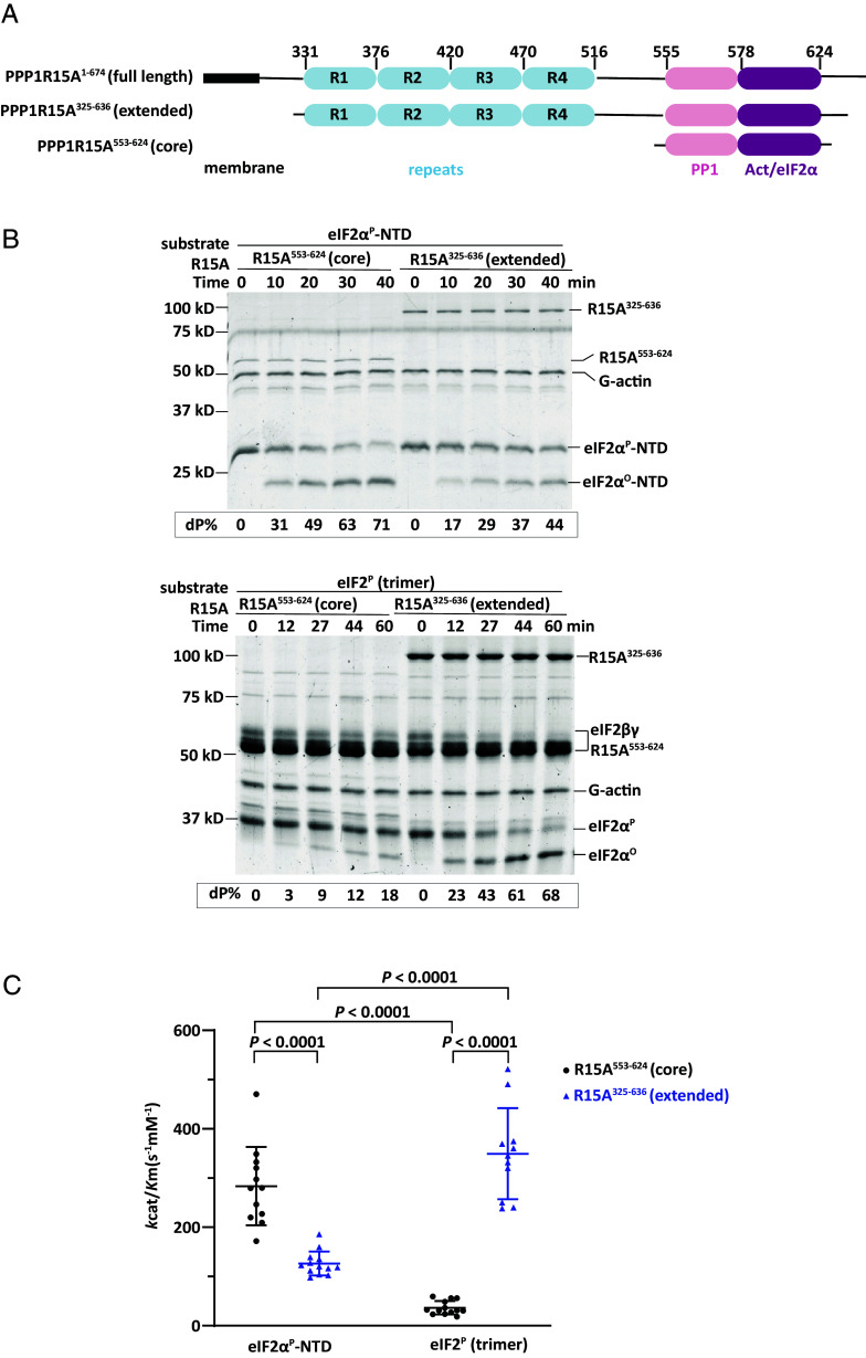
Fig. 1. Different substrate preferences of the core and extended PPP1R15A-containing holophosphatase. (A) Schema of human PPP1R15A. Shown is the N-terminal hydrophobic membrane interacting domain, the repeats at the central domain, and the C-terminal functional core region that recruits PP1 and G-actin. Elements present in the bacterially expressed N-terminally extended and core PPP1R15A used experimentally are shown alongside the corresponding residue numbers (based on UniProt O75807). (B) Coomassie-stained sodium dodecyl sulfate–polyacrylamide gel electrophoresis (SDS-PAGE) PhosTag gels of dephosphorylation reactions of the core substrate (eIF2αP-NTD) or the physiological substrate (eIF2P trimer) by the core holophosphatase (comprised of PPP1R15A533-624, G-actin, and PP1A) or the extended holophosphatase (comprised of PPP1R15A325-636, G-actin, and PP1A). Shown is a representative of experiments reproduced three times. (C) Graphic display of the mean ± SD of the kcat/Km extracted from all experimental points with the indicated substrates and holophosphatases. P values for the two-tailed parametric t test are shown.
Fig. 1. Different substrate preferences of the core and extended PPP1R15A-containing holophosphatase. (A) Schema of human PPP1R15A. Shown is the N-terminal hydrophobic membrane interacting domain, the repeats at the central domain, and the C-terminal functional core region that recruits PP1 and G-actin. Elements present in the bacterially expressed N-terminally extended and core PPP1R15A used experimentally are shown alongside the corresponding residue numbers (based on UniProt O75807). (B) Coomassie-stained sodium dodecyl sulfate–polyacrylamide gel electrophoresis (SDS-PAGE) PhosTag gels of dephosphorylation reactions of the core substrate (eIF2αP-NTD) or the physiological substrate (eIF2P trimer) by the core holophosphatase (comprised of PPP1R15A533-624, G-actin, and PP1A) or the extended holophosphatase (comprised of PPP1R15A325-636, G-actin, and PP1A). Shown is a representative of experiments reproduced three times. (C) Graphic display of the mean ± SD of the kcat/Km extracted from all experimental points with the indicated substrates and holophosphatases. P values for the two-tailed parametric t test are shown.CC BY · PubMed Central
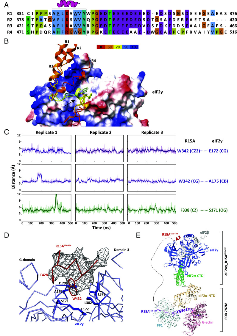
Fig. 2. The structural basis of PPP1R15A binding to the γ subunit of eIF2. (A) Alignment of the four repeats region of human PPP1R15A with conserved residues colored. The helical region that stably interacts with eIF2γ is boxed and the Phe and Trp residues whose sidechains contact eIF2γ are underlined. (B) Overlaid models of individual repeat (R1, R2, R3 or R4) complexed with eIF2γ as predicted by AFM. eIF2γ is shown as electrostatic surface with blue positive-charged and red negative-charged patches. Repeats peptides are shown as cartoon and colored by pLDDT score. The arrow points at the helix from which the Phe and Trp sidechains face the hydrophobic groove of eIF2γ. (C) Plots of time-dependent variation of distances between the atoms of R15A F338 or W342 and eIF2γ residues (as indicated) throughout 500 ns of an all-atom MD simulations performed using the AFM predicted complex structure of eIF2γ and repeat 1 (PPP1R15A331-376). Shown are three replicates of the simulation. (D) A local line diagram view of the crystal structure of the eIF2αγ and R3420-452 peptide complex. The gray mesh around the R15A426-434 (R3 peptide) is the polder omit map contoured at 3σ, and interacted residues in the eIF2γ hydrophobic groove are shown as sticks, into which the conserved Phe428 and Trp432 of R15A project their sidechains. (E) Modeling of the complete holophosphatase and eIF2 trimer complex. The crystal structure of the eIF2αγ and R3420-452 peptide complex is overlaid with the core phosphatase-substrate complex (PDB 7NZM) through an AFM predicted eIF2 trimer. The components are colored as indicated and the long dashed line represents the disordered region between the repeats domain and the PP1 RVxF binding motif of R15A.
Fig. 2. The structural basis of PPP1R15A binding to the γ subunit of eIF2. (A) Alignment of the four repeats region of human PPP1R15A with conserved residues colored. The helical region that stably interacts with eIF2γ is boxed and the Phe and Trp residues whose sidechains contact eIF2γ are underlined. (B) Overlaid models of individual repeat (R1, R2, R3 or R4) complexed with eIF2γ as predicted by AFM. eIF2γ is shown as electrostatic surface with blue positive-charged and red negative-charged patches. Repeats peptides are shown as cartoon and colored by pLDDT score. The arrow points at the helix from which the Phe and Trp sidechains face the hydrophobic groove of eIF2γ. (C) Plots of time-dependent variation of distances between the atoms of R15A F338 or W342 and eIF2γ residues (as indicated) throughout 500 ns of an all-atom MD simulations performed using the AFM predicted complex structure of eIF2γ and repeat 1 (PPP1R15A331-376). Shown are three replicates of the simulation. (D) A local line diagram view of the crystal structure of the eIF2αγ and R3420-452 peptide complex. The gray mesh around the R15A426-434 (R3 peptide) is the polder omit map contoured at 3σ, and interacted residues in the eIF2γ hydrophobic groove are shown as sticks, into which the conserved Phe428 and Trp432 of R15A project their sidechains. (E) Modeling of the complete holophosphatase and eIF2 trimer complex. The crystal structure of the eIF2αγ and R3420-452 peptide complex is overlaid with the core phosphatase-substrate complex (PDB 7NZM) through an AFM predicted eIF2 trimer. The components are colored as indicated and the long dashed line represents the disordered region between the repeats domain and the PP1 RVxF binding motif of R15A.CC BY · PubMed Central
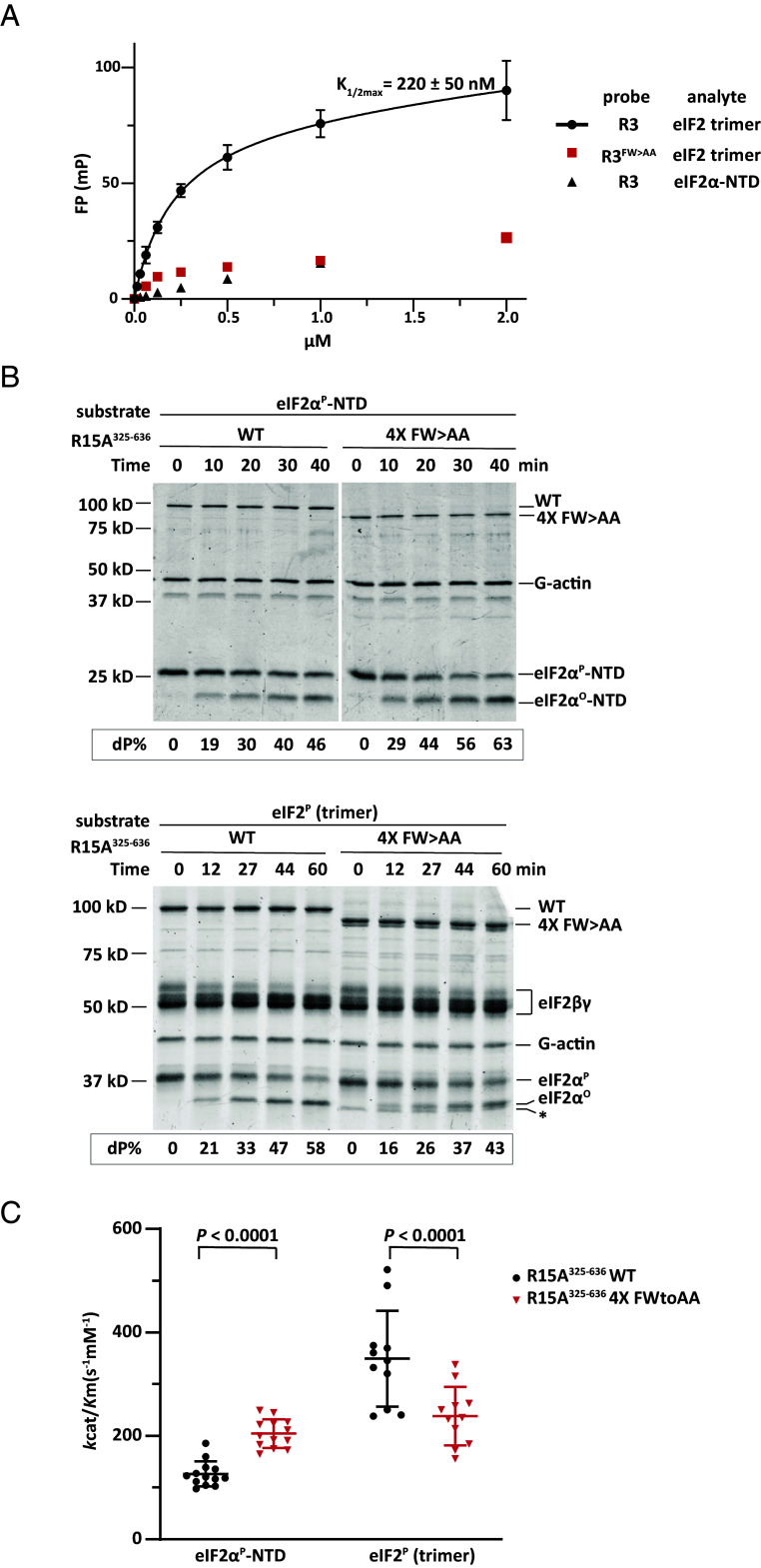
Fig. 3. The eIF2γ-contacting Phe and Trp residues of PPP1R15A contribute to eIF2 binding and eIF2P dephosphorylation in vitro. (A) FP signal as a function of analyte concentrations. Traces from pairings of the wild-type and FW > AA mutant fluorescein labeled R3420-466 peptide (as probes) with eIF2 trimer or eIF2α-NTD (an inert reference) are labeled. The binding of the wild-type probe to eIF2 has been fitted to a one-site binding model and K1/2 max ± 95% CI is indicated. Shown are the mean ± SD from three experiments. (B) Coomassie-stained SDS-PAGE PhosTag gels of dephosphorylation reactions of the core substrate (eIF2αP-NTD) or the physiological substrate (eIF2P trimer) by a holophosphatase comprised of the extended wild type or 4× FW > AA mutant (F338A, W342A, F386A, W389A, F428A, W432A, F479A, W482A) PPP1R15A5325-636, G-actin, and PP1A. Shown is a representative of experiments reproduced three times. (C) Graphic display of the mean ± SD of the kcat/Km extracted from all experimental points with the indicated substrates and holophosphatases. P values for the two-tailed parametric t test are shown.
Fig. 3. The eIF2γ-contacting Phe and Trp residues of PPP1R15A contribute to eIF2 binding and eIF2P dephosphorylation in vitro. (A) FP signal as a function of analyte concentrations. Traces from pairings of the wild-type and FW > AA mutant fluorescein labeled R3420-466 peptide (as probes) with eIF2 trimer or eIF2α-NTD (an inert reference) are labeled. The binding of the wild-type probe to eIF2 has been fitted to a one-site binding model and K1/2 max ± 95% CI is indicated. Shown are the mean ± SD from three experiments. (B) Coomassie-stained SDS-PAGE PhosTag gels of dephosphorylation reactions of the core substrate (eIF2αP-NTD) or the physiological substrate (eIF2P trimer) by a holophosphatase comprised of the extended wild type or 4× FW > AA mutant (F338A, W342A, F386A, W389A, F428A, W432A, F479A, W482A) PPP1R15A5325-636, G-actin, and PP1A. Shown is a representative of experiments reproduced three times. (C) Graphic display of the mean ± SD of the kcat/Km extracted from all experimental points with the indicated substrates and holophosphatases. P values for the two-tailed parametric t test are shown.CC BY · PubMed Central
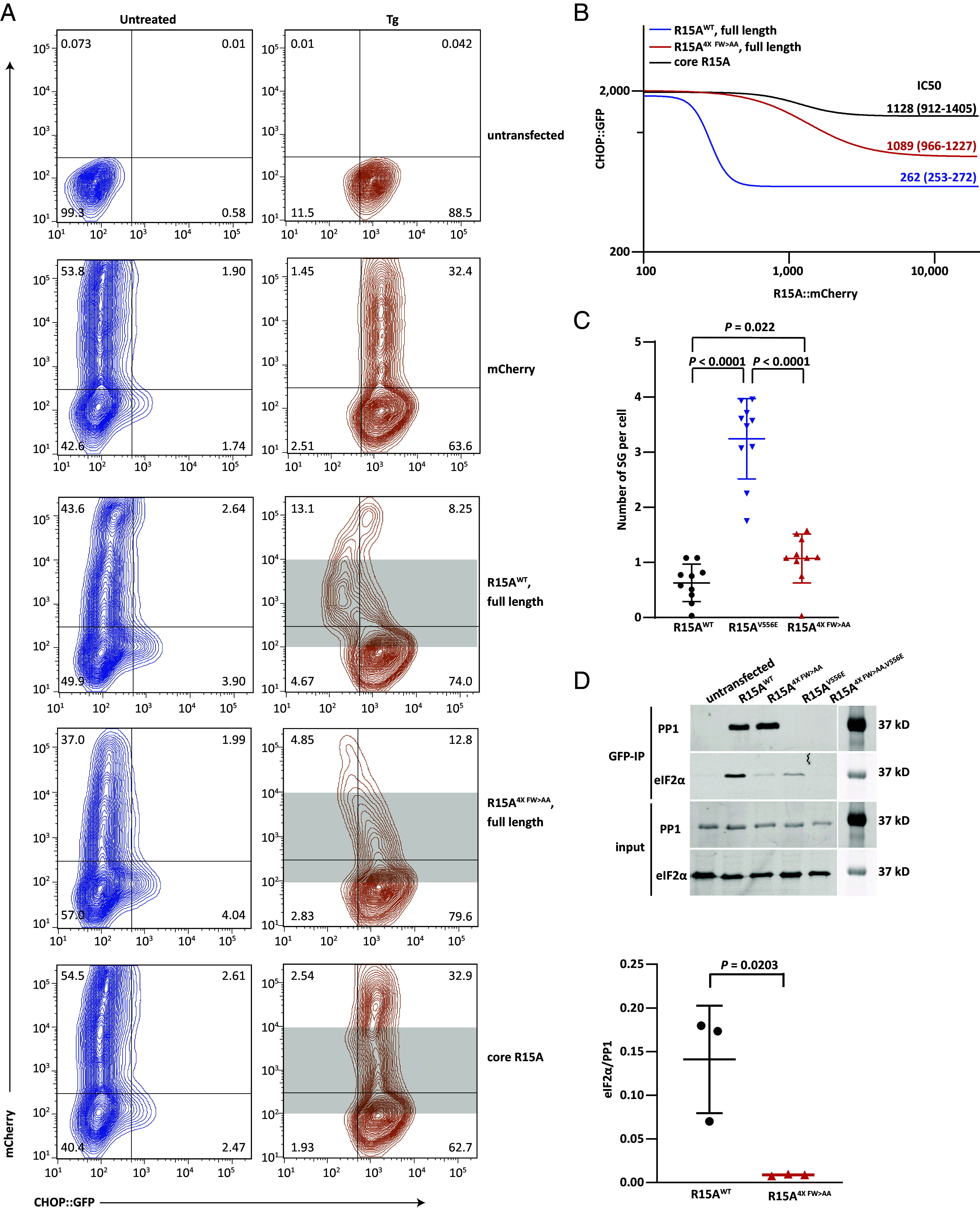
Fig. 4. PPP1R15A contacts with eIF2γ contribute to ISR termination in cells. (A) Dual channel flow cytometry plots of untreated and thapsigargin (Tg)-treated CHOP:GFP transgenic CHO-K1 cells transfected with mCherry alone or human PPP1R15A (wild type or mutants as indicated) with mCherry fused to their C termini. (B) The dual channel fluorescent intensity data from shaded region of plot A was extracted for [inhibitor] (R15A::mCherry) vs. [response] (CHOP::GFP) fitting. IC50 and 95% CI are indicated for each construct. A combination of fluorescence contamination and protein overexpression artifacts obscure interpretation of signals arising in cells expressing very high levels of PPP1R15A-mCherry, that were therefore excluded from the analysis by setting an upper limit to the shaded zone. (C) Plot of the number of stress granules per arsenite-treated cell expressing the indicated PPP1R15A-GFP fusion protein. Shown are all the date points from three experiments performed in triplicate and mean (SD) (P values for the two-tailed parametric t test are shown). (D) Immunoblots of eIF2α (a marker of the eIF2 trimer) and PP1 (a reference for integrity of the holophosphatase) in the input lysate or recovered in complex with GFP-tagged PPP1R15A (from HEK293T cells transfected with plasmids encoding the indicated wild-type and mutant versions of full-length human PPP1R15A-GFP fusion proteins. Shown is a representative of an experiment reproduced three times. Analyzed were 1% (v/v) of input and 20% (v/v) of immunoprecipitation fractions. The panel below displays the ratio of the eIF2α/PP1 signal in all three experiments and the mean ± SD (P values for the two-tailed parametric t test are shown).
Fig. 4. PPP1R15A contacts with eIF2γ contribute to ISR termination in cells. (A) Dual channel flow cytometry plots of untreated and thapsigargin (Tg)-treated CHOP:GFP transgenic CHO-K1 cells transfected with mCherry alone or human PPP1R15A (wild type or mutants as indicated) with mCherry fused to their C termini. (B) The dual channel fluorescent intensity data from shaded region of plot A was extracted for [inhibitor] (R15A::mCherry) vs. [response] (CHOP::GFP) fitting. IC50 and 95% CI are indicated for each construct. A combination of fluorescence contamination and protein overexpression artifacts obscure interpretation of signals arising in cells expressing very high levels of PPP1R15A-mCherry, that were therefore excluded from the analysis by setting an upper limit to the shaded zone. (C) Plot of the number of stress granules per arsenite-treated cell expressing the indicated PPP1R15A-GFP fusion protein. Shown are all the date points from three experiments performed in triplicate and mean (SD) (P values for the two-tailed parametric t test are shown). (D) Immunoblots of eIF2α (a marker of the eIF2 trimer) and PP1 (a reference for integrity of the holophosphatase) in the input lysate or recovered in complex with GFP-tagged PPP1R15A (from HEK293T cells transfected with plasmids encoding the indicated wild-type and mutant versions of full-length human PPP1R15A-GFP fusion proteins. Shown is a representative of an experiment reproduced three times. Analyzed were 1% (v/v) of input and 20% (v/v) of immunoprecipitation fractions. The panel below displays the ratio of the eIF2α/PP1 signal in all three experiments and the mean ± SD (P values for the two-tailed parametric t test are shown).CC BY · PubMed Central
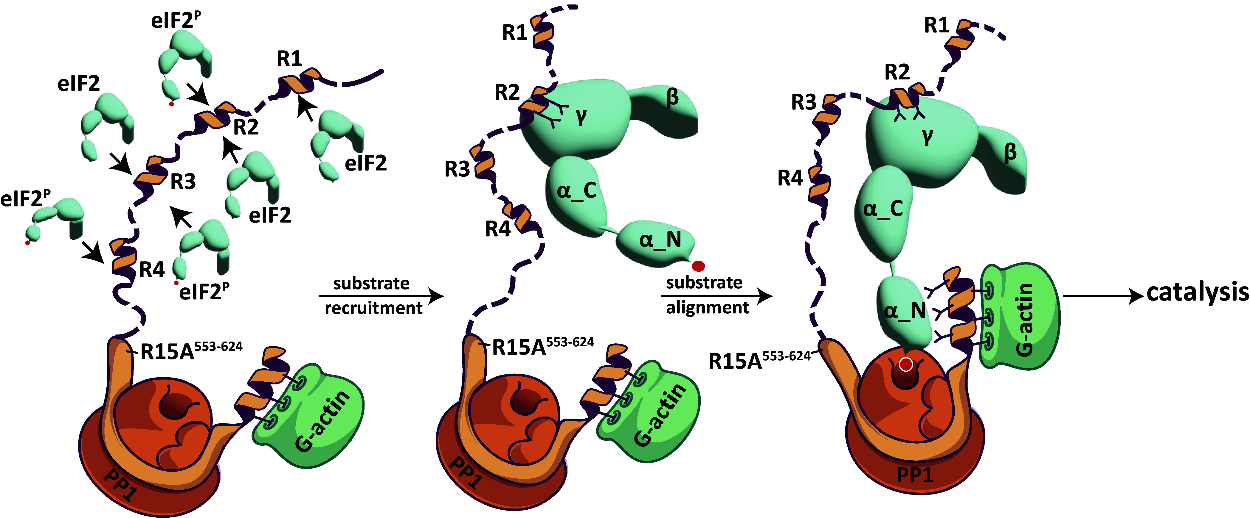
Fig. 5. Cartoon model of the consequences of PPP1R15A’s helical repeats binding to eIF2γ. In the first step (Left-most), PPP1R15A recruits eIF2 by binding of its helical repeats in eIF2γ’s groove. Subsequently (Middle) the helix-groove interactions (exemplified here by repeat #2) stabilize the bulky eIF2 (γ-β-α C-term) lobe to counteract the entropic penalties caused by the flexible hinge between eIF2α-NTD and eIF2α-CTD (SI Appendix, Fig. S2B) and nonproductive interactions of the largely disordered N-terminal extension of PPP1R15A with holoenzyme (SI Appendix, Fig. S2C). This favors establishment of contacts between the eIF2α-NTD and the core holophosphatase (a complex of PP1, G-actin and the conserved CTD of PPP1R15A, PDB 7NZM) to direct insertion of eIF2α pSer51 in the PP1 active site (Right-most) and promote catalysis. Note that the repeat-groove interactions would enrich the local concentration of eIF2P in vicinity of the holophosphatase. However, the net benefit of enriched localized eIF2P concentration might be limited by product inhibition.
Fig. 5. Cartoon model of the consequences of PPP1R15A’s helical repeats binding to eIF2γ. In the first step (Left-most), PPP1R15A recruits eIF2 by binding of its helical repeats in eIF2γ’s groove. Subsequently (Middle) the helix-groove interactions (exemplified here by repeat #2) stabilize the bulky eIF2 (γ-β-α C-term) lobe to counteract the entropic penalties caused by the flexible hinge between eIF2α-NTD and eIF2α-CTD (SI Appendix, Fig. S2B) and nonproductive interactions of the largely disordered N-terminal extension of PPP1R15A with holoenzyme (SI Appendix, Fig. S2C). This favors establishment of contacts between the eIF2α-NTD and the core holophosphatase (a complex of PP1, G-actin and the conserved CTD of PPP1R15A, PDB 7NZM) to direct insertion of eIF2α pSer51 in the PP1 active site (Right-most) and promote catalysis. Note that the repeat-groove interactions would enrich the local concentration of eIF2P in vicinity of the holophosphatase. However, the net benefit of enriched localized eIF2P concentration might be limited by product inhibition.CC BY · PubMed Central

Verification

Claims the paper makes

The claims and statistics of this paper haven’t been checked yet.

Run the check — free, no account needed. It reads the open-access text and checks the sources it cites.

A few free checks a day · papers already verified open instantly · results are public

Self-check

Percentages, sums and reported differences re-computed from the paper’s own numbers.

The self-check needs the paper’s full text.

Citations

Cites

Research works printed but not linked: 55

  1. 10.1016/S1097-2765(03)00105-9 · DOI
  2. 10.1126/science.aad9868 · DOI · Paper page
  3. 10.1126/science.aat5314 · DOI · Paper page
  4. 10.1083/jcb.200308075 · DOI
  5. 10.1083/jcb.153.5.1011 · DOI
  6. 10.1074/jbc.M301107200 · DOI
  7. 10.1093/emboj/cdg112 · DOI
  8. 10.1242/jcs.117614 · DOI
  9. 2015, Structural and Functional Analysis of the GADD34:PP1 eIF2alpha Phosphatase, Cell reports, 11, 1885, 10.1016/j.celrep.2015.05.043 · DOI
  10. J. E. Chambers et al., Actin dynamics tune the integrated stress response by regulating eukaryotic initiation factor 2alpha dephosphorylation. eLife 4 (2015). · DOI
  11. 10.1128/MCB.23.4.1292-1303.2003 · DOI
  12. 10.1073/pnas.0809632106 · DOI · Paper page
  13. 10.7554/eLife.04871 · DOI
  14. 2021, Higher-order phosphatase-substrate contacts terminate the integrated stress response, Nat Struct Mol Biol, 28, 835, 10.1038/s41594-021-00666-7 · DOI
  15. 10.1073/pnas.1501557112 · DOI
  16. 10.1016/j.str.2004.07.010 · DOI
  17. 10.1126/science.aba4904 · DOI
  18. 10.1073/pnas.1400129111 · DOI
  19. 10.1083/jcb.147.7.1431 · DOI
  20. C. Sidrauski , A. M. McGeachy , N. T. Ingolia , P. Walter , The small molecule ISRIB reverses the effects of eIF2alpha phosphorylation on translation and stress granule assembly. eLife 4 (2015). · DOI
  21. 10.1016/j.chom.2012.05.013 · DOI
  22. 10.1073/pnas.89.8.3266 · DOI
  23. 10.1074/jbc.273.33.20737 · DOI
  24. 10.1083/jcb.200307151 · DOI
  25. 10.1101/gad.1250704 · DOI
  26. 10.1084/jem.20122005 · DOI
  27. 10.1126/science.aaa4484 · DOI
  28. 10.1038/nsmb.3443 · DOI
  29. 10.7554/eLife.26109 · DOI
  30. 10.1074/jbc.RA118.002325 · DOI
  31. Y. L. Wong et al., The small molecule ISRIB rescues the stability and activity of Vanishing White Matter Disease eIF2B mutant complexes. eLife 7 (2018). · DOI
  32. 2018, DIALS: implementation and evaluation of a new integration package, Acta Crystallogr D Struct Biol, 74, 85, 10.1107/S2059798317017235 · DOI
  33. 10.1107/S0907444913000061 · DOI
  34. 10.1107/S0907444910045749 · DOI · Paper page
  35. 10.1107/S0021889807021206 · DOI · Paper page
  36. 10.1107/S0907444909052925 · DOI · Paper page
  37. 10.1107/S0907444910007493 · DOI · Paper page
  38. 10.1107/S0907444911001314 · DOI
  39. 2018, Real-space refinement in PHENIX for cryo-EM and crystallography, Acta Crystallogr D Struct Biol, 74, 531, 10.1107/S2059798318006551 · DOI
  40. 10.1107/S0907444909042073 · DOI · Paper page
  41. 10.1002/pro.4792 · DOI
  42. 2019, Real-time cryo-electron microscopy data preprocessing with Warp, Nat Methods, 16, 1146, 10.1038/s41592-019-0580-y · DOI
  43. 10.1038/nmeth.4169 · DOI · Paper page
  44. 10.1101/2021.10.04.463034 · DOI · Paper page
  45. M. J. Abraham et al., GROMACS: High performance molecular simulations through multi-level parallelism from laptops to supercomputers. SoftwareX 1-2, 19–25 (2015). · DOI · Paper page
  46. 2021, Semi-automated Optimization of the CHARMM36 Lipid Force Field to Include Explicit Treatment of Long-Range Dispersion, J Chem Theory Comput, 17, 1562, 10.1021/acs.jctc.0c01326 · DOI
  47. 10.1063/1.2408420 · DOI
  48. 10.1063/1.328693 · DOI
  49. 10.1063/1.470117 · DOI
  50. 10.1002/jcc.540130805 · DOI
  51. 2008, P-LINCS:L A Parallel Linear Constraint Solver for Molecular Simulation, Journal of Chemical Theory and Computation, 4, 116, 10.1021/ct700200b · DOI
  52. 10.1016/0263-7855(96)00018-5 · DOI · Paper page
  53. 10.1002/jcc.20084 · DOI
  54. 10.1016/j.ab.2011.01.016 · DOI
  55. 10.1093/nar/gkq643 · DOI

Metadata

Authors
Yan, Y., Shetty, M., Harding, H. P., George, G., Zyryanova, A., Labbé, K., Mafi, A., Hao, Q., Sidrauski, C., & Ron, D.
Affiliations
  • University of Cambridge (GB)
  • Calico Life Sciences (US)
Corresponding author
  • Yan, Yahui
  • Ron, David
Venue
Proceedings of the National Academy of Sciences
Publisher
openRxiv
Published
2023-12-15
Volume
121
Issue
14
Pages
e2320013121-e2320013121
ISSN
0027-8424 (ISSN-L)
OpenAlex
W4393278999
Cited by
7
References
1
Citations by year
2026: 2 · 2025: 4 · 2024: 1
FWCI
1.57
Type
journal article
Language
en
Open access
Licence
cc-by
Locations
  • Proceedings of the National Academy of Sciences (publishedVersion, cc-by)
  • bioRxiv (Cold Spring Harbor Laboratory) (acceptedVersion, cc-by-nc-nd)
  • PubMed Central (publishedVersion, cc-by)
  • PubMed (publishedVersion)
  • Apollo (University of Cambridge) (submittedVersion, cc-by)
  • Apollo (cc-by)
Funders
  • Wellcome Trust (224407, 224407/Z/21/Z, 200848/Z/16/Z)
  • Diamond Light Source
  • Cambridge Institute for Medical Research, University of Cambridge
  • Department of Biochemistry, University of Cambridge
  • RIKEN
  • Calico Life Sciences
Topics
  • Life Sciences › Biochemistry, Genetics and Molecular Biology › Cell Biology › Endoplasmic Reticulum Stress and Disease
  • Life Sciences › Biochemistry, Genetics and Molecular Biology › Molecular Biology › RNA and protein synthesis mechanisms
  • Life Sciences › Biochemistry, Genetics and Molecular Biology › Molecular Biology › Protein Structure and Dynamics
Keywords
integrated stress response, eIF2α dephosphorylation, PPP1R15A, protein phosphatase 1, eIF2γ, substrate recruitment, hydrophobic groove, catalytic efficiency
MeSH terms
Actins, Animals, Humans, Phosphorylation, Protein Processing, Post-Translational, Eukaryotic Initiation Factor-2, Protein Phosphatase 1

About these checks

Registry, retraction, Cites and statistics results are computed, not judged, with one exception: a printed reference that no index lists is read once by a model before it counts as a citation error. Claims are extracted by a model and kept only when their quote appears verbatim in the text; each cited source is then checked, with a model’s help, for whether it says what the claim relies on. Those verdicts can be wrong — read the claim’s quote and the cited source before relying on one.

Statistics and arithmetic are recomputed, not judged.

Confidence bands are shown only for fields calibrated on our annotated test set. None is calibrated yet, so no band is shown.

What counts as a claim ›How we measure confidence ›How we check this ›

Are you an author, or do you see a problem with this page? Request a review