

The DOI matches its Crossref record; 2 of the 2 references it cites were found in a registry or an index.
Genetic diversity, predictive protein structures, and interaction networks of Cysteine-Rich Receptor-Like Kinases in Arabidopsis thaliana
Open accessPreprint — published as 10.34133/csbj.0043 · Paper page
Abstract
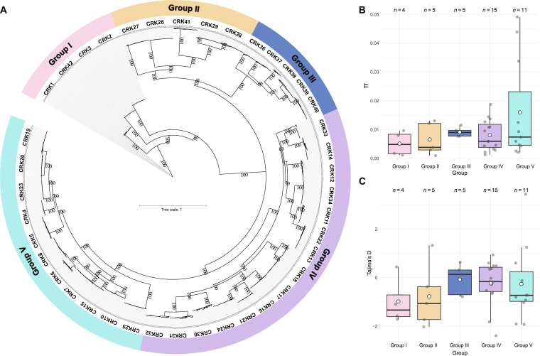
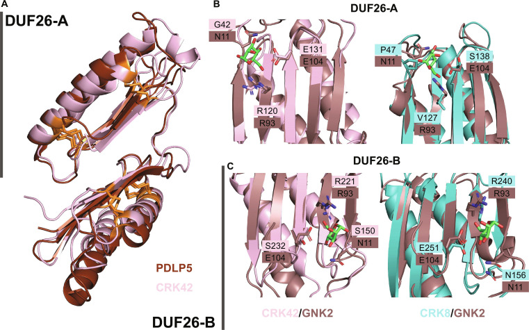
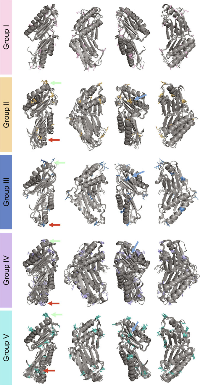
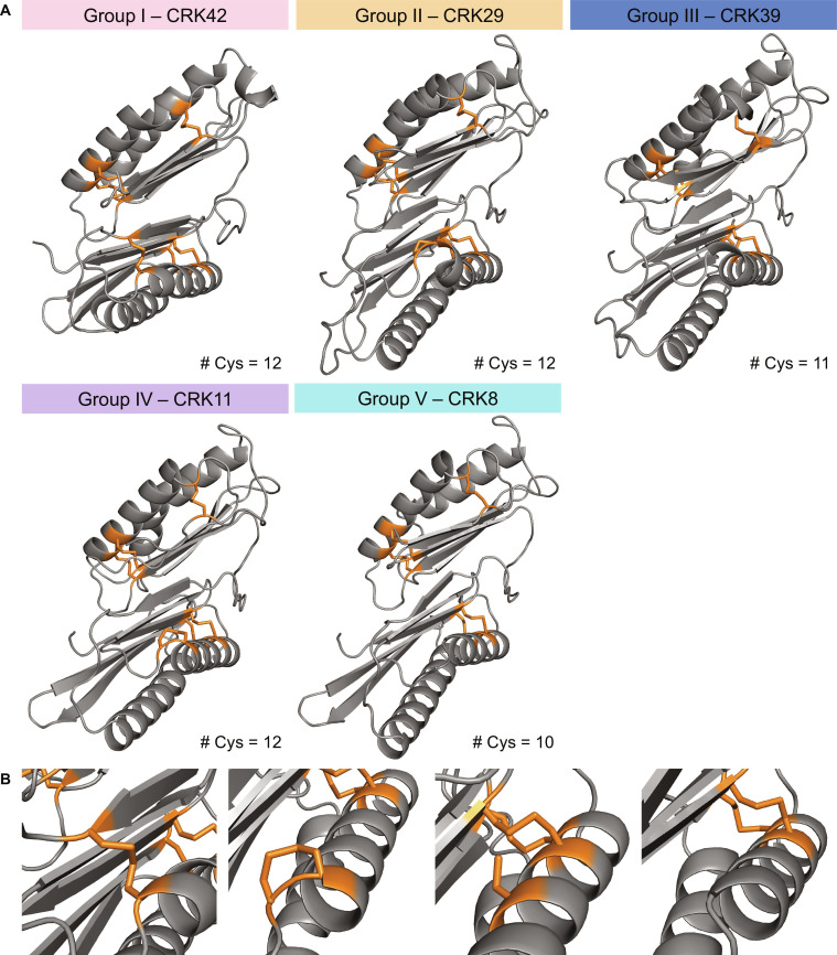
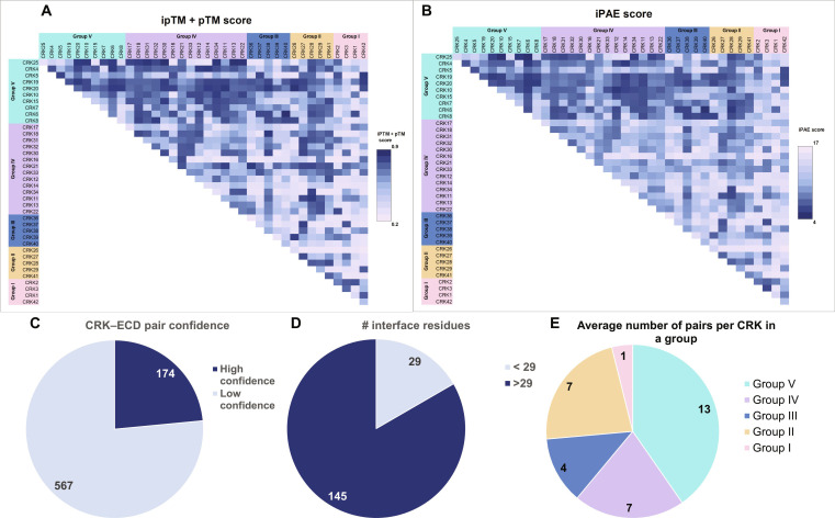
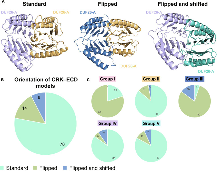
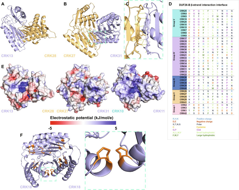
Cysteine-rich receptor-like kinases (CRKs) are a large subfamily of plant receptor-like kinases (RLKs) implicated in immunity and development, yet their ligands, interaction partners, and mechanistic roles remain poorly defined. We combined population-genetic analyses and AlphaFold-based structural prediction to characterise the Arabidopsis thaliana CRK family. Phylogenetic reconstruction from 69 natural accessions resolved five well-supported CRK clades. Nucleotide diversity (π) and neutrality tests revealed heterogeneous diversity across loci, with evidence of both positive and negative selection pressure acting on different CRKs. AlphaFold models of CRK extracellular domains (ECDs) recapitulate the DUF26 structure observed in Plasmodesmata Localizing Protein (PDLP)5/PDLP8 and ginkbilobin-2 but display distinct biochemical properties and disulfide-bond topologies. Pairwise AlphaFold dimer modelling of all 780 CRK-ECD combinations produced 145 high-confidence interaction models; ∼78% of these adopt a shared dimer conformation characterized by an extended intermolecular β-sheet at the interface. Integrating evolutionary and structural approaches reveals clade-specific selective regimes and conserved structural features of CRK ECDs that likely underpin receptor–receptor interactions. Predicted high-confidence dimer interfaces suggest a general mode of CRK-ECD association that can guide targeted biochemical and genetic validation, accelerating functional dissection of this important receptor family.
Overview
The claims and statistics of this paper haven’t been checked yet.
Run the check — free, no account needed. It reads the open-access text and checks the sources it cites.
Integrity signals
Registry
Cites
Statistics
Figures
Verification
Claims the paper makes
The claims and statistics of this paper haven’t been checked yet.
Run the check — free, no account needed. It reads the open-access text and checks the sources it cites.
Self-check
Percentages, sums and reported differences re-computed from the paper’s own numbers.
The self-check needs the paper’s full text.
Citations
Cites
Highly accurate protein structure prediction with AlphaFold
Jumper, J., Evans, R., Pritzel, A., Green, T., Figurnov, M., Ronneberger, O., Tunyasuvunakool, K., Bates, R., Žídek, A., Potapenko, A., et al. (2021) · Nature · doi:10.1038/s41586-021-03819-2 · Cited by 43,764
Varadi, M., Bertoni, D., Magana, P., Paramval, U., Pidruchna, I., Radhakrishnan, M., Tsenkov, M., Nair, S., Mirdita, M., Yeo, J., et al. (2024) · Nucleic Acids Research · doi:10.1093/nar/gkad1011 · Cited by 2,052
Research works printed but not linked: 98
- 10.1104/pp.126.2.473 · DOI
- 10.1104/pp.126.2.473 · DOI
- 2025, More questions than answers: insights into potential cysteine-rich receptor-like kinases redox signalling in Arabidopsis, The Plant Journal, 122, e70176, 10.1111/tpj.70176 · DOI
- 10.1146/annurev-arplant-042916-040957 · DOI · Paper page
- Shiu, S. H. & Bleecker, A. B . Plant receptor-like kinase gene family: diversity, function, and signaling. Science’s STKE : signal transduction knowledge environment Preprint at (2001). · DOI
- 10.1371/journal.pgen.1005373 · DOI
- 10.1073/pnas.181141598 · DOI · Paper page
- 10.1105/tpc.020834 · DOI · Paper page
- 10.1104/pp.126.2.473 · DOI
- 10.1038/s42003-019-0306-9 · DOI
- 10.1104/pp.16.01404 · DOI
- 10.1186/s12864-020-06844-z · DOI
- 2021, Computational prediction method to decipher receptor–glycoligand interactions in plant immunity, The Plant Journal, 105, 1710, 10.1111/tpj.15133 · DOI
- Ali, F . Patterns of Change in Nucleotide Diversity Over Gene Length. Genome Biol Evol 16, (2024). · DOI
- Tatarinova, T. V. et al. Nucleotide diversity analysis highlights functionally important genomic regions. Sci Rep 6, 35730 (2016). · DOI
- 10.1038/s42003-019-0306-9 · DOI
- 10.1038/s44319-024-00122-9 · DOI
- 2024, Extensive immune receptor repertoire diversity in disease-resistant rice landraces, Current Biology, 34, 3983, 10.1016/j.cub.2024.07.061 · DOI
- 10.1146/annurev-arplant-042916-040957 · DOI · Paper page
- 10.1002/pro.4318 · DOI
- 10.1093/nar/gkac928 · DOI
- 10.1104/pp.114.242636 · DOI
- 10.1038/nplants.2016.151 · DOI · Paper page
- 10.1515/BC.2007.030 · DOI
- 2009, Crystal structure of ginkbilobin-2 with homology to the extracellular domain of plant cysteine-rich receptor-like kinases. Proteins: Structure, Function, and Bioinformatics, 77, 247, 10.1002/prot.22494 · DOI
- 10.1074/jbc.M109.063073 · DOI
- 2024, N-glycosylation facilitates the activation of a plant cell-surface receptor, Nature Plants, 10, 2014, 10.1038/s41477-024-01841-6 · DOI
- 2018, Function of N-glycosylation in plants, Plant Science, 274, 70, 10.1016/j.plantsci.2018.05.007 · DOI
- de Oliveira, M. V. V. et al. Specific control of Arabidopsis BAK1/SERK4-regulated cell death by protein glycosylation. Nat Plants 2, 15218 (2016). · DOI
- 10.1016/j.jmb.2017.03.017 · DOI
- 10.1104/pp.16.01404 · DOI
- 10.1101/2021.10.04.463034 · DOI · Paper page
- 10.1110/ps.03234503 · DOI
- 2008, Characterization of protein-protein interfaces, Protein Journal, 27, 59, 10.1007/s10930-007-9108-x · DOI
- Wei, X. , Liu, X. , Zhang, X. , Guo, S. & Shi, J . Structural insights into ligand recognition and receptor activation of plant leucine-rich repeat (LRR) transmembrane receptors. New Crops 1, 100022 (2024). · DOI
- 2014, Detecting Communities Based on Network Topology, Scientific Reports, 4, 1
- 10.1111/nph.18305 · DOI
- Pasion, J. & Hind, S. R . Utilizing Tajima’s D to identify potential microbe-associated molecular patterns in Xanthomonas euvesicatoria and X. perforans. Physiol Mol Plant Pathol 116, 101744 (2021). · DOI
- Steidele, C. E. & Stam, R . Multi-omics approach highlights differences between RLP classes in Arabidopsis thaliana. BMC Genomics 22, 557 (2021). · DOI
- 2025, A species-wide inventory of receptor-like kinases in Arabidopsis thaliana, BMC Biol, 23, 266, 10.1186/s12915-025-02364-y · DOI
- Ngou, B. P. M. , Wyler, M. , Schmid, M. W. , Kadota, Y. & Shirasu, K . Evolutionary trajectory of pattern recognition receptors in plants. Nature Communications 2024 15:1 15, 308- (2024). · DOI
- 10.1104/pp.15.01470 · DOI
- 2023, An Interactome Assay for Detecting Interactions between Extracellular Domains of Receptor Kinases, Methods in Molecular Biology, 2690, 193, 10.1007/978-1-0716-3327-4_18 · DOI
- 10.1105/tpc.113.120071 · DOI
- 10.1107/S1744309107034793 · DOI
- Man, J. , Harrington, T. A. , Lally, K. & Bartlett, M. E . Asymmetric Evolution of Protein Domains in the Leucine-Rich Repeat Receptor-Like Kinase Family of Plant Signaling Proteins. Mol Biol Evol 40, msad220 (2023). · DOI
- Hurst, C. H. et al. Juxta-membrane S-acylation of plant receptor-like kinases is likely fortuitous and does not necessarily impact upon function. Scientific Reports 2019 9:1 9, 12818- (2019). · DOI
- 10.1074/jbc.M109.063073 · DOI
- 10.1104/pp.16.00667 · DOI
- 10.7554/eLife.06633 · DOI · Paper page
- 2016, Altered glycosylation of exported proteins, including surface immune receptors, compromises calcium and downstream signaling responses to microbe-associated molecular patterns in Arabidopsis thaliana, BMC Plant Biol, 16, 31, 10.1186/s12870-016-0718-3 · DOI
- 10.1046/j.1365-313X.1999.00447.x · DOI
- 10.1371/journal.pgen.1005373 · DOI
- 10.1016/j.jprot.2016.02.031 · DOI
- Butera, D. et al. Autoregulation of von Willebrand factor function by a disulfide bond switch. Sci Adv 4, (2018). · DOI
- 10.1073/pnas.052706099 · DOI · Paper page
- 10.1016/B978-0-12-386931-9.00002-7 · DOI
- 10.1016/j.bbrc.2014.02.013 · DOI
- 10.1093/jxb/erad080 · DOI
- 2024, The CRK14 gene encoding a cysteine-rich receptor-like kinase is implicated in the regulation of global proliferative arrest in Arabidopsis thaliana, Genes to Cells, 29, 735, 10.1111/gtc.13139 · DOI
- 10.1038/s41586-020-2032-3 · DOI · Paper page
- 10.1186/1471-2229-10-1 · DOI · Paper page
- 10.1093/jxb/erad236 · DOI
- 10.1016/j.bbrc.2014.02.013 · DOI
- 10.1016/j.jmb.2010.09.027 · DOI
- 2023, Functionally annotating cysteine disulfides and metal binding sites in the plant kingdom using AlphaFold2 predicted structures, Free Radic Biol Med, 194, 220, 10.1016/j.freeradbiomed.2022.12.001 · DOI
- 10.1093/protein/gzg088 · DOI
- Molina, A. et al. Plant cell walls: source of carbohydrate-based signals in plant-pathogen interactions. Curr Opin Plant Biol 82, 102630 (2024). · DOI
- 10.1093/jxb/eraa414 · DOI
- 2018, The Ustilago maydis repetitive effector Rsp3 blocks the antifungal activity of mannose-binding maize proteins, Nature Communications, 9, 1
- 10.1093/jxb/erab328 · DOI
- 10.1101/2025.09.18.677107 · DOI
- 2024, Mechanistic study of SCOOPs recognition by MIK2–BAK1 complex reveals the role of N-glycans in plant ligand–receptor–coreceptor complex formation, Nature Plants, 10, 1984, 10.1038/s41477-024-01836-3 · DOI
- 10.1016/j.cell.2022.07.012 · DOI
- 10.1038/S41586-024-07487-W · DOI · Paper page
- 2023, AlphaFold predictions are valuable hypotheses and accelerate but do not replace experimental structure determination, Nature Methods, 21, 110
- 10.1038/s41588-024-01715-9 · DOI
- 10.1186/1471-2105-10-1 · DOI · Paper page
- 10.1093/gigascience/giab016 · DOI · Paper page
- 10.1093/molbev/mst010 · DOI · Paper page
- 10.1093/bioinformatics/btp348 · DOI · Paper page
- 10.1093/molbev/msaa015 · DOI · Paper page
- 10.1093/nar/gkae268 · DOI · Paper page
- 10.1111/2041-210X.12410 · DOI
- 10.1109/IPDPS.2002.1016585 · DOI
- 10.1093/bioinformatics/bti079 · DOI · Paper page
- 10.1371/journal.pgen.1002764 · DOI · Paper page
- 10.1093/molbev/mst030 · DOI · Paper page
- 10.1093/molbev/msi105 · DOI · Paper page
- 10.1093/bioinformatics/btu531 · DOI
- 10.1093/BIOINFORMATICS/BTZ921 · DOI
- Sievers, F. et al. Fast, scalable generation of high-quality protein multiple sequence alignments using Clustal Omega. Mol Syst Biol 7, 539 (2011). · DOI · Paper page
- 10.1002/PRO.3290 · DOI · Paper page
- 10.1093/nar/gkh340 · DOI · Paper page
- 10.1093/nar/gkae241 · DOI · Paper page
- 10.1038/S41586-021-03819-2 · DOI · Paper page
- 10.1016/j.jmb.2007.05.022 · DOI · Paper page
- 2014, Detecting Communities Based on Network Topology, Scientific Reports, 4, 1
Metadata
- Authors
- Stouthamer, J., Pereira, D., Martin-Ramirez, S., Mutte, S., Mott, G. A., & Smakowska-Luzan, E.
- Affiliations
- Wageningen University & Research (NL)
- University of Toronto Scarborough (CA)
- Venue
- Computational and Structural Biotechnology Journal
- Publisher
- openRxiv
- Published
- 2025-11-29
- Volume
- 35
- Issue
- 1
- Pages
- 0043-0043
- ISSN
- 2001-0370 (ISSN-L)
- PMC
- PMC13058244
- OpenAlex
- W7137929205
- Cited by
- 2
- References
- 2
- Citations by year
- 2026: 2
- FWCI
- 7.36
- Type
- journal article
- Language
- en
- Open access
- gold
- doi.org
- Licence
- Locations
- Computational and Structural Biotechnology Journal (publishedVersion, cc-by)
- bioRxiv (Cold Spring Harbor Laboratory) (acceptedVersion, public-domain)
- PubMed (publishedVersion)
- PubMed Central (publishedVersion, cc-by)
- DOAJ (DOAJ: Directory of Open Access Journals) (submittedVersion)
- Socio-Environmental Systems Modeling (submittedVersion, cc-by)
- Funders
- NWO Talent Programme (Vidi grant VI.Vidi.193.074)
- Natural Sciences and Engineering Research Council of Canada (Discovery Grant Award (RGPIN-2019-06395))
- OVPRI International Research Fund at the University of Toronto Scarborough
- Topics
- Life Sciences › Agricultural and Biological Sciences › Plant Science › Plant Molecular Biology Research
- Life Sciences › Agricultural and Biological Sciences › Plant Science › Plant-Microbe Interactions and Immunity
- Life Sciences › Biochemistry, Genetics and Molecular Biology › Molecular Biology › Protein Structure and Dynamics
- Keywords
- cysteine-rich receptor-like kinases, Arabidopsis thaliana, genetic diversity, extracellular domain structure, protein dimerization, receptor-receptor interactions, positive and negative selection, phylogenetic clades, plant immunity, population genetics
About these checks
Registry, retraction, Cites and statistics results are computed, not judged, with one exception: a printed reference that no index lists is read once by a model before it counts as a citation error. Claims are extracted by a model and kept only when their quote appears verbatim in the text; each cited source is then checked, with a model’s help, for whether it says what the claim relies on. Those verdicts can be wrong — read the claim’s quote and the cited source before relying on one.
Statistics and arithmetic are recomputed, not judged.
Confidence bands are shown only for fields calibrated on our annotated test set. None is calibrated yet, so no band is shown.
What counts as a claim ›How we measure confidence ›How we check this ›
Are you an author, or do you see a problem with this page? Request a review
Data sources
Bibliographic data: Semantic Scholar, OpenAlex, Crossref.
Contains information from the Semantic Scholar Academic Graph Datasets, made available under the ODC Attribution License (ODC-BY 1.0).
Semantic Scholar’s data is described in: Kinney, R., et al. (2023). The Semantic Scholar Open Data Platform. arXiv:2301.10140.
