

The DOI matches its Crossref record; 46 of the 46 references it cites were found in a registry or an index.
Angiopoietin 2 regulates the transformation and integrity of lymphatic endothelial cell junctions
Open accessAbstract
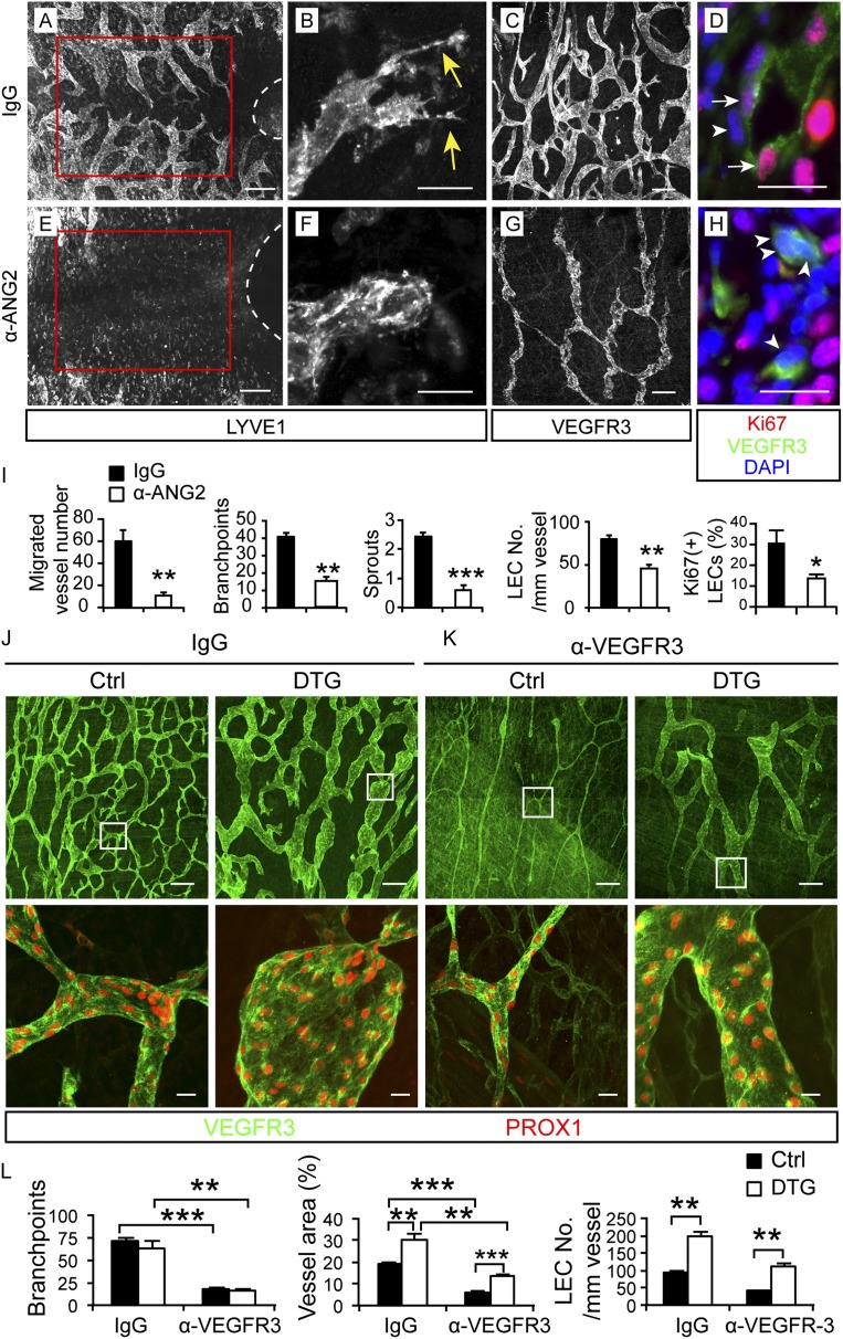
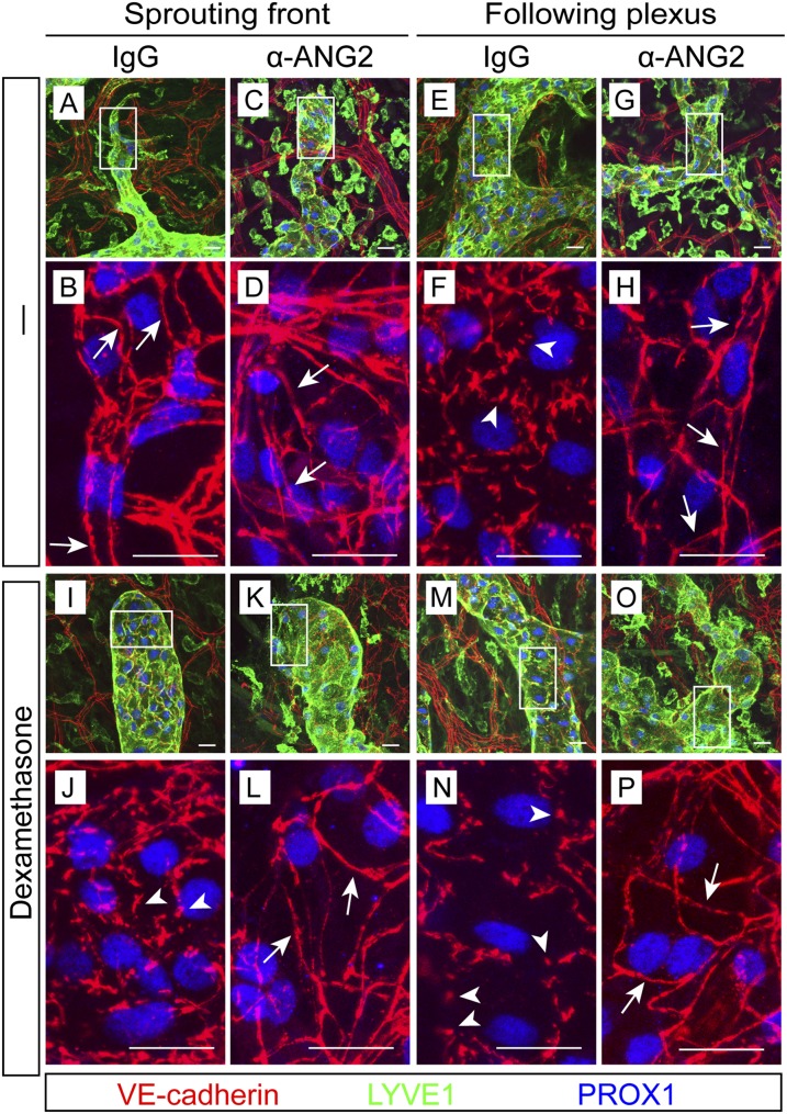
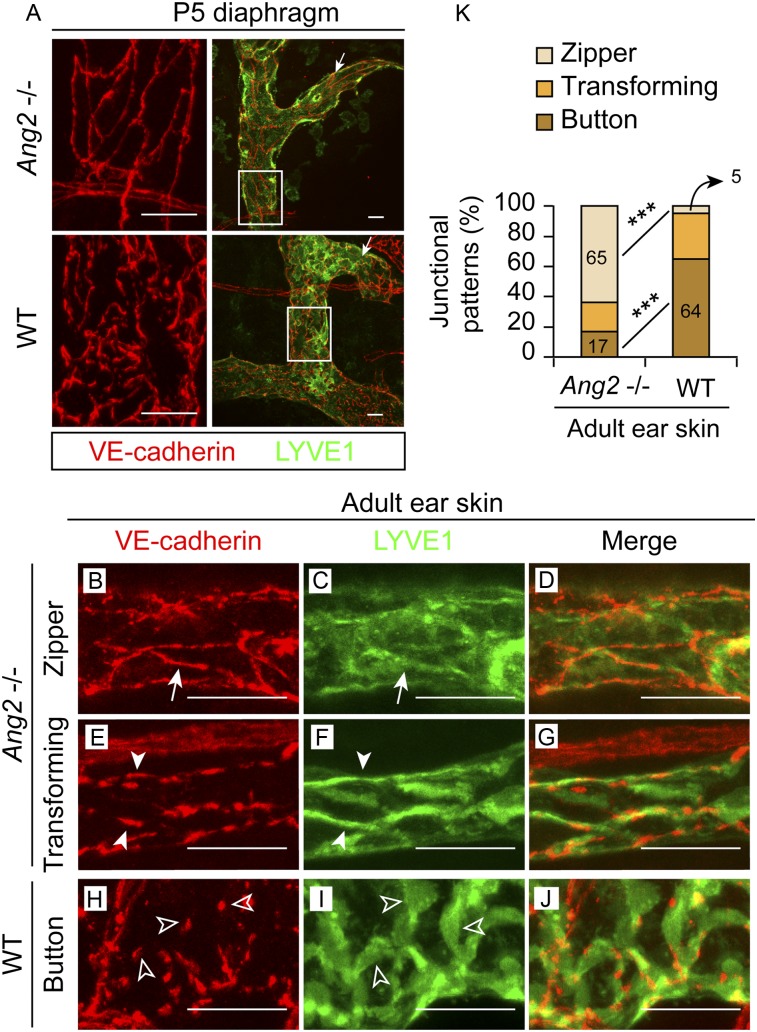
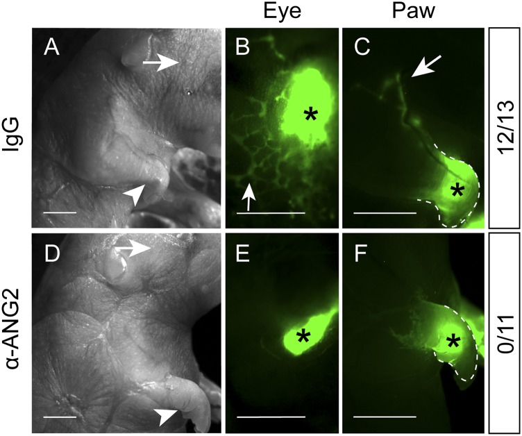
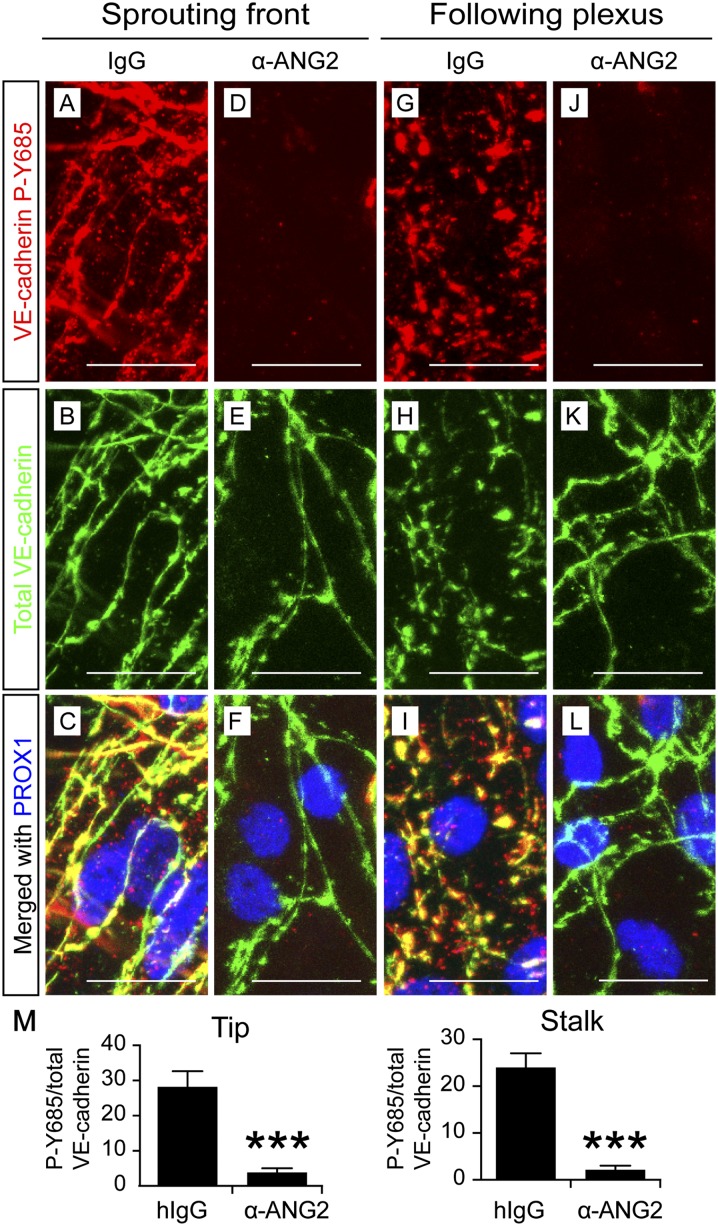
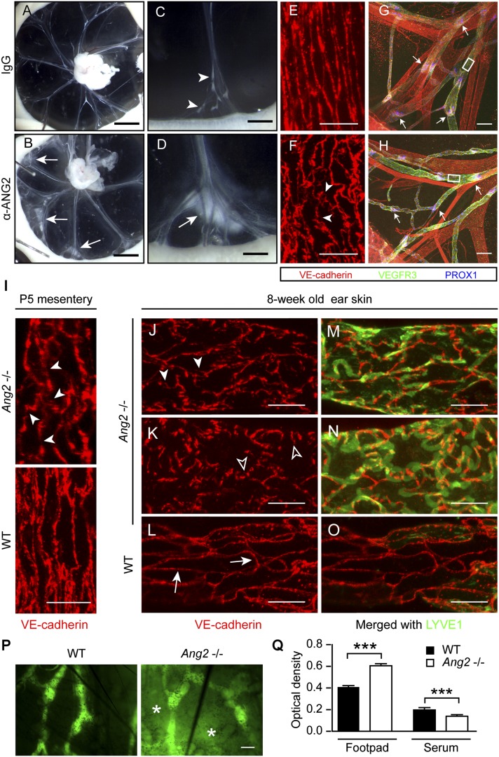
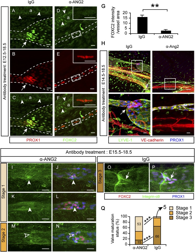
Primitive lymphatic vessels are remodeled into functionally specialized initial and collecting lymphatics during development. Lymphatic endothelial cell (LEC) junctions in initial lymphatics transform from a zipper-like to a button-like pattern during collecting vessel development, but what regulates this process is largely unknown. Angiopoietin 2 ( Ang2 ) deficiency leads to abnormal lymphatic vessels. Here we found that an ANG2-blocking antibody inhibited embryonic lymphangiogenesis, whereas endothelium-specific ANG2 overexpression induced lymphatic hyperplasia. ANG2 inhibition blocked VE-cadherin phosphorylation at tyrosine residue 685 and the concomitant formation of button-like junctions in initial lymphatics. The defective junctions were associated with impaired lymph uptake. In collecting lymphatics, adherens junctions were disrupted, and the vessels leaked upon ANG2 blockade or gene deletion. ANG2 inhibition also suppressed the onset of lymphatic valve formation and subsequent valve maturation. These data identify ANG2 as the first essential regulator of the functionally important interendothelial cell–cell junctions that form during lymphatic development.
Overview
The claims and statistics of this paper haven’t been checked yet.
Run the check — free, no account needed. It reads the open-access text and checks the sources it cites.
Integrity signals
Registry
Cites
Statistics
Figures
Verification
Claims the paper makes
The claims and statistics of this paper haven’t been checked yet.
Run the check — free, no account needed. It reads the open-access text and checks the sources it cites.
Claims other papers make about this one
Each is checked against this paper’s own text: does it say what the citing paper says it does?
The cited source says what the claim attributes to it.
In the citing paper
“During vascular development, Ang2 inhibition blocked VE-cadherin phosphorylation at tyrosine residue 685 and the concomitant formation of button-like junctions in initial lymphatic vessels20”
From Endothelial destabilization by angiopoietin-2 via integrin β1 activation (2015) · cites this paper as [20]
Self-check
Percentages, sums and reported differences re-computed from the paper’s own numbers.
The self-check needs the paper’s full text.
Citations
Cites
Angiopoietin-2, a Natural Antagonist for Tie2 That Disrupts in vivo Angiogenesis
Maisonpierre, P. C., Suri, C., Jones, P. F., Bartunkova, S., Wiegand, S. J., Radziejewski, C., Compton, D., McClain, J., Aldrich, T. H., Papadopoulos, N., et al. (1997) · Science · doi:10.1126/science.277.5322.55 · Cited by 3,570
Requisite Role of Angiopoietin-1, a Ligand for the TIE2 Receptor, during Embryonic Angiogenesis
Suri, C., Jones, P. F., Patan, S., Bartunkova, S., Maisonpierre, P. C., Davis, S. H., Sato, T. N., & Yancopoulos, G. D. (1996) · Cell · doi:10.1016/s0092-8674(00)81813-9 · Cited by 2,872
Vessel Cooption, Regression, and Growth in Tumors Mediated by Angiopoietins and VEGF
Holash, J. A., Maisonpierre, P. C., Compton, D. L., Boland, P., Alexander, C., Zagzag, D., Yancopoulos, G. D., & Wiegand, S. J. (1999) · Science · doi:10.1126/science.284.5422.1994 · Cited by 2,222
Distinct roles of the receptor tyrosine kinases Tie-1 and Tie-2 in blood vessel formation
Sato, T. N., Tozawa, Y., Deutsch, U., Wolburg‐Buchholz, K., Fujiwara, Y., Gendron‐Maguire, M., Gridley, T., Wolburg, H., Risau, W., & Qin, Y. (1995) · Nature · doi:10.1038/376070a0 · Cited by 1,821
Prox1 Function Is Required for the Development of the Murine Lymphatic System
Wigle, J. T., & Oliver, G. C. (1999) · Cell · doi:10.1016/s0092-8674(00)81511-1 · Cited by 1,581
Control of vascular morphogenesis and homeostasis through the angiopoietin–Tie system
Augustin, H. G., Koh, G. Y., Thurston, G., & Alitalo, K. K. (2009) · Nature Reviews Molecular Cell Biology · doi:10.1038/nrm2639 · Cited by 1,512
Self-citationLymphangiogenesis: Molecular Mechanisms and Future Promise
Tammela, T., & Alitalo, K. K. (2010) · Cell · doi:10.1016/j.cell.2010.01.045 · Cited by 1,485
Self-citationKarkkainen, M. J., Haiko, P., Sainio, K., Partanen, J. M., Taipale, J., Petrova, T. V., Jeltsch, M., Jackson, D. G., Talikka, M., Rauvala, H., et al. (2003) · Nature Immunology · doi:10.1038/ni1013 · Cited by 1,441
Self-citationGale, N. W., Thurston, G., Hackett, S. F., Renard, R. A., Wang, Q., McClain, J., Martin, C., Witte, C. L., Witte, M. H., Jackson, D. G., et al. (2002) · Developmental Cell · doi:10.1016/s1534-5807(02)00217-4 · Cited by 1,017
Functionally specialized junctions between endothelial cells of lymphatic vessels
Baluk, P., Fuxe, J., Hashizume, H., Romano, T., Lashnits, E., Butz, S., Vestweber, D., Corada, M., Molendini, C., Dejana, E., et al. (2007) · The Journal of Experimental Medicine · doi:10.1084/jem.20062596 · Cited by 1,006
Self-citation
Cited by
Sorted by citations
Vascular heterogeneity and specialization in development and disease
Potente, M., & Mäkinen, T. (2017) · Nature Reviews Molecular Cell Biology · doi:10.1038/nrm.2017.36 · Cited by 653
Role of Angiopoietin-2 in Vascular Physiology and Pathophysiology
Akwii, R. G., Sajib, M. S., Zahra, F. T., & Mikelis, C. M. (2019) · Cells · doi:10.3390/cells8050471 · Cited by 566
Macrophages: An Inflammatory Link Between Angiogenesis and Lymphangiogenesis
Corliss, B. A., Azimi, M. S., Munson, J. M., Peirce, S. M., & Murfee, W. L. (2016) · Microcirculation · doi:10.1111/micc.12259 · Cited by 356
Lymphatic Vessel Network Structure and Physiology
Breslin, J. W., Yang, Y., Scallan, J. P., Sweat, R. S., Adderley, S., & Murfee, W. L. (2018) · Comprehensive physiology · doi:10.1002/cphy.c180015 · Cited by 420
The effects of sepsis on endothelium and clinical implications
Dolmatova, E. V., Wang, K., Mandavilli, R., & Griendling, K. K. (2020) · Cardiovascular Research · doi:10.1093/cvr/cvaa070 · Cited by 324
Lymphatic System in Cardiovascular Medicine
Aspelund, A., Robciuc, M. R., Karaman, S., Mäkinen, T., & Alitalo, K. K. (2016) · Circulation Research · doi:10.1161/circresaha.115.306544 · Cited by 336
Self-citationEndothelial destabilization by angiopoietin-2 via integrin β1 activation
Hakanpaa, L., Sipila, T., Leppanen, V.-M., Gautam, P., Nurmi, H., Jacquemet, G., Eklund, L., Ivaska, J., Alitalo, K., & Saharinen, P. (2015) · Nature Communications · doi:10.1038/ncomms6962 · Cited by 287
Self-citationLacteal junction zippering protects against diet-induced obesity
Zhang, F., Zarkada, G., Han, J., Li, J., Dubrac, A., Ola, R., Genet, G., Boyé, K., Michon, P., Künzel, S. E., et al. (2018) · Science · doi:10.1126/science.aap9331 · Cited by 237
Lymphatic Malformations: Genetics, Mechanisms and Therapeutic Strategies
Mäkinen, T., Boon, L. M., Vikkula, M., & Alitalo, K. K. (2021) · Circulation Research · doi:10.1161/circresaha.121.318142 · Cited by 237
Self-citationIntestinal lymphatic vasculature: structure, mechanisms and functions
Bernier‐Latmani, J., & Petrova, T. V. (2017) · Nature Reviews Gastroenterology & Hepatology · doi:10.1038/nrgastro.2017.79 · Cited by 219
Self-citationAngiopoietin–Tie signalling in the cardiovascular and lymphatic systems
Eklund, L., Kangas, J., & Saharinen, P. (2016) · Clinical Science · doi:10.1042/cs20160129 · Cited by 210
Takeda, A., Hollmén, M., Đermadi, D., Pan, J., Brulois, K., Kaukonen, R., Lönnberg, T., Boström, P., Koskivuo, I., Irjala, H., et al. (2019) · Immunity · doi:10.1016/j.immuni.2019.06.027 · Cited by 183
Dieterich, L. C., Tacconi, C., Ducoli, L., & Detmar, M. J. (2022) · Physiological Reviews · doi:10.1152/physrev.00039.2021 · Cited by 165
Tumor lymphangiogenesis and new drug development
Dieterich, L. C., & Detmar, M. J. (2015) · Advanced Drug Delivery Reviews · doi:10.1016/j.addr.2015.12.011 · Cited by 145
A Single-Cell Transcriptional Roadmap of the Mouse and Human Lymph Node Lymphatic Vasculature
Xiang, M., Grosso, R. A., Takeda, A., Pan, J., Bekkhus, T., Brulois, K., Đermadi, D., Nordling, S., Vanlandewijck, M., Jalkanen, S., et al. (2020) · Frontiers in Cardiovascular Medicine · doi:10.3389/fcvm.2020.00052 · Cited by 143
Impaired angiopoietin/Tie2 signaling compromises Schlemm’s canal integrity and induces glaucoma
Kim, J., Park, D. Y., Bae, H., Park, D. Y., Kim, D., Lee, C., Song, S., Chung, T.-Y., Lim, D. H., Kubota, Y., et al. (2017) · Journal of Clinical Investigation · doi:10.1172/jci94668 · Cited by 134
Self-citationContext-dependent functions of angiopoietin 2 are determined by the endothelial phosphatase VEPTP
Souma, T., Thomson, B. R., Heinen, S., Carota, I. A., Yamaguchi, S., Onay, T., Liu, P., Ghosh, A. K., Li, C., Eremina, V., et al. (2018) · Proceedings of the National Academy of Sciences · doi:10.1073/pnas.1714446115 · Cited by 138
Mechanisms Ensuring Endothelial Junction Integrity Beyond VE-Cadherin
Duong, C. N., & Vestweber, D. (2020) · Frontiers in Physiology · doi:10.3389/fphys.2020.00519 · Cited by 134
Gut microbiota regulates lacteal integrity by inducing VEGF‐C in intestinal villus macrophages
Suh, S. H., Choe, K., Hong, S. P., Jeong, S., Mäkinen, T., Kim, K. S., Alitalo, K. K., Surh, C. D., Koh, G. Y., & Song, J. (2019) · EMBO Reports · doi:10.15252/embr.201846927 · Cited by 134
Self-citationAngiopoietins and Tie2 in vascular inflammation
Parikh, S. M. (2017) · Current Opinion in Hematology · doi:10.1097/moh.0000000000000361 · Cited by 122
Lymphangiogenesis requires Ang2/Tie/PI3K signaling for VEGFR3 cell-surface expression
Korhonen, E. A., Murtomäki, A., Jha, S. K., Anisimov, A., Pink, A., Zhang, Y., Stritt, S., Liaqat, I., Stanczuk, L., Alderfer, L., et al. (2022) · Journal of Clinical Investigation · doi:10.1172/jci155478 · Cited by 113
Self-citationLymphatic vessel: Origin, heterogeneity, biological functions and therapeutic targets
Hu, Z., Zhao, X., Wu, Z., Qu, B., Yuan, M., Xing, Y., Song, Y., & Wang, Z. (2024) · Signal Transduction and Targeted Therapy · doi:10.1038/s41392-023-01723-x · Cited by 107
Multiple roles of lymphatic vessels in peripheral lymph node development
Bovay, E., Sabine, A., Prat-Luri, B., Kim, S., Son, K., Willrodt, A., Olsson, C., Halin, C., Kiefer, F., Betsholtz, C., et al. (2018) · The Journal of Experimental Medicine · doi:10.1084/jem.20180217 · Cited by 95
Self-citationLymphatic Endothelial Cell Junctions: Molecular Regulation in Physiology and Diseases
Zhang, F., Zarkada, G., Yi, S., & Eichmann, A. (2020) · Frontiers in Physiology · doi:10.3389/fphys.2020.00509 · Cited by 96
Polydom Is an Extracellular Matrix Protein Involved in Lymphatic Vessel Remodeling
Morooka, N., Futaki, S., Nishiuchi, R., Nishino, M., Totani, Y., Shimono, C., Nakano, I., Nakajima, H., Mochizuki, N., & Sekiguchi, K. (2017) · Circulation Research · doi:10.1161/circresaha.116.308825 · Cited by 90
Development and aging of the lymphatic vascular system
González-Loyola, A., & Petrova, T. V. (2021) · Advanced Drug Delivery Reviews · doi:10.1016/j.addr.2020.12.005 · Cited by 89
Self-citationJung, M., Ören, B., Mora, J., Mertens, C., Dziumbla, S., Popp, R., Weigert, A., Grossmann, N., Fleming, I., & Brüne, B. (2016) · Science Signaling · doi:10.1126/scisignal.aaf3241 · Cited by 87
The Angiopoietin-Tie2 Signaling Axis in Systemic Inflammation
Parikh, S. M. (2017) · Journal of the American Society of Nephrology · doi:10.1681/asn.2017010069 · Cited by 90
Bile Acids, Their Receptors, and the Gut Microbiota
Poland, J. C., & Flynn, C. R. (2021) · Physiology · doi:10.1152/physiol.00028.2020 · Cited by 93
Homeostatic maintenance of the lymphatic vasculature
Stritt, S., Koltowska, K., & Mäkinen, T. (2021) · Trends in Molecular Medicine · doi:10.1016/j.molmed.2021.07.003 · Cited by 79
Cytokines regulating lymphangiogenesis
Sáinz‐Jaspeado, M., & Claesson, L. (2018) · Current Opinion in Immunology · doi:10.1016/j.coi.2018.04.003 · Cited by 79
Distinct roles of VE‐cadherin for development and maintenance of specific lymph vessel beds
Hägerling, R., Hoppe, E., Dierkes, C., Stehling, M., Mäkinen, T., Butz, S., Vestweber, D., & Kiefer, F. (2018) · The EMBO Journal · doi:10.15252/embj.201798271 · Cited by 77
Regulation of Lymphatic Function in Obesity
Kataru, R. P., Park, H. J., Baik, J. E., Li, C., Shin, J., & Mehrara, B. J. (2020) · Frontiers in Physiology · doi:10.3389/fphys.2020.00459 · Cited by 81
The lymphatics in kidney health and disease
Donnan, M. D., Kenig-Kozlovsky, Y., & Quaggin, S. E. (2021) · Nature Reviews Nephrology · doi:10.1038/s41581-021-00438-y · Cited by 68
The lymphatic system and pancreatic cancer
Fink, D. M., Steele, M. M., & Hollingsworth, M. A. (2015) · Cancer Letters · doi:10.1016/j.canlet.2015.11.048 · Cited by 72
Geng, X., Yanagida, K., Akwii, R. G., Choi, D., Chen, L., Ho, Y., Cha, B., Mahamud, M. R., de Ruiz, K. B., Ichise, H., et al. (2020) · JCI Insight · doi:10.1172/jci.insight.137652 · Cited by 61
Buttons and Zippers: Endothelial Junctions in Lymphatic VesselsPreprint
Bałuk, P., & McDonald, D. M. (2022) · Cold Spring Harbor Perspectives in Medicine · doi:10.1101/cshperspect.a041178 · Cited by 61
VE-cadherin in Vascular Development
Lagendijk, A. K., & Hogan, B. M. (2015) · Current topics in developmental biology/Current Topics in Developmental Biology · doi:10.1016/bs.ctdb.2014.11.024 · Cited by 57
Endothelial Cell Responses to Biomechanical Forces in Lymphatic Vessels
Sabine, A., Demir, C. S., & Petrova, T. V. (2016) · Antioxidants and Redox Signaling · doi:10.1089/ars.2016.6685 · Cited by 55
Self-citationInfection-induced lymphatic zippering restricts fluid transport and viral dissemination from skin
Churchill, M. J., du Bois, H., Heim, T. A., Mudianto, T., Steele, M. M., Nolz, J. C., & Lund, A. W. (2022) · The Journal of Experimental Medicine · doi:10.1084/jem.20211830 · Cited by 51
Zhou, H. J., Qin, L., Jiang, Q., Murray, K. N., Zhang, H., Li, B., Lin, Q., Graham, M. M., Liu, X., Grutzendler, J., et al. (2021) · Nature Communications · doi:10.1038/s41467-020-20774-0 · Cited by 55
Characterization of ANGPT2 mutations associated with primary lymphedema
Leppänen, V.-M., Brouillard, P., Korhonen, E. A., Sipilä, T., Jha, S. K., Revencu, N., Labarque, V., Fastré, E., Schlögel, M., Ravoet, M., et al. (2020) · Science Translational Medicine · doi:10.1126/scitranslmed.aax8013 · Cited by 49
Self-citationProulx, S. T., Ma, Q., Andina, D., Leroux, J., & Detmar, M. J. (2017) · JCI Insight · doi:10.1172/jci.insight.90861 · Cited by 46
Molecular and metabolic orchestration of the lymphatic vasculature in physiology and pathology
Montenegro-Navarro, N., García-Báez, C., & García‐Caballero, M. (2023) · Nature Communications · doi:10.1038/s41467-023-44133-x · Cited by 45
Tie1 is required for lymphatic valve and collecting vessel development
Qu, X., Zhou, B., & Baldwin, H. S. (2015) · Developmental Biology · doi:10.1016/j.ydbio.2014.12.021 · Cited by 43
Lin, C.-C., Chen, P., Lein, M., Tsao, C.-W., Huang, C.-C., Wang, S., Tang, C., & Tung, K. (2016) · Oncotarget · doi:10.18632/oncotarget.7014 · Cited by 40
Molecular Mechanisms Controlling Lymphatic Endothelial Junction Integrity
Norden, P. R., & Kume, T. (2021) · Frontiers in Cell and Developmental Biology · doi:10.3389/fcell.2020.627647 · Cited by 38
VEGFR3 is required for button junction formation in lymphatic vessels
Jannaway, M., Iyer, D., Mastrogiacomo, D. M., Li, K., Sung, D. C., Yang, Y., Kahn, M. L., & Scallan, J. P. (2023) · Cell Reports · doi:10.1016/j.celrep.2023.112777 · Cited by 35
Dynamic cytoskeletal regulation of cell shape supports resilience of lymphatic endothelium
Schoofs, H., Daubel, N., Schnabellehner, S., Grönloh, M. L. B., Martínez, S. P., Halme, A., Marks, A. M., Jeansson, M., Barcos, S., Brakebusch, C., et al. (2025) · Nature · doi:10.1038/s41586-025-08724-6 · Cited by 37
Gengenbacher, N., Singhal, M., Mogler, C., Hai, L., Milde, L., Pari, A. A. A., Besemfelder, E., Fricke, C., Baumann, D., Gehrs, S., et al. (2020) · Cancer Discovery · doi:10.1158/2159-8290.cd-20-0122 · Cited by 36
Self-citationTzeng, H.-E., Chang, A., Tsai, C., Wang, S., & Tang, C. (2016) · Oncotarget · doi:10.18632/oncotarget.9570 · Cited by 32
Du, Y., Cao, M., Liu, Y., He, Y., Yang, C., Wu, M., Zhang, G., & Gao, F. (2016) · OncoImmunology · doi:10.1080/2162402x.2016.1232235 · Cited by 32
Le, C. T., Laidlaw, G., Morehouse, C. A., Naiman, B. M., Brohawn, P. Z., Mustelin, T., Connor, J. R., & McDonald, D. M. (2015) · American Journal Of Pathology · doi:10.1016/j.ajpath.2015.07.010 · Cited by 30
Decreased lymphatic HIF-2α accentuates lymphatic remodeling in lymphedema
Jiang, X., Tian, W., Granucci, E. J., Tu, A. B., Kim, D., Dahms, P. A., Pasupneti, S., Peng, G., Kim, Y., Lim, A. H., et al. (2020) · Journal of Clinical Investigation · doi:10.1172/jci136164 · Cited by 28
Drosos, I., Pavlaki, M., Carrillo, M. O., Kourkouli, A., Buschmann, K., Konstantinou, F., Gogiraju, R., Bochenek, M. L., Chalikias, G. K., Tortopidis, C., et al. (2019) · Journal of Clinical Medicine · doi:10.3390/jcm8071000 · Cited by 28
Lymphatic and blood systems: Identical or fraternal twins?
Morfoisse, F., & Noël, A. (2019) · The International Journal of Biochemistry & Cell Biology · doi:10.1016/j.biocel.2019.105562 · Cited by 27
Edema and lymphatic clearance: molecular mechanisms and ongoing challenges
Breslin, J. W. (2023) · Clinical Science · doi:10.1042/cs20220314 · Cited by 26
Heterogeneity of tumor lymphangiogenesis: Progress and prospects
Hu, X., & Luo, J. (2018) · Cancer Science · doi:10.1111/cas.13738 · Cited by 26
Qin, T., Xu, G., Qi, J., Yang, G., Zhang, K., Liu, H., Xu, L., Xiang, R., Xiao, G., Cao, H., et al. (2015) · The Journal of Pathology · doi:10.1002/path.4577 · Cited by 25
Pirouzpanah, S., Varshosaz, P., Fakhrjou, A., & Montazeri, V. (2019) · Scientific Reports · doi:10.1038/s41598-019-51050-x · Cited by 24
Biochemical and mechanical signals in the lymphatic vasculature
Geng, X., Ho, Y., & Srinivasan, R. S. (2021) · Cellular and Molecular Life Sciences · doi:10.1007/s00018-021-03886-8 · Cited by 25
Kataru, R. P., Baik, J. E., Park, H. J., Ly, C. L., Shin, J., Schwartz, N., Lu, T. T., Ortega, S., & Mehrara, B. J. (2021) · Science Signaling · doi:10.1126/scisignal.abc0836 · Cited by 24
Rasip1 controls lymphatic vessel lumen maintenance by regulating endothelial cell junctions
Liu, X., Gu, X., Ma, W., Oxendine, M., Gil, H. J., Davis, G. E., Cleaver, O. B., & Oliver, G. C. (2018) · Development · doi:10.1242/dev.165092 · Cited by 23
Lymphatic Vascular PermeabilityPreprint
Scallan, J. P., & Jannaway, M. (2022) · Cold Spring Harbor Perspectives in Medicine · doi:10.1101/cshperspect.a041274 · Cited by 22
Interplay between Gut Lymphatic Vessels and Microbiota
Solari, E., Marcozzi, C., Negrini, D., & Moriondo, A. (2021) · Cells · doi:10.3390/cells10102584 · Cited by 22
Lee, E., Chan, S., Lee, Y., Polacheck, W. J., Kwak, S., Wen, A., Nguyen, D.-H. T., Kutys, M. L., Alimperti, S., Kolarzyk, A. M., et al. (2023) · Proceedings of the National Academy of Sciences · doi:10.1073/pnas.2308941120 · Cited by 27
Bile acid‐activated receptors in innate and adaptive immunity: targeted drugs and biological agents
Cheung, K. C. P., Ma, J., Loiola, R. A., Chen, X., & Jia, W. P. (2023) · European Journal of Immunology · doi:10.1002/eji.202250299 · Cited by 22
Lee, H. K., Lee, S. M., & Lee, D.-I. (2021) · International Journal of Molecular Sciences · doi:10.3390/ijms222111628 · Cited by 19
Angiopoietin-2 Blockade Promotes Survival of Corneal Transplants
Zhang, L., Li, G., Sessa, R., Kang, G. J., Shi, M., Ge, S., Gong, A. J., Wen, Y., Chintharlapalli, S. R., & Chen, L. (2017) · Investigative Ophthalmology & Visual Science · doi:10.1167/iovs.16-20485 · Cited by 18
The role of microRNA‐93 regulating angiopoietin2 in the formation of malignant pleural effusion
Qian, Q., Sun, W., Zhu, W., Liu, Y., Ge, A., Ma, Y., Zhang, Y., Zeng, X., & Huang, M. (2017) · Cancer Medicine · doi:10.1002/cam4.1000 · Cited by 17
Serafin, D. S., Harris, N. R., Bálint, L., Douglas, E. S., & Caron, K. M. (2024) · Nature Communications · doi:10.1038/s41467-024-51918-1 · Cited by 16
Exploring Angiopoietin-2: Clinical Insights and Experimental Perspectives in Kidney Diseases
Luo, A.-J., Chang, F., & Lin, S. (2024) · Kidney International Reports · doi:10.1016/j.ekir.2024.09.001 · Cited by 16
Osteopathy in Complex Lymphatic Anomalies
Ernesto Solorzano, Z., Alejo, A. L., Ball, H. C., Magoline, J. A., Khalil, Y., Kelly, M., & Safadi, F. F. (2022) · International Journal of Molecular Sciences · doi:10.3390/ijms23158258 · Cited by 16
Kempe, S., Fois, G., Brunner, C., Hoffmann, T. K., Hahn, J., & Greve, J. (2019) · Microcirculation · doi:10.1111/micc.12592 · Cited by 16
MicroRNA-1 protects the endothelium in acute lung injury
Korde, A., Haslip, M., Pednekar, P., Khan, A., Chioccioli, M., Mehta, S. S., López‐Giráldez, F., Bermejo, S. D., Rojas, M. M., Cruz, C. S. D., et al. (2023) · JCI Insight · doi:10.1172/jci.insight.164816 · Cited by 16
Saharinen, P., Jeltsch, M., Santoyo, M. M., Leppänen, V., & Alitalo, K. K. (2015) · Springer eBooks · doi:10.1007/978-3-319-11888-8_16 · Cited by 18
Self-citationTighter lymphatic junctions prevent obesity
McDonald, D. M. (2018) · Science · doi:10.1126/science.aau5583 · Cited by 15
A single-cell transcriptional roadmap of the mouse and human lymph node lymphatic vasculaturePreprint
Xiang, M., Grosso, R. A., Takeda, A., Pan, J., Bekkhus, T., Brulois, K., Đermadi, D., Nordling, S., Vanlandewijck, M., Jalkanen, S., et al. (2020) · bioRxiv (Cold Spring Harbor Laboratory) · doi:10.1101/2019.12.31.892166 · Cited by 15
Lampejo, A. O., Jo, M., Murfee, W. L., & Breslin, J. W. (2022) · Journal of Vascular Research · doi:10.1159/000525787 · Cited by 15
Fowler, J. W. M., Song, L., Tam, K., & Flach, R. J. R. (2024) · Frontiers in Cardiovascular Medicine · doi:10.3389/fcvm.2024.1412857 · Cited by 12
Regulating Lymphatic Vasculature in Fibrosis: Understanding the Biology to Improve the Modeling
Ruliffson, B. N. K., & Whittington, C. F. (2023) · Advanced Biology · doi:10.1002/adbi.202200158 · Cited by 10
Zhao, T., Zhi‐xin, Y., You‐wen, T., Yang, F., Sun, H., Mao, F., Zhu, W., Xu, W., Qian, H., & Yan, Y. (2021) · Clinical and Translational Medicine · doi:10.1002/ctm2.384 · Cited by 11
RetractedIn and out: Leishmania metastasis by hijacking lymphatic system and migrating immune cells
Jha, B., Reverte, M., Ronet, C., Prével, F., Morgenthaler, F. D., Desponds, C., Lye, L., Owens, K. L., Scarpellino, L., Dubey, L. K., et al. (2022) · Frontiers in Cellular and Infection Microbiology · doi:10.3389/fcimb.2022.941860 · Cited by 10
Self-citationAssessing functional status of cardiac lymphatics: From macroscopic imaging to molecular profiling
Jankowska-Steifer, E. A., Ratajska, A., Czarnowska, E., Badurek, I., Matryba, P., Niderla‐Bielińska, J., Ciszek, B., & Bråkenhielm, E. (2020) · Trends in Cardiovascular Medicine · doi:10.1016/j.tcm.2020.06.006 · Cited by 10
Peluzzo, A. M., Bkhache, M., Hoàng, L. N., Autieri, M. V., & Liu, X. (2023) · Frontiers in Physiology · doi:10.3389/fphys.2023.1198052 · Cited by 9
Biallelic ANGPT2 loss-of-function causes severe early-onset non-immune hydrops fetalis
Smeland, M. F., Brouillard, P., Prescott, T., Boon, L. M., Hvingel, B. H., Nordbakken, C. V., Nystad, M., Holla, Ø. L., & Vikkula, M. (2021) · Journal of Medical Genetics · doi:10.1136/jmedgenet-2021-108179 · Cited by 9
Lung Lymphatics in Edema, Inflammation, and Thrombosis
Chou, C., & Reed, H. O. (2025) · Arteriosclerosis Thrombosis and Vascular Biology · doi:10.1161/atvbaha.125.322486 · Cited by 8
VE-cadherin junction dynamics in initial lymphatic vessels promotes lymph node metastasis
Sáinz‐Jaspeado, M., Ring, S., Proulx, S. T., Richards, M., Martinsson, P., Li, X., Claesson, L., Ulvmar, M. H., & Jin, Y. (2023) · Life Science Alliance · doi:10.26508/lsa.202302168 · Cited by 8
Communication Of Cancer Cells And Lymphatic Vessels In Cancer: Focus On Bladder Cancer
Wu, Z., Ding, W., Cai, J., Bashir, G., Li, Y., & Wu, S. (2019) · OncoTargets and Therapy · doi:10.2147/ott.s219111 · Cited by 7
Russell, P. S., Itkin, M. G., Windsor, J. A., & Phillips, A. R. J. (2023) · Comprehensive physiology · doi:10.1002/cphy.c220029 · Cited by 13
Russell, P. S., Itkin, M. G., Windsor, J. A., & Phillips, A. R. J. (2023) · Comprehensive physiology · doi:10.1002/j.2040-4603.2023.tb00276.x · Cited by 6
Network patterning, morphogenesis and growth in lymphatic vascular development
Grimm, L., & Hogan, B. M. (2020) · Current topics in developmental biology/Current Topics in Developmental Biology · doi:10.1016/bs.ctdb.2020.10.007 · Cited by 6
Cardiac lymphatic dysfunction and repair in cardiovascular disease
Bråkenhielm, E. (2026) · Nature Reviews Cardiology · doi:10.1038/s41569-026-01271-1 · Cited by 6
Xia, Q., Dong, H., Guo, Y., Fang, K., Hu, M., Xu, L., Lu, F., & Gong, J. (2022) · Frontiers in Endocrinology · doi:10.3389/fendo.2022.1007856 · Cited by 5
Park, M.-R., Lee, H.-J., Jang, H., Kim, N. H., Lee, J., Jeong, Y. T., Kim, I.-H., Choi, S.-H., Seo, K. S., & Kim, D. (2023) · Journal of Lipid Research · doi:10.1016/j.jlr.2023.100387 · Cited by 5
Microanatomical organization of hepatic venous lymphatic system in humans
Umemura, K., Shimoda, H., Ishido, K., Kimura, N., Wakiya, T., Kagiya, T., Sato, K., Mitsuhashi, Y., Watanabe, S., Narita, H., et al. (2023) · PLoS ONE · doi:10.1371/journal.pone.0286316 · Cited by 5
Angiopoietins and TIE Receptors in Lymphangiogenesis and Tumor Metastasis
He, Y. (2019) · Springer eBooks · doi:10.1007/978-3-319-33673-2_43 · Cited by 5
Dahms, P. A., & Lyons, T. R. (2024) · Journal of Mammary Gland Biology and Neoplasia · doi:10.1007/s10911-023-09554-w · Cited by 4
The Role of VEGF in Controlling Vascular Permeability
Venkatraman, L., & Claesson, L. (2019) · Springer eBooks · doi:10.1007/978-3-319-33673-2_37 · Cited by 4
Adaptive Immunity and the Tumor Microenvironment
., H., & Lotze (2020) · Cancer treatment and research · doi:10.1007/978-3-030-38862-1_4 · Cited by 4
Metadata
- Authors
- Zheng, W., Nurmi, H., Appak, S., Sabine, A., Bovay, E., Korhonen, E. A., Orsenigo, F., Lohela, M., D’Amico, G., Holopainen, T., Leow, C. C., Dejana, E., Petrova, T. V., Augustin, H. G., & Alitalo, K.
- Affiliations
- University of Helsinki (FI)
- Centre Hospitalier Universitaire Vaudois (CH)
- University of Milan (IT)
- German Cancer Research Center (DE)
- Corresponding author
- Alitalo, Kari
- ORCID
- Venue
- Genes & Development
- Publisher
- Cold Spring Harbor Laboratory
- Published
- 2014-07-15
- Volume
- 28
- Issue
- 14
- Pages
- 1592-1603
- ISSN
- 0890-9369 (ISSN-L)
- 1549-5477
- PMC
- PMC4102766
- OpenAlex
- W2066164950
- Cited by
- 134
- References
- 46
- Citations by year
- 2026: 9 · 2025: 9 · 2024: 7 · 2023: 16 · 2022: 13 · 2021: 12 · 2020: 15 · 2019: 12 · 2018: 12 · 2017: 10 · 2016: 9 · 2015: 9 · 2014: 1
- Type
- posted content
- Language
- en
- Open access
- hybrid
- doi.org
- Licence
- cc-by-nc
- Locations
- Genes & Development (publishedVersion, cc-by-nc)
- Europe PMC (PubMed Central) (publishedVersion, cc-by-nc)
- PubMed Central (publishedVersion, cc-by-nc)
- PubMed (publishedVersion)
- SERVAL (Université de Lausanne) (publishedVersion, cc-by-nc)
- IRIS (submittedVersion, cc-by-nc)
- Archivio Istituzionale della Ricerca (Universita Degli Studi Di Milano) (submittedVersion, other-oa)
- HighWire Press Open Archive (submittedVersion)
- Funders
- European Commission
- Fondation Leducq (11CVD03)
- Academy of Finland
- Biomedicum Helsinki-säätiö
- Orionin Tutkimussäätiö
- FP7 Ideas: European Research Council (268804)
- Topics
- Health Sciences › Medicine › Oncology › Lymphatic System and Diseases
- Health Sciences › Medicine › Surgery › Lymphatic Disorders and Treatments
- Health Sciences › Medicine › Surgery › Vascular Malformations and Hemangiomas
- Keywords
- angiopoietin-2, lymphatic endothelial cells, lymphatic development, lymphangiogenesis, lymphoid hyperplasia, VE-cadherin phosphorylation, adherens junctions, lymphatic leakage, lymphatic absorption
- MeSH terms
- Animals, Embryo, Mammalian, Intercellular Junctions, Lymphoid Tissue, Mice, Inbred C57BL, Phosphorylation, Cadherins, Gene Deletion, Lymphangiogenesis, Angiopoietin-2, Endothelial Cells, Mice
About these checks
Registry, retraction, Cites and statistics results are computed, not judged, with one exception: a printed reference that no index lists is read once by a model before it counts as a citation error. Claims are extracted by a model and kept only when their quote appears verbatim in the text; each cited source is then checked, with a model’s help, for whether it says what the claim relies on. Those verdicts can be wrong — read the claim’s quote and the cited source before relying on one.
Statistics and arithmetic are recomputed, not judged.
Confidence bands are shown only for fields calibrated on our annotated test set. None is calibrated yet, so no band is shown.
What counts as a claim ›How we measure confidence ›How we check this ›
Are you an author, or do you see a problem with this page? Request a review
