

The DOI matches its Crossref record; 46 of the 46 references it cites were found in a registry or an index.
Angiopoietin 2 regulates the transformation and integrity of lymphatic endothelial cell junctions
Open accessAbstract
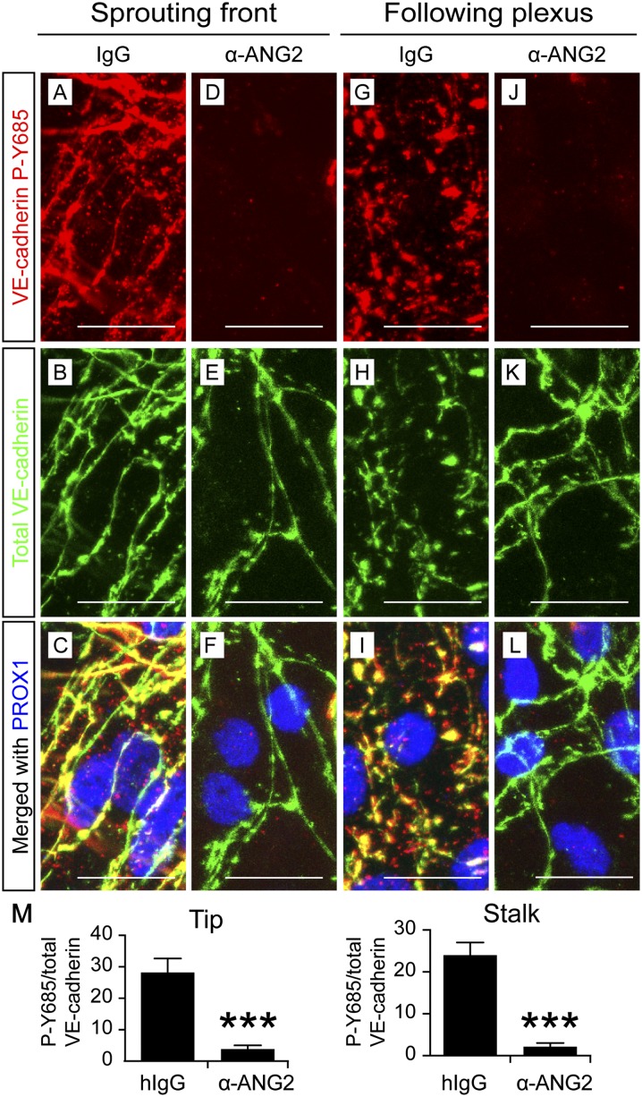
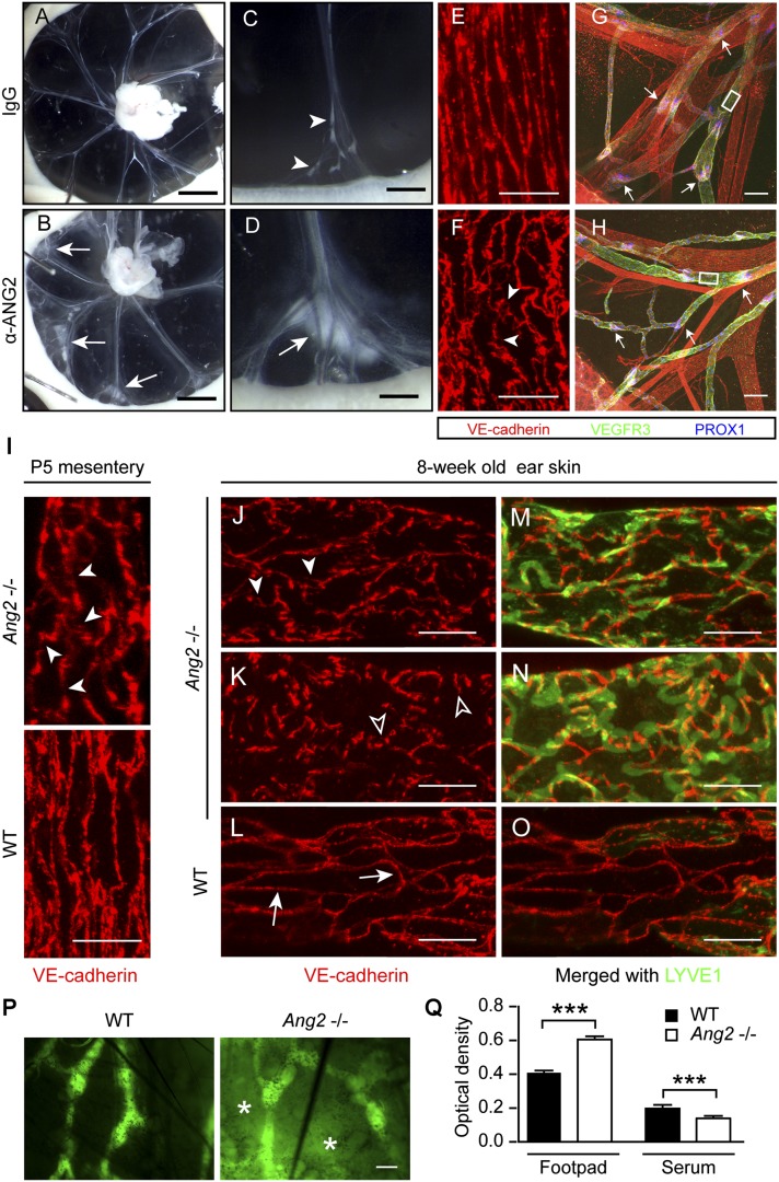
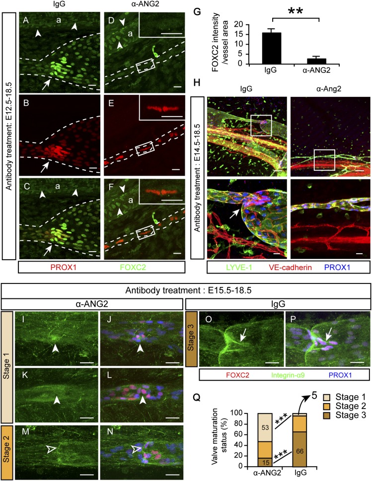
Primitive lymphatic vessels are remodeled into functionally specialized initial and collecting lymphatics during development. Lymphatic endothelial cell (LEC) junctions in initial lymphatics transform from a zipper-like to a button-like pattern during collecting vessel development, but what regulates this process is largely unknown. Angiopoietin 2 ( Ang2 ) deficiency leads to abnormal lymphatic vessels. Here we found that an ANG2-blocking antibody inhibited embryonic lymphangiogenesis, whereas endothelium-specific ANG2 overexpression induced lymphatic hyperplasia. ANG2 inhibition blocked VE-cadherin phosphorylation at tyrosine residue 685 and the concomitant formation of button-like junctions in initial lymphatics. The defective junctions were associated with impaired lymph uptake. In collecting lymphatics, adherens junctions were disrupted, and the vessels leaked upon ANG2 blockade or gene deletion. ANG2 inhibition also suppressed the onset of lymphatic valve formation and subsequent valve maturation. These data identify ANG2 as the first essential regulator of the functionally important interendothelial cell–cell junctions that form during lymphatic development.
Overview
The claims and statistics of this paper haven’t been checked yet.
Run the check — free, no account needed. It reads the open-access text and checks the sources it cites.
Integrity signals
Registry
Cites
Statistics
Figures
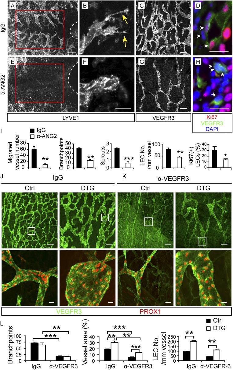
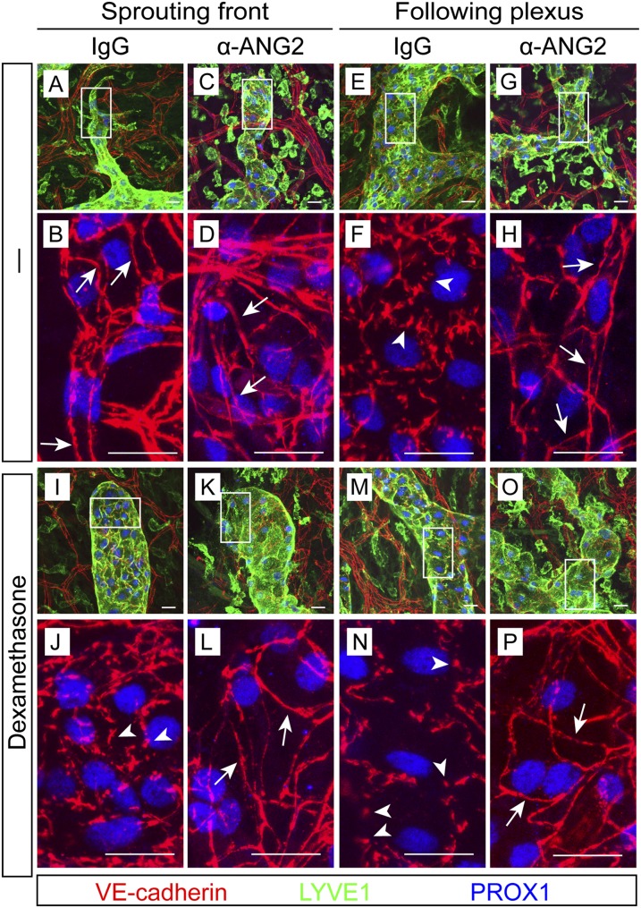
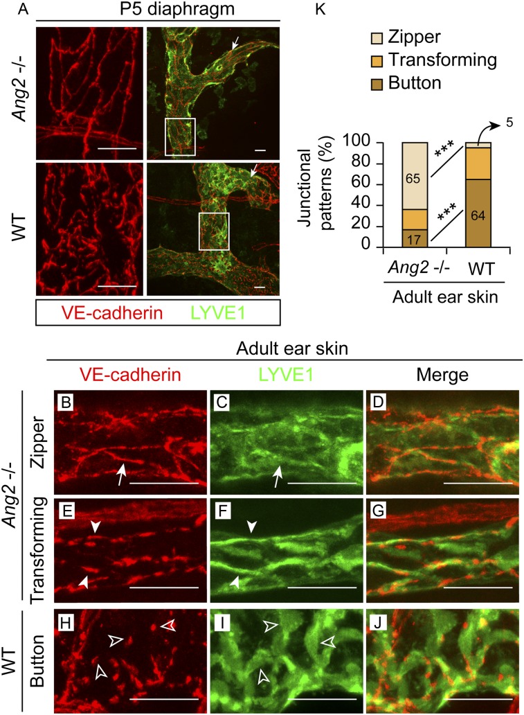
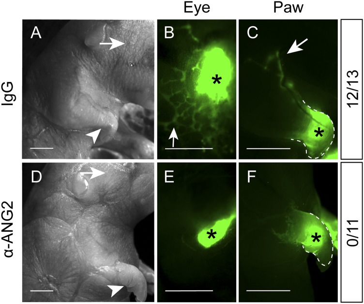
Verification
Claims the paper makes
The claims and statistics of this paper haven’t been checked yet.
Run the check — free, no account needed. It reads the open-access text and checks the sources it cites.
Claims other papers make about this one
Each is checked against this paper’s own text: does it say what the citing paper says it does?
The cited source says what the claim attributes to it.
In the citing paper
“During vascular development, Ang2 inhibition blocked VE-cadherin phosphorylation at tyrosine residue 685 and the concomitant formation of button-like junctions in initial lymphatic vessels20”
From Endothelial destabilization by angiopoietin-2 via integrin β1 activation (2015) · cites this paper as [20]
Self-check
Percentages, sums and reported differences re-computed from the paper’s own numbers.
The self-check needs the paper’s full text.
Citations
Cites
Angiopoietin-2, a Natural Antagonist for Tie2 That Disrupts in vivo Angiogenesis
Maisonpierre, P. C., Suri, C., Jones, P. F., Bartunkova, S., Wiegand, S. J., Radziejewski, C., Compton, D., McClain, J., Aldrich, T. H., Papadopoulos, N., et al. (1997) · Science · doi:10.1126/science.277.5322.55 · Cited by 3,570
Requisite Role of Angiopoietin-1, a Ligand for the TIE2 Receptor, during Embryonic Angiogenesis
Suri, C., Jones, P. F., Patan, S., Bartunkova, S., Maisonpierre, P. C., Davis, S. H., Sato, T. N., & Yancopoulos, G. D. (1996) · Cell · doi:10.1016/s0092-8674(00)81813-9 · Cited by 2,872
Vessel Cooption, Regression, and Growth in Tumors Mediated by Angiopoietins and VEGF
Holash, J. A., Maisonpierre, P. C., Compton, D. L., Boland, P., Alexander, C., Zagzag, D., Yancopoulos, G. D., & Wiegand, S. J. (1999) · Science · doi:10.1126/science.284.5422.1994 · Cited by 2,222
Distinct roles of the receptor tyrosine kinases Tie-1 and Tie-2 in blood vessel formation
Sato, T. N., Tozawa, Y., Deutsch, U., Wolburg‐Buchholz, K., Fujiwara, Y., Gendron‐Maguire, M., Gridley, T., Wolburg, H., Risau, W., & Qin, Y. (1995) · Nature · doi:10.1038/376070a0 · Cited by 1,821
Prox1 Function Is Required for the Development of the Murine Lymphatic System
Wigle, J. T., & Oliver, G. C. (1999) · Cell · doi:10.1016/s0092-8674(00)81511-1 · Cited by 1,581
Control of vascular morphogenesis and homeostasis through the angiopoietin–Tie system
Augustin, H. G., Koh, G. Y., Thurston, G., & Alitalo, K. K. (2009) · Nature Reviews Molecular Cell Biology · doi:10.1038/nrm2639 · Cited by 1,512
Self-citationLymphangiogenesis: Molecular Mechanisms and Future Promise
Tammela, T., & Alitalo, K. K. (2010) · Cell · doi:10.1016/j.cell.2010.01.045 · Cited by 1,485
Self-citationKarkkainen, M. J., Haiko, P., Sainio, K., Partanen, J. M., Taipale, J., Petrova, T. V., Jeltsch, M., Jackson, D. G., Talikka, M., Rauvala, H., et al. (2003) · Nature Immunology · doi:10.1038/ni1013 · Cited by 1,441
Self-citationGale, N. W., Thurston, G., Hackett, S. F., Renard, R. A., Wang, Q., McClain, J., Martin, C., Witte, C. L., Witte, M. H., Jackson, D. G., et al. (2002) · Developmental Cell · doi:10.1016/s1534-5807(02)00217-4 · Cited by 1,017
Functionally specialized junctions between endothelial cells of lymphatic vessels
Baluk, P., Fuxe, J., Hashizume, H., Romano, T., Lashnits, E., Butz, S., Vestweber, D., Corada, M., Molendini, C., Dejana, E., et al. (2007) · The Journal of Experimental Medicine · doi:10.1084/jem.20062596 · Cited by 1,006
Self-citation
Cited by
Sorted by citations
Vascular heterogeneity and specialization in development and disease
Potente, M., & Mäkinen, T. (2017) · Nature Reviews Molecular Cell Biology · doi:10.1038/nrm.2017.36 · Cited by 653
Role of Angiopoietin-2 in Vascular Physiology and Pathophysiology
Akwii, R. G., Sajib, M. S., Zahra, F. T., & Mikelis, C. M. (2019) · Cells · doi:10.3390/cells8050471 · Cited by 566
Macrophages: An Inflammatory Link Between Angiogenesis and Lymphangiogenesis
Corliss, B. A., Azimi, M. S., Munson, J. M., Peirce, S. M., & Murfee, W. L. (2016) · Microcirculation · doi:10.1111/micc.12259 · Cited by 356
Lymphatic Vessel Network Structure and Physiology
Breslin, J. W., Yang, Y., Scallan, J. P., Sweat, R. S., Adderley, S., & Murfee, W. L. (2018) · Comprehensive physiology · doi:10.1002/cphy.c180015 · Cited by 420
The effects of sepsis on endothelium and clinical implications
Dolmatova, E. V., Wang, K., Mandavilli, R., & Griendling, K. K. (2020) · Cardiovascular Research · doi:10.1093/cvr/cvaa070 · Cited by 324
Metadata
- Authors
- Zheng, W., Nurmi, H., Appak, S., Sabine, A., Bovay, E., Korhonen, E. A., Orsenigo, F., Lohela, M., D’Amico, G., Holopainen, T., Leow, C. C., Dejana, E., Petrova, T. V., Augustin, H. G., & Alitalo, K.
- Affiliations
- University of Helsinki (FI)
- Centre Hospitalier Universitaire Vaudois (CH)
- University of Milan (IT)
- German Cancer Research Center (DE)
- Corresponding author
- Alitalo, Kari
- ORCID
- Venue
- Genes & Development
- Publisher
- Cold Spring Harbor Laboratory
- Published
- 2014-07-15
- Volume
- 28
- Issue
- 14
- Pages
- 1592-1603
- ISSN
- 0890-9369 (ISSN-L)
- 1549-5477
- PMC
- PMC4102766
- OpenAlex
- W2066164950
- Cited by
- 134
- References
- 46
- Citations by year
- 2026: 9 · 2025: 9 · 2024: 7 · 2023: 16 · 2022: 13 · 2021: 12 · 2020: 15 · 2019: 12 · 2018: 12 · 2017: 10 · 2016: 9 · 2015: 9 · 2014: 1
- Type
- posted content
- Language
- en
- Open access
- hybrid
- doi.org
- Licence
- cc-by-nc
- Locations
- Genes & Development (publishedVersion, cc-by-nc)
- Europe PMC (PubMed Central) (publishedVersion, cc-by-nc)
- PubMed Central (publishedVersion, cc-by-nc)
- PubMed (publishedVersion)
- SERVAL (Université de Lausanne) (publishedVersion, cc-by-nc)
- IRIS (submittedVersion, cc-by-nc)
- Archivio Istituzionale della Ricerca (Universita Degli Studi Di Milano) (submittedVersion, other-oa)
- HighWire Press Open Archive (submittedVersion)
- Funders
- European Commission
- Fondation Leducq (11CVD03)
- Academy of Finland
- Biomedicum Helsinki-säätiö
- Orionin Tutkimussäätiö
- FP7 Ideas: European Research Council (268804)
- Topics
- Health Sciences › Medicine › Oncology › Lymphatic System and Diseases
- Health Sciences › Medicine › Surgery › Lymphatic Disorders and Treatments
- Health Sciences › Medicine › Surgery › Vascular Malformations and Hemangiomas
- Keywords
- angiopoietin-2, lymphatic endothelial cells, lymphatic development, lymphangiogenesis, lymphoid hyperplasia, VE-cadherin phosphorylation, adherens junctions, lymphatic leakage, lymphatic absorption
- MeSH terms
- Animals, Embryo, Mammalian, Intercellular Junctions, Lymphoid Tissue, Mice, Inbred C57BL, Phosphorylation, Cadherins, Gene Deletion, Lymphangiogenesis, Angiopoietin-2, Endothelial Cells, Mice
About these checks
Registry, retraction, Cites and statistics results are computed, not judged, with one exception: a printed reference that no index lists is read once by a model before it counts as a citation error. Claims are extracted by a model and kept only when their quote appears verbatim in the text; each cited source is then checked, with a model’s help, for whether it says what the claim relies on. Those verdicts can be wrong — read the claim’s quote and the cited source before relying on one.
Statistics and arithmetic are recomputed, not judged.
Confidence bands are shown only for fields calibrated on our annotated test set. None is calibrated yet, so no band is shown.
What counts as a claim ›How we measure confidence ›How we check this ›
Are you an author, or do you see a problem with this page? Request a review
