Cited by 204

The DOI matches its Crossref record; 5 of the 5 references it cites were found in a registry or an index.

dFOXO‐independent effects of reduced insulin‐like signaling in Drosophila

Slack, Cathy, Giannakou, Maria E., Foley, Andrea, Goss, Martin, Partridge, Linda·2011·Aging Cell

Abstract

Summary: The insulin/insulin‐like growth factor‐like signaling (IIS) pathway in metazoans has evolutionarily conserved roles in growth control, metabolic homeostasis, stress responses, reproduction, and lifespan. Genetic manipulations that reduce IIS in the nematode worm Caenorhabditis elegans , the fruit fly Drosophila melanogaster , and the mouse have been shown not only to produce substantial increases in lifespan but also to ameliorate several age‐related diseases. In C. elegans , the multitude of phenotypes produced by the reduction in IIS are all suppressed in the absence of the worm FOXO transcription factor, DAF‐16, suggesting that they are all under common regulation. It is not yet clear in other animal models whether the activity of FOXOs mediate all of the physiological effects of reduced IIS, especially increased lifespan. We have addressed this issue by examining the effects of reduced IIS in the absence of dFOXO in Drosophila , using a newly generated null allele of dfoxo . We found that the removal of dFOXO almost completely blocks IIS‐dependent lifespan extension. However, unlike in C. elegans , removal of dFOXO does not suppress the body size, fecundity, or oxidative stress resistance phenotypes of IIS‐compromised flies. In contrast, IIS‐dependent xenobiotic resistance is fully dependent on dFOXO activity. Our results therefore suggest that there is evolutionary divergence in the downstream mechanisms that mediate the effects of IIS. They also imply that in Drosophila , additional factors act alongside dFOXO to produce IIS‐dependent responses in body size, fecundity, and oxidative stress resistance and that these phenotypes are not causal in IIS‐mediated extension of lifespan.

Overview

The claims and statistics of this paper haven’t been checked yet.

Run the check — free, no account needed. It reads the open-access text and checks the sources it cites.

A few free checks a day · papers already verified open instantly · results are public

Integrity signals

Registry

Crossref · 10.1111/j.1474-9726.2011.00707.x

Cites

5 of 5 references verified0 citation errors · 0 not checkable

Statistics

Figures

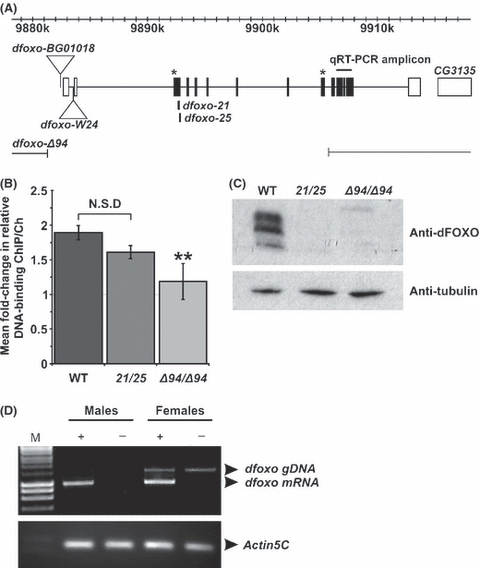
Fig. 1 Molecular characterization of the dfoxoΔ94 deletion. (A) Schematic representation of the dfoxo locus. Coding exons are in black and noncoding exons are in white. The position of the P-element insertion (dfoxoBG01018) used to generate the dfoxoΔ94 deletion by imprecise excision and the breakpoints of the dfoxoΔ94 deletion are shown along with the positions of the dfoxo21, dfoxo25, and dfoxow24 mutations. The position of the amplicon amplified by RT-PCR to detect dfoxo mRNA expression is also indicated along with the position of the sequences encoding the peptides from which the dFOXO antibody was generated (marked by *). (B) Quantitative PCR (qPCR) on the Lnk promoter normalized to a control genomic region located 3′ to the Lnk gene to determine the proportion of DNA recovered after chromatin immunoprecipitation (ChIP) using a specific anti-dFOXO antibody from chromatin extracts prepared from wild-type, dfoxo21/dfoxo25 and dfoxoΔ94 homozygous mutant flies. Relative DNA binding was calculated as the proportion of chromatin recovered in the ChIP divided by that in the total chromatin preparation. Data are presented as mean fold change in DNA binding at the Lnk 5′ region compared with the control Lnk 3′ region ± SEM of two (for dfoxo21/dfoxo25) or three biological repeats. (**P <0.05, t-test). (C) Western blot analysis of dFOXO protein expression in extracts prepared from wild-type, dfoxo21/dfoxo25 (21/25) heterozygotes and dfoxoΔ94 (Δ94/Δ94) homozygous mutant flies. Blots were probed with anti-tubulin as a control for protein loading. (D) RT-PCR analysis of dfoxo transcript expression in 7-day-old male and female wild-type (+) and dfoxoΔ94 homozygous mutant (−) flies. M indicates the marker lane. Amplification of the actin5C transcript was used as a control for the RT-PCR protocol.
Fig. 1 Molecular characterization of the dfoxoΔ94 deletion. (A) Schematic representation of the dfoxo locus. Coding exons are in black and noncoding exons are in white. The position of the P-element insertion (dfoxoBG01018) used to generate the dfoxoΔ94 deletion by imprecise excision and the breakpoints of the dfoxoΔ94 deletion are shown along with the positions of the dfoxo21, dfoxo25, and dfoxow24 mutations. The position of the amplicon amplified by RT-PCR to detect dfoxo mRNA expression is also indicated along with the position of the sequences encoding the peptides from which the dFOXO antibody was generated (marked by *). (B) Quantitative PCR (qPCR) on the Lnk promoter normalized to a control genomic region located 3′ to the Lnk gene to determine the proportion of DNA recovered after chromatin immunoprecipitation (ChIP) using a specific anti-dFOXO antibody from chromatin extracts prepared from wild-type, dfoxo21/dfoxo25 and dfoxoΔ94 homozygous mutant flies. Relative DNA binding was calculated as the proportion of chromatin recovered in the ChIP divided by that in the total chromatin preparation. Data are presented as mean fold change in DNA binding at the Lnk 5′ region compared with the control Lnk 3′ region ± SEM of two (for dfoxo21/dfoxo25) or three biological repeats. (**P <0.05, t-test). (C) Western blot analysis of dFOXO protein expression in extracts prepared from wild-type, dfoxo21/dfoxo25 (21/25) heterozygotes and dfoxoΔ94 (Δ94/Δ94) homozygous mutant flies. Blots were probed with anti-tubulin as a control for protein loading. (D) RT-PCR analysis of dfoxo transcript expression in 7-day-old male and female wild-type (+) and dfoxoΔ94 homozygous mutant (−) flies. M indicates the marker lane. Amplification of the actin5C transcript was used as a control for the RT-PCR protocol.CC BY · PubMed Central
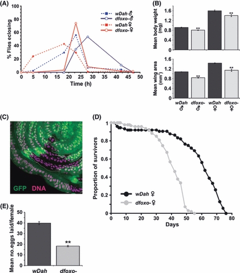
Fig. 2 Phenotypic analysis of dfoxoΔ94 homozygous mutant flies. (A) Egg-to-adult development time is delayed in both males and females homozygous for the dfoxoΔ94 deletion (dfoxo−) compared with wDahomey controls. Only the eclosion period of the adult flies is shown. Data are shown as percentage of flies eclosing. (For males, n = 114 for wDahomey and n = 106 for dfoxo−. For females, n = 107 for wDahomey and n = 116 for dfoxo−). (B) Homozygous dfoxoΔ94 flies (dfoxo−) have significantly reduced adult body weights and smaller wing sizes than wDahomey controls. Data are represented as means ± SEM (n = 10 for each measurement, **P <0.05, anova). (C) Clonal analysis of the effects of dfoxoΔ94 mutation on cell size and proliferation in the developing wing disk. dfoxoΔ94 mutant cells were generated by mitotic recombination and are marked by the absence of GFP. No obvious differences were observed in clone size nor in the size of in dfoxoΔ94 mutant cells within the clone (GFP-negative cells) compared with adjacent heterozygous cells (GFP-positive cells) outside of the clone. (D) Survival curves of female flies homozygous mutant for dfoxo− mutants (n = 99, median survival = 41 days, maximum survival = 48 days) and wDahomey controls (n = 94, median survival = 61 days, maximum survival = 73 days). dfoxo− mutants are significantly shorter lived than controls (P <0.0001; Log-rank test). Representative of two independent experiments. (E) Average number of eggs laid per dfoxo− mutant 7-day-old female compared with age-matched wDahomey controls. Data are presented as the mean number of eggs laid per female over a 24-h period ± SEM. Eggs were counted from ten separate vials, and each vial contained ten females. dfoxo− females laid significantly fewer eggs than wDahomey controls. (**P <0.05, anova).
Fig. 2 Phenotypic analysis of dfoxoΔ94 homozygous mutant flies. (A) Egg-to-adult development time is delayed in both males and females homozygous for the dfoxoΔ94 deletion (dfoxo−) compared with wDahomey controls. Only the eclosion period of the adult flies is shown. Data are shown as percentage of flies eclosing. (For males, n = 114 for wDahomey and n = 106 for dfoxo−. For females, n = 107 for wDahomey and n = 116 for dfoxo−). (B) Homozygous dfoxoΔ94 flies (dfoxo−) have significantly reduced adult body weights and smaller wing sizes than wDahomey controls. Data are represented as means ± SEM (n = 10 for each measurement, **P <0.05, anova). (C) Clonal analysis of the effects of dfoxoΔ94 mutation on cell size and proliferation in the developing wing disk. dfoxoΔ94 mutant cells were generated by mitotic recombination and are marked by the absence of GFP. No obvious differences were observed in clone size nor in the size of in dfoxoΔ94 mutant cells within the clone (GFP-negative cells) compared with adjacent heterozygous cells (GFP-positive cells) outside of the clone. (D) Survival curves of female flies homozygous mutant for dfoxo− mutants (n = 99, median survival = 41 days, maximum survival = 48 days) and wDahomey controls (n = 94, median survival = 61 days, maximum survival = 73 days). dfoxo− mutants are significantly shorter lived than controls (P <0.0001; Log-rank test). Representative of two independent experiments. (E) Average number of eggs laid per dfoxo− mutant 7-day-old female compared with age-matched wDahomey controls. Data are presented as the mean number of eggs laid per female over a 24-h period ± SEM. Eggs were counted from ten separate vials, and each vial contained ten females. dfoxo− females laid significantly fewer eggs than wDahomey controls. (**P <0.05, anova).CC BY · PubMed Central
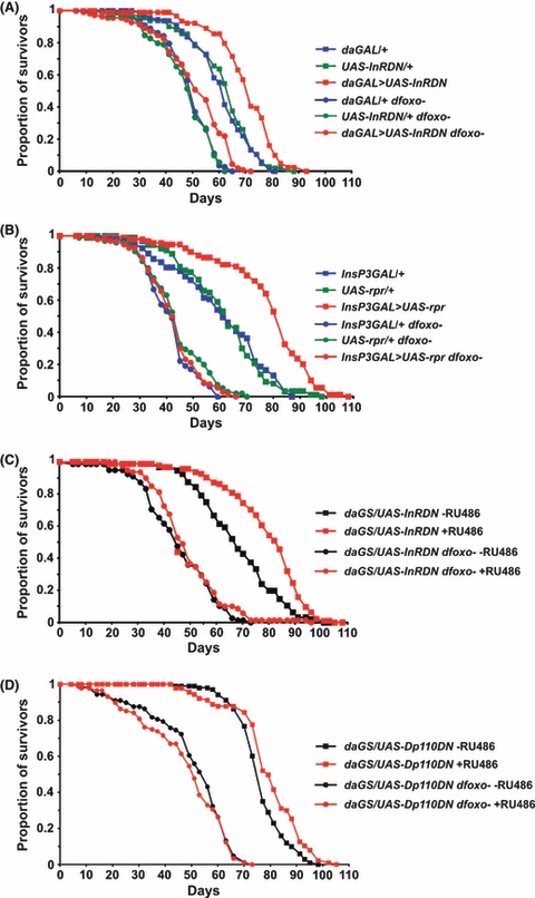
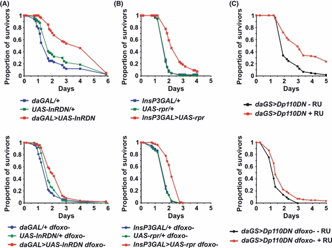
Fig. 3 Effects of dfoxo removal on the survivorship of IIS-compromised flies. (A) Survival curves of female flies overexpressing a dominant negative version of the insulin receptor (daGAL > UAS-InRDN) and their genetic controls (daGAL/+ and UAS-InRDN/+) in both wild-type and dfoxo− backgrounds (representative of two independent experiments). In a wild-type background: for daGAL > UAS-InRDN median survival = 71 days, maximum survival = 82 days, n = 93; for daGAL/+ median survival = 61 days, maximum survival = 77 days, n = 114; for UAS-InRDN/+ median survival = 64 days, maximum survival = 75, n = 115. The survival of daGAL > UAS-InRDN flies was significantly different from each of the controls (P <0.0001; log-rank test). No significant difference in survival was detected between the two controls (P = 0.6, log-rank test). In a dfoxo− background: for daGAL > UAS-InRDN median survival = 53 days, maximum survival = 64 days, n = 114; for daGAL/+ median survival = 49 days, maximum survival = 59 days, n = 109; for UAS-InRDN/+ median survival = 49 days, maximum survival = 57 days, n = 106. The survival of daGAL > UAS-InRDN dfoxo− flies is significantly different from each of the controls (P <0.001; log-rank test). No significant difference in survival was detected between the two controls (P = 0.5, log-rank test). (B) Survival curves of female flies with late ablation of the median neurosecretary cells (InsP3GAL > UAS-rpr) and their genetic controls (InsP3GAL/+ and UAS-rpr/+) in both wild-type and dfoxo− backgrounds. In a wild-type background: for InsP3GAL > UAS-rpr median survival = 82 days, maximum survival = 100 days, n = 99; for InsP3/+ median survival = 60 days, maximum survival = 82 days, n = 94; for UAS-rpr/+ median survival = 62 days, maximum survival = 82 days, n = 93. The survival of InsP3GAL > UAS-rpr flies was significantly different from each of the controls (P <10−12, log-rank test). No significant difference in survival was detected between the two controls (P = 0.8, log-rank test). In a dfoxo− background: for InsP3GAL > UAS-rpr median survival = 44 days, maximum survival = 58 days, n = 94; for InsP3/+ median survival = 41 days, maximum survival = 56 days, n = 108; for UAS-rpr/+ median survival = 44 days, maximum survival = 59 days, n = 95. The survival of InsP3GAL > UAS-rpr flies was not significantly different from either of the controls (P>0.2, log-rank test). (C) Survival curves of female daGS > UAS-InRDN flies induced to ubiquitously express the dominant negative insulin receptor by feeding RU486-containing food from day 3 of adulthood (wild-type +RU486: median survival = 83 days, maximum survival = 95 days, n = 94; dfoxo− +RU486: median survival = 46 days, maximum survival = 70 days, n = 81) compared with uninduced controls (wild-type −RU486: median survival = 67 days, maximum survival = 90 days, n = 97; dfoxo−−RU486: median survival = 44 days, maximum survival = 65 days, n = 79). The survival of wild-type daGS > UAS-InRDN +RU486 was significantly different from the −RU486 control (P <10−6, log-rank test). The survival of dfoxo− daGS > UAS-InRDN +RU486 was not significantly different from the −RU486 control (P = 0.2, log-rank test). (D) Survival curves of female daGS > UAS-dp110DN flies induced to ubiquitously overexpress a dominant negative form of Dp110 by feeding RU486-containing food from day 3 of adulthood (wild-type +RU486: median survival = 80 days, maximum survival = 97 days, n = 91; dfoxo− +RU486: median survival = 51 days, maximum survival = 65 days, n = 95) compared with uninduced controls (wild-type −RU486: median survival = 75 days, maximum survival = 92 days, n = 103; dfoxo−−RU486: median survival = 54 days, maximum survival = 65 days, n = 97). The survival of wild-type daGS > UAS-dp110DN +RU486 was significantly different from the −RU486 control (P<0.001, log-rank test). The survival of dfoxo− daGS > UAS-dp110DN +RU486 was not significantly different from the −RU486 control (P = 0.4, log-rank test). Representative of two independent experiments.
Fig. 3 Effects of dfoxo removal on the survivorship of IIS-compromised flies. (A) Survival curves of female flies overexpressing a dominant negative version of the insulin receptor (daGAL > UAS-InRDN) and their genetic controls (daGAL/+ and UAS-InRDN/+) in both wild-type and dfoxo− backgrounds (representative of two independent experiments). In a wild-type background: for daGAL > UAS-InRDN median survival = 71 days, maximum survival = 82 days, n = 93; for daGAL/+ median survival = 61 days, maximum survival = 77 days, n = 114; for UAS-InRDN/+ median survival = 64 days, maximum survival = 75, n = 115. The survival of daGAL > UAS-InRDN flies was significantly different from each of the controls (P <0.0001; log-rank test). No significant difference in survival was detected between the two controls (P = 0.6, log-rank test). In a dfoxo− background: for daGAL > UAS-InRDN median survival = 53 days, maximum survival = 64 days, n = 114; for daGAL/+ median survival = 49 days, maximum survival = 59 days, n = 109; for UAS-InRDN/+ median survival = 49 days, maximum survival = 57 days, n = 106. The survival of daGAL > UAS-InRDN dfoxo− flies is significantly different from each of the controls (P <0.001; log-rank test). No significant difference in survival was detected between the two controls (P = 0.5, log-rank test). (B) Survival curves of female flies with late ablation of the median neurosecretary cells (InsP3GAL > UAS-rpr) and their genetic controls (InsP3GAL/+ and UAS-rpr/+) in both wild-type and dfoxo− backgrounds. In a wild-type background: for InsP3GAL > UAS-rpr median survival = 82 days, maximum survival = 100 days, n = 99; for InsP3/+ median survival = 60 days, maximum survival = 82 days, n = 94; for UAS-rpr/+ median survival = 62 days, maximum survival = 82 days, n = 93. The survival of InsP3GAL > UAS-rpr flies was significantly different from each of the controls (P <10−12, log-rank test). No significant difference in survival was detected between the two controls (P = 0.8, log-rank test). In a dfoxo− background: for InsP3GAL > UAS-rpr median survival = 44 days, maximum survival = 58 days, n = 94; for InsP3/+ median survival = 41 days, maximum survival = 56 days, n = 108; for UAS-rpr/+ median survival = 44 days, maximum survival = 59 days, n = 95. The survival of InsP3GAL > UAS-rpr flies was not significantly different from either of the controls (P>0.2, log-rank test). (C) Survival curves of female daGS > UAS-InRDN flies induced to ubiquitously express the dominant negative insulin receptor by feeding RU486-containing food from day 3 of adulthood (wild-type +RU486: median survival = 83 days, maximum survival = 95 days, n = 94; dfoxo− +RU486: median survival = 46 days, maximum survival = 70 days, n = 81) compared with uninduced controls (wild-type −RU486: median survival = 67 days, maximum survival = 90 days, n = 97; dfoxo−−RU486: median survival = 44 days, maximum survival = 65 days, n = 79). The survival of wild-type daGS > UAS-InRDN +RU486 was significantly different from the −RU486 control (P <10−6, log-rank test). The survival of dfoxo− daGS > UAS-InRDN +RU486 was not significantly different from the −RU486 control (P = 0.2, log-rank test). (D) Survival curves of female daGS > UAS-dp110DN flies induced to ubiquitously overexpress a dominant negative form of Dp110 by feeding RU486-containing food from day 3 of adulthood (wild-type +RU486: median survival = 80 days, maximum survival = 97 days, n = 91; dfoxo− +RU486: median survival = 51 days, maximum survival = 65 days, n = 95) compared with uninduced controls (wild-type −RU486: median survival = 75 days, maximum survival = 92 days, n = 103; dfoxo−−RU486: median survival = 54 days, maximum survival = 65 days, n = 97). The survival of wild-type daGS > UAS-dp110DN +RU486 was significantly different from the −RU486 control (P<0.001, log-rank test). The survival of dfoxo− daGS > UAS-dp110DN +RU486 was not significantly different from the −RU486 control (P = 0.4, log-rank test). Representative of two independent experiments.CC BY · PubMed Central
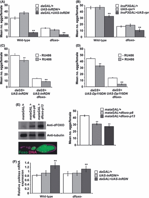
Fig. 4 Effects of dfoxo removal on the female fecundity. (A–D) Average number of eggs laid per 7-day-old female or after 7 days of RU486 treatment. Data are presented as the mean number of eggs laid per female over a 24-h period ± SEM. Eggs were counted from ten separate vials, and each vial contained ten females. (A) Females with constitutive and ubiquitous expression of the dominant negative insulin receptor. In both wild-type and dfoxo− backgrounds, daGAL > UAS-InRDN females laid significantly fewer eggs than both daGAL/+ and UAS-InRDN/+ controls (**P <0.05, t-test). There was no significant difference between daGAL > UAS-InRDN and daGAL > UAS-InRDN dfoxo− flies. (B) Females with late ablation of the median neurosecretary cells. In both wild-type and dfoxo− backgrounds, InsP3GAL > UAS-rpr laid significantly fewer eggs than both InsP3GAL/+ and UAS-rpr/+ controls (**P <0.05, t-test). (C) Females induced to ubiquitously overexpress the dominant negative insulin receptor by feeding RU486-containing food from day 3 of adulthood. In both wild-type and dfoxo− backgrounds, daGS > UAS-InRDN females induced with RU486 (+RU486) laid significantly fewer eggs uninduced controls (−RU486) (**P <0.05, t-test). (D) Females induced to ubiquitously overexpress dominant negative Dp110 by feeding RU486-containing food from day 3 of adulthood. In both wild-type and dfoxo− backgrounds, daGS > UAS-Dp110DN females induced with RU486 (+RU486) laid significantly fewer eggs uninduced controls (−RU486) (**P <0.05, t-test). (E) dFOXO protein expression in the germline. Western blot analysis of dFOXO protein expression in ovaries overexpressing two independent dFOXO transgenes (dfoxo-p8 and dfoxo-p13) within the germline under the control of the mataGAL4 driver. Blots were probed with anti-tubulin as a control for protein loading. Ovaries from these females overexpressing dFOXO protein in the germline look structurally wild-type, but egg production is significantly reduced. Eggs were collected from 7-day-old females over a 24-h period and counted. Data are presented as the mean number of eggs laid per female over these 24-h periods ± SEM (**P <0.05, anova). (F) Quantitative RT-PCR analysis of yolkless mRNA expression in female flies of the indicated genotypes normalized to actin5C. Data are presented as means ± SEM (n = 5). yolkless expression is upregulated in daGAL > UAS-InRDN females compared with controls (**P <0.05, t-test) in both wild-type and dfoxo− backgrounds.
Fig. 4 Effects of dfoxo removal on the female fecundity. (A–D) Average number of eggs laid per 7-day-old female or after 7 days of RU486 treatment. Data are presented as the mean number of eggs laid per female over a 24-h period ± SEM. Eggs were counted from ten separate vials, and each vial contained ten females. (A) Females with constitutive and ubiquitous expression of the dominant negative insulin receptor. In both wild-type and dfoxo− backgrounds, daGAL > UAS-InRDN females laid significantly fewer eggs than both daGAL/+ and UAS-InRDN/+ controls (**P <0.05, t-test). There was no significant difference between daGAL > UAS-InRDN and daGAL > UAS-InRDN dfoxo− flies. (B) Females with late ablation of the median neurosecretary cells. In both wild-type and dfoxo− backgrounds, InsP3GAL > UAS-rpr laid significantly fewer eggs than both InsP3GAL/+ and UAS-rpr/+ controls (**P <0.05, t-test). (C) Females induced to ubiquitously overexpress the dominant negative insulin receptor by feeding RU486-containing food from day 3 of adulthood. In both wild-type and dfoxo− backgrounds, daGS > UAS-InRDN females induced with RU486 (+RU486) laid significantly fewer eggs uninduced controls (−RU486) (**P <0.05, t-test). (D) Females induced to ubiquitously overexpress dominant negative Dp110 by feeding RU486-containing food from day 3 of adulthood. In both wild-type and dfoxo− backgrounds, daGS > UAS-Dp110DN females induced with RU486 (+RU486) laid significantly fewer eggs uninduced controls (−RU486) (**P <0.05, t-test). (E) dFOXO protein expression in the germline. Western blot analysis of dFOXO protein expression in ovaries overexpressing two independent dFOXO transgenes (dfoxo-p8 and dfoxo-p13) within the germline under the control of the mataGAL4 driver. Blots were probed with anti-tubulin as a control for protein loading. Ovaries from these females overexpressing dFOXO protein in the germline look structurally wild-type, but egg production is significantly reduced. Eggs were collected from 7-day-old females over a 24-h period and counted. Data are presented as the mean number of eggs laid per female over these 24-h periods ± SEM (**P <0.05, anova). (F) Quantitative RT-PCR analysis of yolkless mRNA expression in female flies of the indicated genotypes normalized to actin5C. Data are presented as means ± SEM (n = 5). yolkless expression is upregulated in daGAL > UAS-InRDN females compared with controls (**P <0.05, t-test) in both wild-type and dfoxo− backgrounds.CC BY · PubMed Central
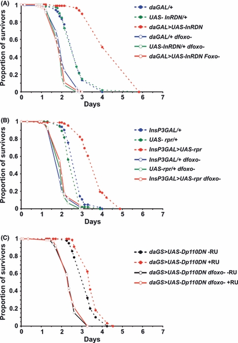
Fig. 5 Functions for dFOXO during IIS-mediated oxidative stress resistance. (A) Survival curves in response to 20 mm paraquat of female flies overexpressing a dominant negative version of the insulin receptor (daGAL > UAS-InRDN) and their genetic controls (daGAL/+ and UAS-InRDN/+) in both wild-type and dfoxo− backgrounds (representative of two independent experiments). In a wild-type background (top panel): for daGAL > UAS-InRDN median survival = 3.5 days, maximum survival = 4.9 days, n = 100; for daGAL/+ median survival = 1.5 days, maximum survival = 1.5 days, n = 90; for UAS-InRDN/+ median survival = 1.5 days, maximum survival = 4.9, n = .80. The survival of daGAL > UAS-InRDN flies was significantly different from each of the controls (P <10−6; Log-rank test). In a dfoxo− background (bottom panel): for daGAL > UAS-InRDN median survival = 1.9 days, maximum survival = 3.5 days, n = 49; for daGAL/+ median survival = 1.5 days, maximum survival = 2.9 days, n = 80; for UAS-InRDN/+ median survival = 1.5 days, maximum survival = 2.4 days, n = 70. The survival of daGAL > UAS-InRDN dfoxo− flies was significantly different from each of the controls (P <0.05; log-rank test). (B) Survival curves in response to 20 mm paraquat of female flies with late ablation of the median neurosecretary cells (InsP3GAL > UAS-rpr) and their genetic controls (InsP3GAL/+ and UAS-rpr/+) in both wild-type and dfoxo− backgrounds. In a wild-type background (top panel): for InsP3GAL > UAS-rpr median survival = 2.1 days, maximum survival = 3.9 days, n = 95; for InsP3GAL/+ median survival = 1.5 days, maximum survival = 2.1 days, n = 88; for UAS-rpr/+ median survival = 2.1 days, maximum survival = 1.9, n = 99. The survival of InsP3GAL > UAS-rpr flies was significantly different from each of the controls (P <10−15; log-rank test). In a dfoxo− background (bottom panel): for InsP3GAL > UAS-rpr median survival = 2.1 days, maximum survival = 2.5 days, n = 78; for InsP3GAL/+ median survival = 1.5 days, maximum survival = 2.1 days, n = 77; for UAS-Rpr/+ median survival = 1.5 days, maximum survival = 2.1 days, n = 76. The survival of InsP3GAL > UAS-rpr dfoxo− flies was significantly different from each of the controls (P <10−12; log-rank test). (C) Survival curves in response to 20 mm paraquat of female flies induced to overexpress dominant negative Dp110 (daGS > UAS-Dp110DN) using RU486 (+RU486) and their uninduced controls (−RU486) in both wild-type and dfoxo− backgrounds. In a wild-type background (top panel): for +RU486 median survival = 2.6 days, maximum survival = 6.5 days, n = 100; for −RU486 median survival = 1.6 days, maximum survival = 3.6 days, n = 100. The survival of +RU486 flies was significantly different from the uninduced controls (P <10−9; log-rank test). In a dfoxo− background (bottom panel): for +RU486 median survival = 1.3 days, maximum survival = 2.6 days, n = 85; for −RU486 median survival = 1.1 days, maximum survival = 2.2 days, n = 0.84. The survival of +RU486 flies was significantly different from the uninduced controls (P <0.001); log-rank test).
Fig. 5 Functions for dFOXO during IIS-mediated oxidative stress resistance. (A) Survival curves in response to 20 mm paraquat of female flies overexpressing a dominant negative version of the insulin receptor (daGAL > UAS-InRDN) and their genetic controls (daGAL/+ and UAS-InRDN/+) in both wild-type and dfoxo− backgrounds (representative of two independent experiments). In a wild-type background (top panel): for daGAL > UAS-InRDN median survival = 3.5 days, maximum survival = 4.9 days, n = 100; for daGAL/+ median survival = 1.5 days, maximum survival = 1.5 days, n = 90; for UAS-InRDN/+ median survival = 1.5 days, maximum survival = 4.9, n = .80. The survival of daGAL > UAS-InRDN flies was significantly different from each of the controls (P <10−6; Log-rank test). In a dfoxo− background (bottom panel): for daGAL > UAS-InRDN median survival = 1.9 days, maximum survival = 3.5 days, n = 49; for daGAL/+ median survival = 1.5 days, maximum survival = 2.9 days, n = 80; for UAS-InRDN/+ median survival = 1.5 days, maximum survival = 2.4 days, n = 70. The survival of daGAL > UAS-InRDN dfoxo− flies was significantly different from each of the controls (P <0.05; log-rank test). (B) Survival curves in response to 20 mm paraquat of female flies with late ablation of the median neurosecretary cells (InsP3GAL > UAS-rpr) and their genetic controls (InsP3GAL/+ and UAS-rpr/+) in both wild-type and dfoxo− backgrounds. In a wild-type background (top panel): for InsP3GAL > UAS-rpr median survival = 2.1 days, maximum survival = 3.9 days, n = 95; for InsP3GAL/+ median survival = 1.5 days, maximum survival = 2.1 days, n = 88; for UAS-rpr/+ median survival = 2.1 days, maximum survival = 1.9, n = 99. The survival of InsP3GAL > UAS-rpr flies was significantly different from each of the controls (P <10−15; log-rank test). In a dfoxo− background (bottom panel): for InsP3GAL > UAS-rpr median survival = 2.1 days, maximum survival = 2.5 days, n = 78; for InsP3GAL/+ median survival = 1.5 days, maximum survival = 2.1 days, n = 77; for UAS-Rpr/+ median survival = 1.5 days, maximum survival = 2.1 days, n = 76. The survival of InsP3GAL > UAS-rpr dfoxo− flies was significantly different from each of the controls (P <10−12; log-rank test). (C) Survival curves in response to 20 mm paraquat of female flies induced to overexpress dominant negative Dp110 (daGS > UAS-Dp110DN) using RU486 (+RU486) and their uninduced controls (−RU486) in both wild-type and dfoxo− backgrounds. In a wild-type background (top panel): for +RU486 median survival = 2.6 days, maximum survival = 6.5 days, n = 100; for −RU486 median survival = 1.6 days, maximum survival = 3.6 days, n = 100. The survival of +RU486 flies was significantly different from the uninduced controls (P <10−9; log-rank test). In a dfoxo− background (bottom panel): for +RU486 median survival = 1.3 days, maximum survival = 2.6 days, n = 85; for −RU486 median survival = 1.1 days, maximum survival = 2.2 days, n = 0.84. The survival of +RU486 flies was significantly different from the uninduced controls (P <0.001); log-rank test).CC BY · PubMed Central
Fig. 6 Functions for dFOXO during IIS-mediated dichlorodiphenyltrichloroethane (DDT) resistance. (A) Survival curves in response to DDT of female flies overexpressing a dominant negative version of the insulin receptor (daGAL > UAS-InRDN) and their genetic controls (daGAL/+ and UAS-InRDN/+) in both wild-type and dfoxo− backgrounds. In a wild-type background: for daGAL > UAS-InRDN median survival = 4.9 days, maximum survival = 4.9 days, n = 76; for daGAL/+ median survival = 2.4 days, maximum survival = 3.5 days, n = 97; for UAS-InRDN/+ median survival = 2.4 days, maximum survival = 3.5, n = 100. The survival of daGAL > UAS-InRDN flies was significantly different from each of the controls (P <10−24; log-rank test). In a dfoxo− background: for daGAL > UAS-InRDN median survival = 1.8 days, maximum survival = 2.4 days, n = 55; for daGAL/+ median survival = 1.8 days, maximum survival = 2.4 days, n = 89; for UAS-InRDN/+ median survival = 1.8 days, maximum survival = 2.1 days, n = 65. The survival of daGAL > UAS-InRDN dfoxo− flies was not significantly different from each of the controls (P>0.1; log-rank test). (B) Survival curves in response to DDT of female flies with late ablation of the median neurosecretary cells (InsP3GAL > UAS-rpr) and their genetic controls (InsP3GAL/+ and UAS-rpr/+) in both wild-type and dfoxo− backgrounds. In a wild-type background: for InsP3GAL > UAS-rpr median survival = 3.6 days, maximum survival = 4.5 days, n = 92; for InsP3GAL/+ median survival = 2.6 days, maximum survival = 3.1 days, n = 83; for UAS-rpr/+ median survival = 2.6 days, maximum survival = 2.6 days, n = 94. In a dfoxo− background: for InsP3GAL > UAS-rpr median survival = 1.9 days, maximum survival = 2.6 days, n = 90; for InsP3GAL/+ median survival = 1.6 days, maximum survival = 2.6 days, n = 90; for UAS-Rpr/+ median survival = 1.6 days, maximum survival = 2.3 days, n = 94. The survival of InsP3GAL > UAS-rpr dfoxo− flies was not significantly different from each of the controls (P >0.08; log-rank test). (C) Survival curves in response to DDT of female flies induced to overexpress dominant negative Dp110 (daGS > UAS-Dp110DN) using RU486 (+RU486) and their uninduced controls (−RU486) in both wild-type and dfoxo− backgrounds. In a wild-type background (top panel): for +RU486 median survival = 3.3 days, maximum survival = 4.0 days, n = 88; for −RU486 median survival = 2.9 days, maximum survival = 4.0 days, n = 99. The survival of +RU486 flies was significantly different from the uninduced controls (P <0.001; log-rank test). In a dfoxo− background (bottom panel): for +RU486 median survival = 2.3 days, maximum survival = 2.9 days, n = 76; for −RU486 median survival = 2.3 days, maximum survival = 2.9 days, n = 85. The survival of +RU486 flies was not significantly different from the uninduced controls (P = 0.6; log-rank test).
Fig. 6 Functions for dFOXO during IIS-mediated dichlorodiphenyltrichloroethane (DDT) resistance. (A) Survival curves in response to DDT of female flies overexpressing a dominant negative version of the insulin receptor (daGAL > UAS-InRDN) and their genetic controls (daGAL/+ and UAS-InRDN/+) in both wild-type and dfoxo− backgrounds. In a wild-type background: for daGAL > UAS-InRDN median survival = 4.9 days, maximum survival = 4.9 days, n = 76; for daGAL/+ median survival = 2.4 days, maximum survival = 3.5 days, n = 97; for UAS-InRDN/+ median survival = 2.4 days, maximum survival = 3.5, n = 100. The survival of daGAL > UAS-InRDN flies was significantly different from each of the controls (P <10−24; log-rank test). In a dfoxo− background: for daGAL > UAS-InRDN median survival = 1.8 days, maximum survival = 2.4 days, n = 55; for daGAL/+ median survival = 1.8 days, maximum survival = 2.4 days, n = 89; for UAS-InRDN/+ median survival = 1.8 days, maximum survival = 2.1 days, n = 65. The survival of daGAL > UAS-InRDN dfoxo− flies was not significantly different from each of the controls (P>0.1; log-rank test). (B) Survival curves in response to DDT of female flies with late ablation of the median neurosecretary cells (InsP3GAL > UAS-rpr) and their genetic controls (InsP3GAL/+ and UAS-rpr/+) in both wild-type and dfoxo− backgrounds. In a wild-type background: for InsP3GAL > UAS-rpr median survival = 3.6 days, maximum survival = 4.5 days, n = 92; for InsP3GAL/+ median survival = 2.6 days, maximum survival = 3.1 days, n = 83; for UAS-rpr/+ median survival = 2.6 days, maximum survival = 2.6 days, n = 94. In a dfoxo− background: for InsP3GAL > UAS-rpr median survival = 1.9 days, maximum survival = 2.6 days, n = 90; for InsP3GAL/+ median survival = 1.6 days, maximum survival = 2.6 days, n = 90; for UAS-Rpr/+ median survival = 1.6 days, maximum survival = 2.3 days, n = 94. The survival of InsP3GAL > UAS-rpr dfoxo− flies was not significantly different from each of the controls (P >0.08; log-rank test). (C) Survival curves in response to DDT of female flies induced to overexpress dominant negative Dp110 (daGS > UAS-Dp110DN) using RU486 (+RU486) and their uninduced controls (−RU486) in both wild-type and dfoxo− backgrounds. In a wild-type background (top panel): for +RU486 median survival = 3.3 days, maximum survival = 4.0 days, n = 88; for −RU486 median survival = 2.9 days, maximum survival = 4.0 days, n = 99. The survival of +RU486 flies was significantly different from the uninduced controls (P <0.001; log-rank test). In a dfoxo− background (bottom panel): for +RU486 median survival = 2.3 days, maximum survival = 2.9 days, n = 76; for −RU486 median survival = 2.3 days, maximum survival = 2.9 days, n = 85. The survival of +RU486 flies was not significantly different from the uninduced controls (P = 0.6; log-rank test).CC BY · PubMed Central
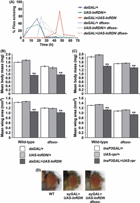
Fig. 7 Effects of dFOXO removal on IIS-mediated developmental delay and growth inhibition. (A) Egg-to-adult development time is delayed in females overexpressing the dominant negative insulin receptor (daGAL > UAS-InRDN) irrespective of the presence or absence of dfoxo. Only the eclosion period of the adult flies is shown. Data are shown as percentage of flies eclosing. (n = 118 for daGAL > UAS-InRDN, n = 110 for daGAL > UAS-InRDN dfoxo−; n = 120 for daGAL/+, n = 102 for daGAL/+ dfoxo−; n = 107 for UAS-InRDN/+, n = 82 for UAS-InRDN/+ dfoxo−). (B) Body weights and wing areas as indicators of adult fly body size in females overexpressing the dominant negative insulin receptor (daGAL > UAS-InRDN). Data are presented as means ± SEM (n = 10 for each measurement). daGAL > UAS-InRDN females in both wild-type and dfoxo− backgrounds are significantly reduced in body size compared with control flies (daGAL/+ and UAS-InRDN/+) (**P <0.05, t-test). (C) Body weights and wing areas as indicators of adult fly body size in females with late ablation of the median neurosecretary cells (InsP3GAL > UAS-rpr). Data are presented as means ± SEM (n = 10 for each measurement). InsP3GAL > UAS-rpr females in both wild-type and dfoxo− backgrounds are significantly reduced in body size compared with control flies (InsP3GAL/+ and UAS-rpr/+) (**P <0.05, t-test). (D) Removal of dFOXO rescued the growth inhibition effects of overexpressing the dominant negative insulin receptor in a tissue-restricted manner. UAS-InRDN was specifically expressed in the developing eye using eyGAL. This resulted in a smaller eye compared with control (middle panel compared with left panel). This tissue-restricted growth inhibition was fully rescued in homozygous dfoxoΔ94 (dfoxo−) mutant flies (right panel).
Fig. 7 Effects of dFOXO removal on IIS-mediated developmental delay and growth inhibition. (A) Egg-to-adult development time is delayed in females overexpressing the dominant negative insulin receptor (daGAL > UAS-InRDN) irrespective of the presence or absence of dfoxo. Only the eclosion period of the adult flies is shown. Data are shown as percentage of flies eclosing. (n = 118 for daGAL > UAS-InRDN, n = 110 for daGAL > UAS-InRDN dfoxo−; n = 120 for daGAL/+, n = 102 for daGAL/+ dfoxo−; n = 107 for UAS-InRDN/+, n = 82 for UAS-InRDN/+ dfoxo−). (B) Body weights and wing areas as indicators of adult fly body size in females overexpressing the dominant negative insulin receptor (daGAL > UAS-InRDN). Data are presented as means ± SEM (n = 10 for each measurement). daGAL > UAS-InRDN females in both wild-type and dfoxo− backgrounds are significantly reduced in body size compared with control flies (daGAL/+ and UAS-InRDN/+) (**P <0.05, t-test). (C) Body weights and wing areas as indicators of adult fly body size in females with late ablation of the median neurosecretary cells (InsP3GAL > UAS-rpr). Data are presented as means ± SEM (n = 10 for each measurement). InsP3GAL > UAS-rpr females in both wild-type and dfoxo− backgrounds are significantly reduced in body size compared with control flies (InsP3GAL/+ and UAS-rpr/+) (**P <0.05, t-test). (D) Removal of dFOXO rescued the growth inhibition effects of overexpressing the dominant negative insulin receptor in a tissue-restricted manner. UAS-InRDN was specifically expressed in the developing eye using eyGAL. This resulted in a smaller eye compared with control (middle panel compared with left panel). This tissue-restricted growth inhibition was fully rescued in homozygous dfoxoΔ94 (dfoxo−) mutant flies (right panel).CC BY · PubMed Central
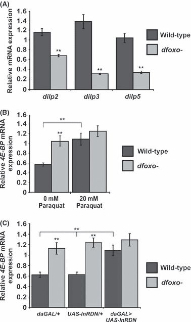
Fig. 8 Gene expression changes in the absence of dFOXO activity. (A) Quantitative RT-PCR analysis of dilp-2, dilp-3, and dilp-5 mRNA expression in female heads isolated from flies of the indicated genotypes normalized to actin5C. Expression of all three dilp transcripts is significantly decreased in dfoxo− flies compared with controls. Data are presented as means ± SEM (n = 5; P <0.05, t-test). (B) Quantitative RT-PCR analysis of 4E-BP mRNA expression normalized to actin5C in female flies treated with 20 mm paraquat. In wild-type flies, 4E-BP mRNA expression was significantly upregulated in response to paraquat treatment. In dfoxoΔ94 homozygous mutant flies, 4E-BP expression was upregulated in comparison with wild-type flies but no further increase in 4E-BP expression was observed when dfoxoΔ94 mutants were treated with paraquat. Data are presented as means ± SEM (n = 5; P <0.05, t-test). (C) Quantitative RT-PCR analysis of 4E-BP mRNA expression in female flies of the indicated genotypes normalized to actin5C. In a wild-type background, 4E-BP mRNA expression was significantly upregulated in daGAL > UAS-InRDN females compared with each of the genetic controls (P <0.05, t-test). In dfoxoΔ94 homozygous mutants, 4E-BP mRNA expression is upregulated in both controls and experimental flies (P <0.05, t-test). However, no further increases in expression were detected in daGAL > UAS-InRDN in a dfoxo mutant background over wild-type background.
Fig. 8 Gene expression changes in the absence of dFOXO activity. (A) Quantitative RT-PCR analysis of dilp-2, dilp-3, and dilp-5 mRNA expression in female heads isolated from flies of the indicated genotypes normalized to actin5C. Expression of all three dilp transcripts is significantly decreased in dfoxo− flies compared with controls. Data are presented as means ± SEM (n = 5; P <0.05, t-test). (B) Quantitative RT-PCR analysis of 4E-BP mRNA expression normalized to actin5C in female flies treated with 20 mm paraquat. In wild-type flies, 4E-BP mRNA expression was significantly upregulated in response to paraquat treatment. In dfoxoΔ94 homozygous mutant flies, 4E-BP expression was upregulated in comparison with wild-type flies but no further increase in 4E-BP expression was observed when dfoxoΔ94 mutants were treated with paraquat. Data are presented as means ± SEM (n = 5; P <0.05, t-test). (C) Quantitative RT-PCR analysis of 4E-BP mRNA expression in female flies of the indicated genotypes normalized to actin5C. In a wild-type background, 4E-BP mRNA expression was significantly upregulated in daGAL > UAS-InRDN females compared with each of the genetic controls (P <0.05, t-test). In dfoxoΔ94 homozygous mutants, 4E-BP mRNA expression is upregulated in both controls and experimental flies (P <0.05, t-test). However, no further increases in expression were detected in daGAL > UAS-InRDN in a dfoxo mutant background over wild-type background.CC BY · PubMed Central

Verification

Claims the paper makes

The claims and statistics of this paper haven’t been checked yet.

Run the check — free, no account needed. It reads the open-access text and checks the sources it cites.

A few free checks a day · papers already verified open instantly · results are public

Claims other papers make about this one

Each is checked against this paper’s own text: does it say what the citing paper says it does?

Self-check

Percentages, sums and reported differences re-computed from the paper’s own numbers.

The self-check needs the paper’s full text.

Citations

Cites

Show all 5 references

Research works printed but not linked: 48

  1. 10.1093/gerona/62.10.1071 · DOI
  2. 10.1016/S0092-8674(00)80799-0 · DOI
  3. 10.1093/gerona/61.6.562 · DOI
  4. 10.1073/pnas.0405775102 · DOI · Paper page
  5. 10.1371/journal.pone.0003721 · DOI
  6. 10.1111/j.1474-9726.2010.00558.x · DOI
  7. 10.1016/j.cmet.2008.02.012 · DOI · Paper page
  8. 10.1371/journal.pone.0015171 · DOI
  9. 10.1126/science.1057991 · DOI
  10. 10.1126/science.1124646 · DOI · Paper page
  11. 10.1126/science.1074240 · DOI · Paper page
  12. 10.1016/j.cub.2006.08.052 · DOI
  13. 10.1016/j.tibs.2007.02.007 · DOI
  14. 10.1126/science.1098219 · DOI
  15. 10.1111/j.1474-9726.2007.00290.x · DOI
  16. 10.1111/j.1474-9726.2007.00362.x · DOI
  17. 10.1038/nature08619 · DOI
  18. 10.1038/sj.onc.1209086 · DOI
  19. 10.1111/j.1748-1716.2007.01780.x · DOI
  20. 10.1371/journal.pgen.1000857 · DOI
  21. 10.1096/fasebj.13.11.1385 · DOI
  22. 10.1073/pnas.0809144106 · DOI
  23. 10.1016/j.ydbio.2007.11.006 · DOI
  24. 10.1038/nature02549 · DOI
  25. 10.1016/S0960-9822(02)01043-6 · DOI
  26. 10.1186/1475-4924-2-20 · DOI
  27. 10.1038/366461a0 · DOI · Paper page
  28. 10.1038/sj.ejhg.5201766 · DOI
  29. 10.1126/science.1111410 · DOI
  30. 10.1073/pnas.90.19.8905 · DOI
  31. 10.1186/gb-2007-8-7-r132 · DOI
  32. 10.1111/j.1474-9726.2008.00373.x · DOI
  33. 10.1038/nature01789 · DOI · Paper page
  34. 10.1111/j.1474-9726.2008.00420.x · DOI
  35. 10.1038/ncb1493 · DOI
  36. 10.1038/onc.2008.28 · DOI
  37. 10.1101/gad.1098703 · DOI
  38. 10.1016/j.jinsphys.2004.12.013 · DOI
  39. 10.1016/j.ceb.2008.02.005 · DOI · Paper page
  40. 10.1038/msb.2010.54 · DOI
  41. 10.1371/journal.pgen.1000881 · DOI
  42. 10.1126/science.1081447 · DOI
  43. 10.1093/genetics/148.2.703 · DOI
  44. 10.1016/j.mad.2009.05.004 · DOI
  45. 10.1534/genetics.104.027748 · DOI
  46. 10.2741/2047 · DOI
  47. 10.1111/j.1474-9726.2009.00504.x · DOI
  48. 10.1073/pnas.0801030105 · DOI · Paper page

Cited by

Sorted by citations

Metadata

Authors
Slack, C., Giannakou, M. E., Foley, A., Goss, M., & Partridge, L.
Venue
Aging Cell
Publisher
Wiley
Published
2011-05-06
Volume
10
Issue
5
Pages
735-748
ISSN
  • 1474-9718
  • 1474-9726
Cited by
204
References
5
Language
en

About these checks

Registry, retraction, Cites and statistics results are computed, not judged, with one exception: a printed reference that no index lists is read once by a model before it counts as a citation error. Claims are extracted by a model and kept only when their quote appears verbatim in the text; each cited source is then checked, with a model’s help, for whether it says what the claim relies on. Those verdicts can be wrong — read the claim’s quote and the cited source before relying on one.

Statistics and arithmetic are recomputed, not judged.

Confidence bands are shown only for fields calibrated on our annotated test set. None is calibrated yet, so no band is shown.

What counts as a claim ›How we measure confidence ›How we check this ›

Are you an author, or do you see a problem with this page? Request a review