Cited by 56

The DOI matches its Crossref record; 3 of the 3 references it cites were found in a registry or an index.

Exosomal MiR‐500a‐3p promotes cisplatin resistance and stemness via negatively regulating FBXW7 in gastric cancer

Lin, Hao, Zhang, Liang, Zhang, Caihua, Liu, Pengpeng·2020·Journal of Cellular and Molecular Medicine

Abstract

Chemoresistance has been a major challenge in advanced gastric cancer (GC) therapy. Exosomal transfer of oncogenic miRNAs implicates important effects in mediating recipient cell chemoresistance by transmitting active molecules. In this study, we found that microRNA‐500a‐3p was highly expressed in cisplatin (DDP) resistant GC cells (MGC803/DDP and MKN45/DDP) and their secreted exosomes than that in the corresponding parental cells. MGC803/DDP‐derived exosomes enhance DDP resistance and stemness properties of MGC803 recipient cells via exosomal delivery of miR‐500a‐3p in vitro and in vivo through targeting FBXW7. However, reintroduction of FBXW7 in MGC803 cells reverses miR‐500a‐3p‐mediated DDP resistance as well as stemness properties. Furthermore, elevated miR‐500a‐3p in the plasma exosomes of GC patients is correlated with DDP resistance and thereby results in poor progression‐free prognosis. Our finding highlights the potential of exosomal miR‐500a‐3p as an potential modality for the prediction and treatment of GC with chemoresistance.

Overview

The claims and statistics of this paper haven’t been checked yet.

Run the check — free, no account needed. It reads the open-access text and checks the sources it cites.

A few free checks a day · papers already verified open instantly · results are public

Integrity signals

Registry

Crossref · 10.1111/jcmm.15524

Cites

3 of 3 references verified0 citation errors · 0 not checkable

Statistics

Figures

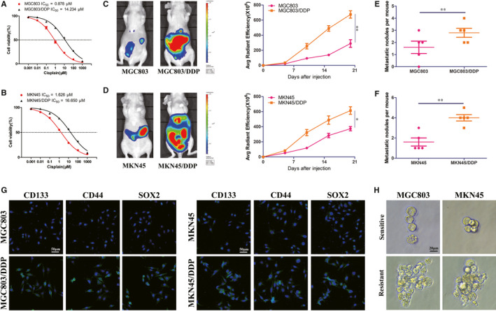
FIGURE 1 DDP resistant MGC803 and MKN45 cells display stem cell‐like features. A, B, Proliferation assay analysis and IC50 value of DDP resistant GC cells (MGC803/DDP and MKN45/DDP) and corresponding parental cells (MGC803 and MKN45). C, D, Representative images showed the bioluminescent signals of abdominal tumorigenesis by indicated GC cells under DDP treatment (left panel). Comparison of bioluminescent signals on the day 21 was analysed (right panel), five nude mice in each group. E, F, Comparison of abdominal metastatic nodules by indicated GC cells under DDP treatment. G, Expression level of stemness markers CD133, CD44 and SOX2 in indicated GC cells by confocal microscopy. H, Sphere formation assay in indicated GC cells. *P < 0.05, **P < 0.01
FIGURE 1 DDP resistant MGC803 and MKN45 cells display stem cell‐like features. A, B, Proliferation assay analysis and IC50 value of DDP resistant GC cells (MGC803/DDP and MKN45/DDP) and corresponding parental cells (MGC803 and MKN45). C, D, Representative images showed the bioluminescent signals of abdominal tumorigenesis by indicated GC cells under DDP treatment (left panel). Comparison of bioluminescent signals on the day 21 was analysed (right panel), five nude mice in each group. E, F, Comparison of abdominal metastatic nodules by indicated GC cells under DDP treatment. G, Expression level of stemness markers CD133, CD44 and SOX2 in indicated GC cells by confocal microscopy. H, Sphere formation assay in indicated GC cells. *P < 0.05, **P < 0.01CC BY · PubMed Central
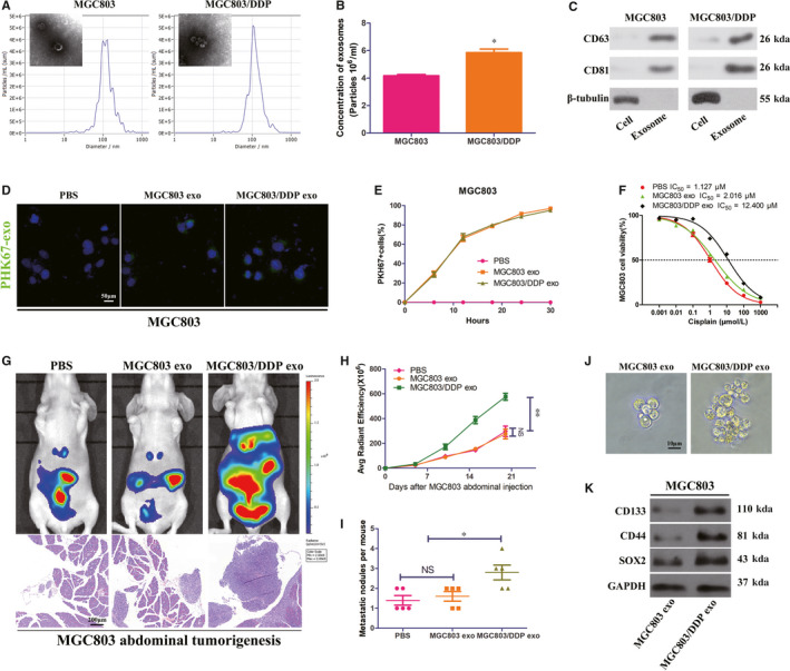
FIGURE 2 MGC803/DDP‐derived exosomes disseminate DDP resistance and promote CSCs properties in recipient MGC803 cells. A, Electron microscopy and NTA images of exosomes isolated from MGC803 and MGC803/DDP cells medium. B, C, Identical quantity of MGC803 and MGC803/DDP cells were cultured 30 h and then their exosomes concentrations and positive markers (CD63, HSP81) were analysed by NTA (B) and Western blot (C). D, MGC803 cells were incubated with PHK67‐labelled exosomes from MGC803 and MGC803/DDP cells for 30 h, and the internalization of exosomes was detected by confocal microscopy. E, Uptake efficiency of PHK67‐labelled exosomes was examined by confocal microscopy. F, Proliferation assay analysis and IC50 value of MGC803 cells co‐cultured with PBS, MGC830 exosomes or MGC803/DDP exosomes under DDP treatment. G, Representative bioluminescent images and microscopy observations showed effects of PBS, MGC803 exosomes or MGC803/DDP exosomes on abdominal tumorigenesis under DDP treatment. H, I, Comparison of bioluminescent signals and abdominal metastatic nodules after indicated treatment, five nude mice in each group. J, K, Effect of MGC803 exosomes and MGC803/DDP exosomes on sphere formation (J) and expression levels of stemness markers (K) in MCG803 cells by Western blot. *P < 0.05, **P < 0.01
FIGURE 2 MGC803/DDP‐derived exosomes disseminate DDP resistance and promote CSCs properties in recipient MGC803 cells. A, Electron microscopy and NTA images of exosomes isolated from MGC803 and MGC803/DDP cells medium. B, C, Identical quantity of MGC803 and MGC803/DDP cells were cultured 30 h and then their exosomes concentrations and positive markers (CD63, HSP81) were analysed by NTA (B) and Western blot (C). D, MGC803 cells were incubated with PHK67‐labelled exosomes from MGC803 and MGC803/DDP cells for 30 h, and the internalization of exosomes was detected by confocal microscopy. E, Uptake efficiency of PHK67‐labelled exosomes was examined by confocal microscopy. F, Proliferation assay analysis and IC50 value of MGC803 cells co‐cultured with PBS, MGC830 exosomes or MGC803/DDP exosomes under DDP treatment. G, Representative bioluminescent images and microscopy observations showed effects of PBS, MGC803 exosomes or MGC803/DDP exosomes on abdominal tumorigenesis under DDP treatment. H, I, Comparison of bioluminescent signals and abdominal metastatic nodules after indicated treatment, five nude mice in each group. J, K, Effect of MGC803 exosomes and MGC803/DDP exosomes on sphere formation (J) and expression levels of stemness markers (K) in MCG803 cells by Western blot. *P < 0.05, **P < 0.01CC BY · PubMed Central
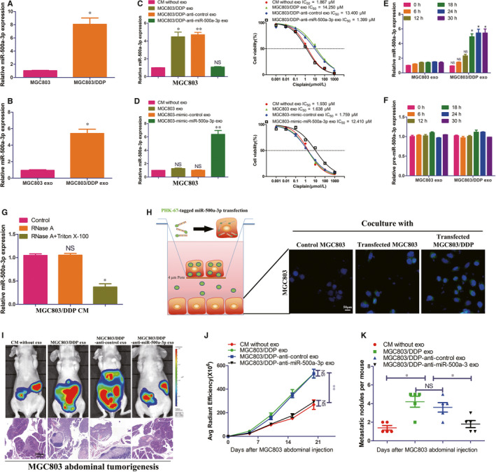
FIGURE 3 MGC803/DDP‐derived exosomes enhance DDP resistance of MGC803 recipient cells via exosome‐mediated delivery of miR‐500a‐3p in vitro and in vivo. A, B, Real‐time qRT‐PCR analysis of miR‐500a‐3p expression in MGC803, MGC803/DDP and their corresponding exosomes. C, D, Real‐time qRT‐PCR analysis of miR‐500a‐3p expression in MGC803 cells treated by indicated conditions and their corresponding proliferation assay analysis as well as IC50 value under DDP treatment. E, F, Real‐time qRT‐PCR analysis of miR‐500a‐3p or pre‐miR‐500a‐3p expression in MGC803 cells treated by MGC803 exosomes or MGC803/DDP exosomes at indicated time. G, Real‐time qRT‐PCR analysis of miR‐500a‐3p expression in the culture medium (CM) of MGC803/DDP after treatment with RNase (2 μg/mL) alone or combined with Triton X‐100 (0.1%) for 20 min. H, MGC803 or MGC803/DDP cells transfected with the PHK67‐miR‐500a‐3p mimic (green fluorescence) were placed in the upper chamber and coincubated with MGC803 cells seeded in the lower chamber in a transwell system with a 0.4 μm pore membrane. After coincubation for 30 h, MGC803 cells in the lower chamber were examined by the fluorescence microscope. I, Representative bioluminescent images and microscopy observations showed effects of CM without exosomes or indicated MGC803 cell‐derived exosomes on abdominal tumorigenesis under DDP treatment. J, K, Comparison of bioluminescent signals and abdominal metastatic nodules after indicated treatment, five nude mice in each group. *P < 0.05, **P < 0.01
FIGURE 3 MGC803/DDP‐derived exosomes enhance DDP resistance of MGC803 recipient cells via exosome‐mediated delivery of miR‐500a‐3p in vitro and in vivo. A, B, Real‐time qRT‐PCR analysis of miR‐500a‐3p expression in MGC803, MGC803/DDP and their corresponding exosomes. C, D, Real‐time qRT‐PCR analysis of miR‐500a‐3p expression in MGC803 cells treated by indicated conditions and their corresponding proliferation assay analysis as well as IC50 value under DDP treatment. E, F, Real‐time qRT‐PCR analysis of miR‐500a‐3p or pre‐miR‐500a‐3p expression in MGC803 cells treated by MGC803 exosomes or MGC803/DDP exosomes at indicated time. G, Real‐time qRT‐PCR analysis of miR‐500a‐3p expression in the culture medium (CM) of MGC803/DDP after treatment with RNase (2 μg/mL) alone or combined with Triton X‐100 (0.1%) for 20 min. H, MGC803 or MGC803/DDP cells transfected with the PHK67‐miR‐500a‐3p mimic (green fluorescence) were placed in the upper chamber and coincubated with MGC803 cells seeded in the lower chamber in a transwell system with a 0.4 μm pore membrane. After coincubation for 30 h, MGC803 cells in the lower chamber were examined by the fluorescence microscope. I, Representative bioluminescent images and microscopy observations showed effects of CM without exosomes or indicated MGC803 cell‐derived exosomes on abdominal tumorigenesis under DDP treatment. J, K, Comparison of bioluminescent signals and abdominal metastatic nodules after indicated treatment, five nude mice in each group. *P < 0.05, **P < 0.01CC BY · PubMed Central
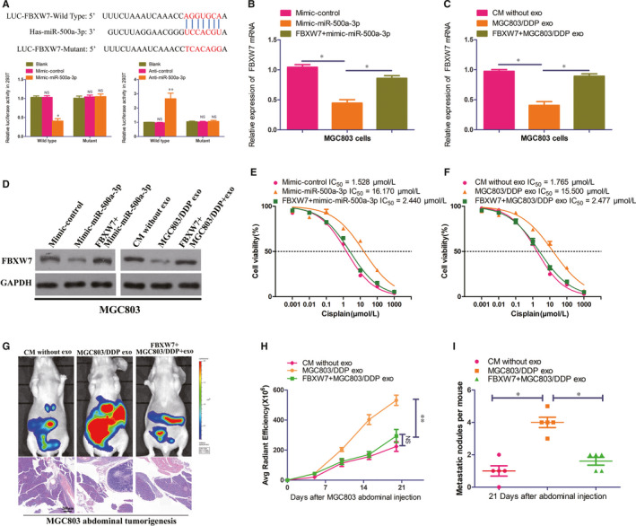
FIGURE 4 MGC803/DDP‐derived exosomal miR‐500a‐3p confers DDP resistance in recipient MGC803 cells via inhibiting FBXW7. A, Sequences of miR‐500a‐3p and the potential miR‐500a‐3p‐binding sites at the 3′UTR of FBXW7, including nucleotides mutated in FBXW7‐3′‐UTR. Seed sequences are marked. Also shown effects of Blank, mimic or anti–miR‐500a‐3p and corresponding control on the luciferase activity of FBXW7 3′UTR‐Wild‐Type and FBXW7 3′UTR‐Mutant by dual‐luciferase reported assay in 293T cells. B‐D, Expression of FBXW7 in MGC803 or FBXW7 overexpressed MGC803 cells transfected with mimic‐control or mimic‐miR‐500a‐3p by real‐time qRT‐PCR (B) and Western blot analysis (D). Expression of FBXW7 in MGC803 or FBXW7 overexpressed MGC803 cells co‐cultured with CM without exosomes or MGC803/DDP exosomes by real‐time qRT‐PCR (C) and Western blot analysis (D). E, Proliferation assay analysis and IC50 value in MGC803 or FBXW7 overexpressed MGC803 cells transfected with mimic‐control or mimic‐miR‐500a‐3p. F, Proliferation assay analysis and IC50 value in MGC803 or FBXW7 overexpressed MGC803 cells co‐cultured with CM without exosomes or MGC803/DDP exosomes. G, Representative bioluminescent images and microscopy observations showed effects of CM without exosomes, MGC803/DDP exosomes on abdominal tumorigenesis by MGC803 or FBXW7 overexpressed MGC803 cells abdominal tumorigenesis under DDP treatment. H, J, Comparison of bioluminescent signals and abdominal metastatic nodules after indicated treatment, five nude mice in each group. *P < 0.05 and **P < 0.01
FIGURE 4 MGC803/DDP‐derived exosomal miR‐500a‐3p confers DDP resistance in recipient MGC803 cells via inhibiting FBXW7. A, Sequences of miR‐500a‐3p and the potential miR‐500a‐3p‐binding sites at the 3′UTR of FBXW7, including nucleotides mutated in FBXW7‐3′‐UTR. Seed sequences are marked. Also shown effects of Blank, mimic or anti–miR‐500a‐3p and corresponding control on the luciferase activity of FBXW7 3′UTR‐Wild‐Type and FBXW7 3′UTR‐Mutant by dual‐luciferase reported assay in 293T cells. B‐D, Expression of FBXW7 in MGC803 or FBXW7 overexpressed MGC803 cells transfected with mimic‐control or mimic‐miR‐500a‐3p by real‐time qRT‐PCR (B) and Western blot analysis (D). Expression of FBXW7 in MGC803 or FBXW7 overexpressed MGC803 cells co‐cultured with CM without exosomes or MGC803/DDP exosomes by real‐time qRT‐PCR (C) and Western blot analysis (D). E, Proliferation assay analysis and IC50 value in MGC803 or FBXW7 overexpressed MGC803 cells transfected with mimic‐control or mimic‐miR‐500a‐3p. F, Proliferation assay analysis and IC50 value in MGC803 or FBXW7 overexpressed MGC803 cells co‐cultured with CM without exosomes or MGC803/DDP exosomes. G, Representative bioluminescent images and microscopy observations showed effects of CM without exosomes, MGC803/DDP exosomes on abdominal tumorigenesis by MGC803 or FBXW7 overexpressed MGC803 cells abdominal tumorigenesis under DDP treatment. H, J, Comparison of bioluminescent signals and abdominal metastatic nodules after indicated treatment, five nude mice in each group. *P < 0.05 and **P < 0.01CC BY · PubMed Central
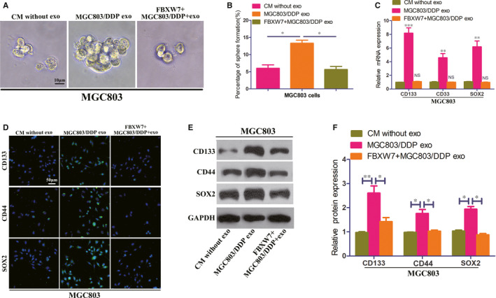
FIGURE 5 FBXW7 reverse the DDP resistance of exosomal miR‐500a‐3p by inhibiting CSCs properties in MGC803 cells. A, B, Effect of CM without exosomes, MGC803/DDP exosomes on sphere formation in MGC803 or FBXW7 overexpressed MGC803 cells. C, Relative mRNA expression of stemness markers CD133, CD44 and SOX2 in MGC803 or FBXW7 overexpressed MGC803 cells co‐cultured with CM without exosomes or MGC803/DDP exosomes. D‐F, Expression level of stemness markers in indicated GC cells by confocal microscopy (D) and Western blot (E, F). *P < 0.05, **P < 0.01 and ***P < 0.001
FIGURE 5 FBXW7 reverse the DDP resistance of exosomal miR‐500a‐3p by inhibiting CSCs properties in MGC803 cells. A, B, Effect of CM without exosomes, MGC803/DDP exosomes on sphere formation in MGC803 or FBXW7 overexpressed MGC803 cells. C, Relative mRNA expression of stemness markers CD133, CD44 and SOX2 in MGC803 or FBXW7 overexpressed MGC803 cells co‐cultured with CM without exosomes or MGC803/DDP exosomes. D‐F, Expression level of stemness markers in indicated GC cells by confocal microscopy (D) and Western blot (E, F). *P < 0.05, **P < 0.01 and ***P < 0.001CC BY · PubMed Central
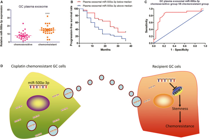
FIGURE 6 Plasma exosomal miR‐500a‐3p is related to DDP resistance in III stage GC patients. A, Plasma exosomal miR‐500a‐3p level was detected in III stage GC patients responding or not responding to DDP treatment by real‐time qRT‐PCR. B, Kaplan‐Meier analysis of 3 y progression‐free survival rate in III stage GC patients from high (n = 30) and low (n = 25) miR‐500a‐3p groups, according to the median exosomal miR‐500a‐3p level in pre‐gastrectomy plasma. C, ROC curve analysis of plasma exosomal miR‐500a‐3p expression for discriminating the DDP resistant group (n = 30) from the sensitive group (n = 25). AUC, area under the curve. D, Schematic diagram of the potential roles of exosomal miR‐500a‐3p in GC DDP resistance. Briefly, the DDP‐resistant GC cell‐secreted exosomes containing miR‐500a‐3p can be taken up by surrounding DDP sensitive GC cells and subsequently down‐regulate FBXW7 to enhance DDP resistance as well as CSCs properties of the recipient GC cells. ***P < 0.001
FIGURE 6 Plasma exosomal miR‐500a‐3p is related to DDP resistance in III stage GC patients. A, Plasma exosomal miR‐500a‐3p level was detected in III stage GC patients responding or not responding to DDP treatment by real‐time qRT‐PCR. B, Kaplan‐Meier analysis of 3 y progression‐free survival rate in III stage GC patients from high (n = 30) and low (n = 25) miR‐500a‐3p groups, according to the median exosomal miR‐500a‐3p level in pre‐gastrectomy plasma. C, ROC curve analysis of plasma exosomal miR‐500a‐3p expression for discriminating the DDP resistant group (n = 30) from the sensitive group (n = 25). AUC, area under the curve. D, Schematic diagram of the potential roles of exosomal miR‐500a‐3p in GC DDP resistance. Briefly, the DDP‐resistant GC cell‐secreted exosomes containing miR‐500a‐3p can be taken up by surrounding DDP sensitive GC cells and subsequently down‐regulate FBXW7 to enhance DDP resistance as well as CSCs properties of the recipient GC cells. ***P < 0.001CC BY · PubMed Central

Verification

Claims the paper makes

The claims and statistics of this paper haven’t been checked yet.

Run the check — free, no account needed. It reads the open-access text and checks the sources it cites.

A few free checks a day · papers already verified open instantly · results are public

Claims other papers make about this one

Each is checked against this paper’s own text: does it say what the citing paper says it does?

Self-check

Percentages, sums and reported differences re-computed from the paper’s own numbers.

The self-check needs the paper’s full text.

Citations

Cites

Research works printed but not linked: 44

  1. 10.3322/caac.21492 · DOI · Paper page
  2. 10.2174/1871520615666150803125121 · DOI
  3. 10.2217/fon.10.21 · DOI
  4. 10.1016/S0140-6736(81)91928-0 · DOI
  5. 10.4251/wjgo.v8.i9.673 · DOI
  6. 10.1007/s00018-019-03035-2 · DOI
  7. 10.1016/j.ceb.2014.05.004 · DOI · Paper page
  8. 10.1016/j.molmed.2015.08.002 · DOI
  9. 10.1038/nri855 · DOI
  10. 10.1159/000438594 · DOI
  11. 10.3389/fimmu.2014.00403 · DOI
  12. 10.1089/scd.2013.0633 · DOI
  13. 10.1016/j.cell.2016.01.043 · DOI · Paper page
  14. 10.1186/s12943-019-0991-5 · DOI · Paper page
  15. 10.1016/j.cell.2009.01.002 · DOI · Paper page
  16. 10.1038/ncb1596 · DOI · Paper page
  17. 10.1038/onc.2016.229 · DOI
  18. 10.1186/s13046-018-0677-7 · DOI
  19. 10.1007/s00280-015-2739-2 · DOI
  20. 10.1139/cjpp‐2019‐0477 · DOI
  21. 10.1016/j.biopha.2020.109896 · DOI
  22. 10.1038/nrd1691 · DOI · Paper page
  23. 10.3748/wjg.v22.i30.6876 · DOI · Paper page
  24. 10.1016/j.biocel.2018.08.011 · DOI
  25. 10.1016/j.matbio.2017.09.001 · DOI
  26. 10.1016/j.jconrel.2014.07.042 · DOI · Paper page
  27. 10.1074/jbc.M112.340588 · DOI · Paper page
  28. 10.3390/cancers8120111 · DOI
  29. 10.1038/s41556-018-0256-3 · DOI
  30. 10.1080/2162402X.2017.1376153 · DOI
  31. 10.1016/j.it.2017.08.002 · DOI
  32. 10.3389/fonc.2019.01113 · DOI
  33. 10.1186/s12943-017-0718-4 · DOI
  34. 10.1186/s12943-018-0897-7 · DOI · Paper page
  35. 10.1186/s13059-018-1604-0 · DOI
  36. 10.1016/j.canlet.2019.05.035 · DOI
  37. 10.1016/j.biopha.2017.04.018 · DOI
  38. 10.4149/neo_2018_170516N355 · DOI
  39. 10.1186/s13046-017-0568-3 · DOI
  40. 10.3390/cancers11020246 · DOI
  41. 10.1016/j.celrep.2019.12.032 · DOI
  42. 10.1186/s12876-017-0616-7 · DOI
  43. 10.1007/s00432-012-1154-x · DOI
  44. 10.1002/ijc.29168 · DOI

Cited by

Sorted by citations

See all 56

Metadata

Authors
Lin, H., Zhang, L., Zhang, C., & Liu, P.
Venue
Journal of Cellular and Molecular Medicine
Publisher
Wiley
Published
2020-06-25
Volume
24
Issue
16
Pages
8930-8941
ISSN
  • 1582-1838
  • 1582-4934
Cited by
56
References
3
Language
en

About these checks

Registry, retraction, Cites and statistics results are computed, not judged, with one exception: a printed reference that no index lists is read once by a model before it counts as a citation error. Claims are extracted by a model and kept only when their quote appears verbatim in the text; each cited source is then checked, with a model’s help, for whether it says what the claim relies on. Those verdicts can be wrong — read the claim’s quote and the cited source before relying on one.

Statistics and arithmetic are recomputed, not judged.

Confidence bands are shown only for fields calibrated on our annotated test set. None is calibrated yet, so no band is shown.

What counts as a claim ›How we measure confidence ›How we check this ›

Are you an author, or do you see a problem with this page? Request a review