

The DOI matches its Crossref record; 5 of the 5 references it cites were found in a registry or an index.
Single‐cell segmentation in bacterial biofilms with an optimized deep learning method enables tracking of cell lineages and measurements of growth rates
Abstract
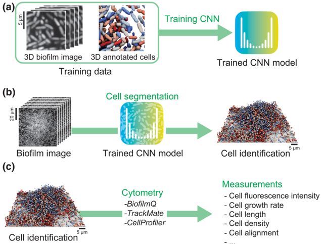
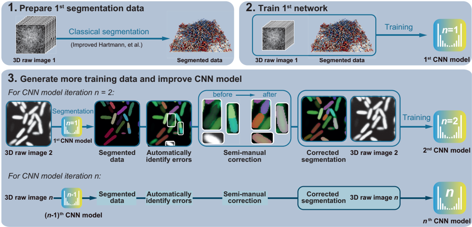
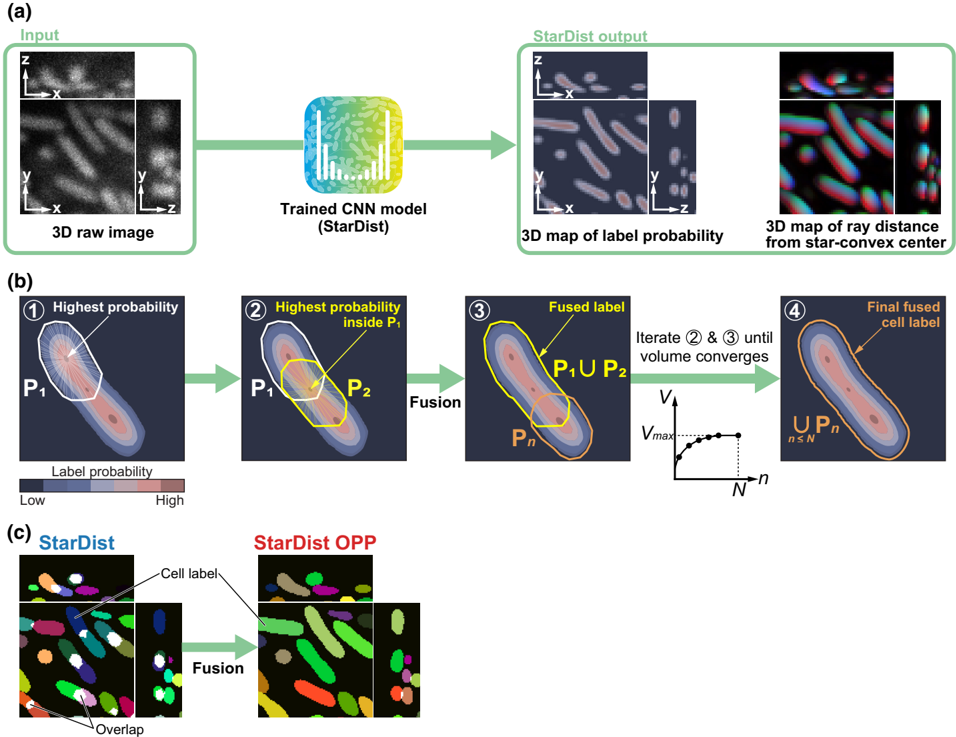
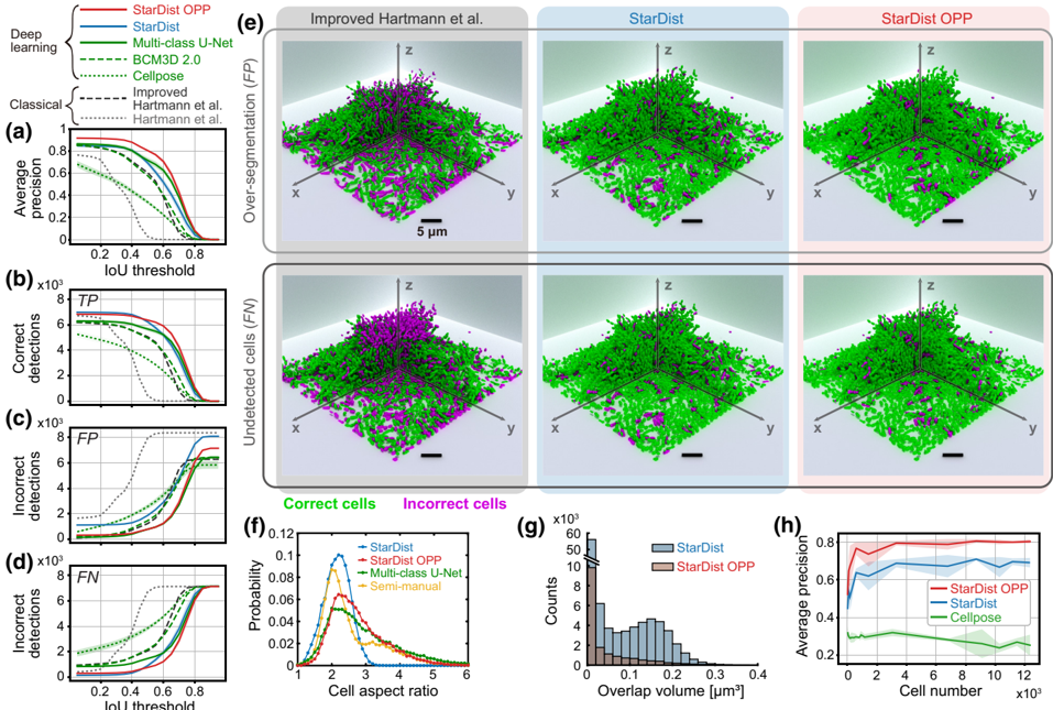
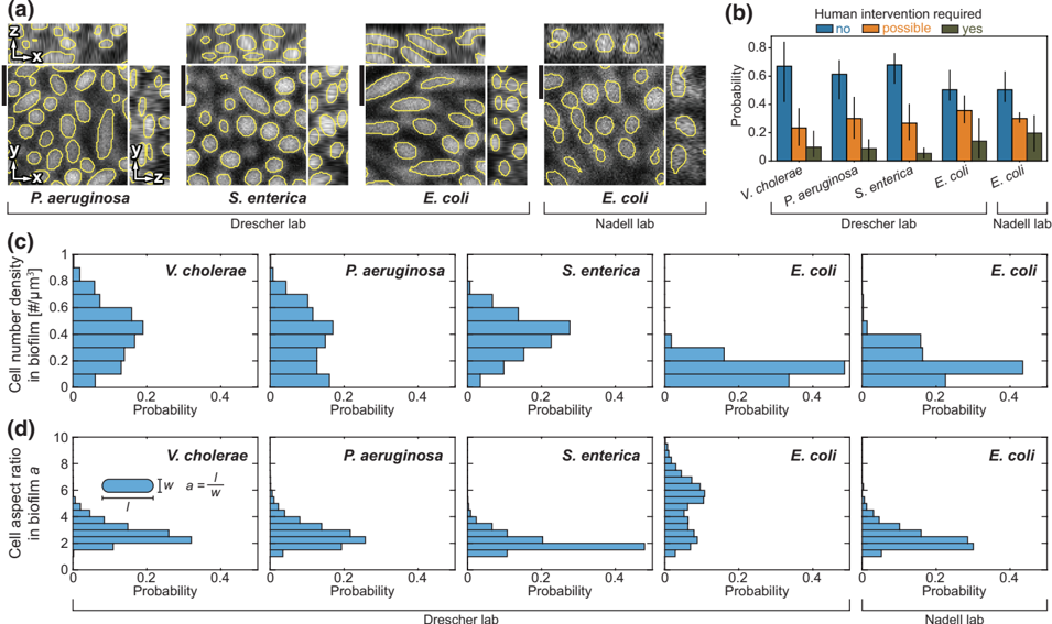
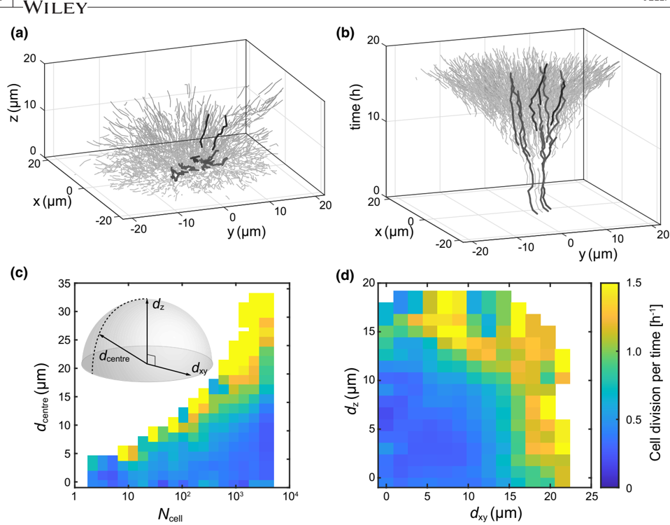
Bacteria often grow into matrix‐encased three‐dimensional (3D) biofilm communities, which can be imaged at cellular resolution using confocal microscopy. From these 3D images, measurements of single‐cell properties with high spatiotemporal resolution are required to investigate cellular heterogeneity and dynamical processes inside biofilms. However, the required measurements rely on the automated segmentation of bacterial cells in 3D images, which is a technical challenge. To improve the accuracy of single‐cell segmentation in 3D biofilms, we first evaluated recent classical and deep learning segmentation algorithms. We then extended StarDist, a state‐of‐the‐art deep learning algorithm, by optimizing the post‐processing for bacteria, which resulted in the most accurate segmentation results for biofilms among all investigated algorithms. To generate the large 3D training dataset required for deep learning, we developed an iterative process of automated segmentation followed by semi‐manual correction, resulting in >18,000 annotated Vibrio cholerae cells in 3D images. We demonstrate that this large training dataset and the neural network with optimized post‐processing yield accurate segmentation results for biofilms of different species and on biofilm images from different microscopes. Finally, we used the accurate single‐cell segmentation results to track cell lineages in biofilms and to perform spatiotemporal measurements of single‐cell growth rates during biofilm development.
Overview
The claims and statistics of this paper haven’t been checked yet.
Run the check — free, no account needed. It reads the open-access text and checks the sources it cites.
Integrity signals
Registry
Cites
Statistics
Figures
Verification
Claims the paper makes
The claims and statistics of this paper haven’t been checked yet.
Run the check — free, no account needed. It reads the open-access text and checks the sources it cites.
Claims other papers make about this one
Each is checked against this paper’s own text: does it say what the citing paper says it does?
In the citing paper
“Quantitative methods, such as machine learning (ML)- and artificial intelligence (AI)-based models31-33 for image analysis and processing, may help minimize bias and improve reproducibility in biofilm quantification of wound SEM images.”
From SEMTWIST Quantification of Biofilm Infection in Human Chronic Wound Using Scanning Electron Microscopy and Machine Learning (2025) · cites this paper as [32]
Self-check
Percentages, sums and reported differences re-computed from the paper’s own numbers.
The self-check needs the paper’s full text.
Citations
Cites
LeCun, Y., Bengio, Y., & Hinton, G. (2015) · Nature · doi:10.1038/nature14539 · Cited by 84,126
Domain randomization for transferring deep neural networks from simulation to the real world
Tobin, J., Fong, R., Ray, A., Schneider, J., Zaremba, W., & Abbeel, P. (2017) · 2017 IEEE/RSJ International Conference on Intelligent Robots and Systems (IROS) · doi:10.1109/iros.2017.8202133 · Cited by 3,124 · cited 1×
“Nevertheless, there is always a gap between the real and synthetic data, which only can be bridged with elaborate randomization approaches (Tobin et al., 2017).”
BackgroundNucleus segmentation across imaging experiments: the 2018 Data Science Bowl
Caicedo, J. C., Goodman, A., Karhohs, K. W., Cimini, B. A., Ackerman, J., Haghighi, M., Heng, C., Becker, T., Doan, M., McQuin, C., et al. (2019) · Nature Methods · doi:10.1038/s41592-019-0612-7 · Cited by 809
Van Valen, D. A., Kudo, T., Lane, K. M., Macklin, D. N., Quach, N. T., DeFelice, M. M., Maayan, I., Tanouchi, Y., Ashley, E. A., & Covert, M. W. (2016) · PLoS Computational Biology · doi:10.1371/journal.pcbi.1005177 · Cited by 557
Weighted boxes fusion: Ensembling boxes from different object detection models
Solovyev, R., Wang, W., & Gabruseva, T. (2021) · Image and Vision Computing · doi:10.1016/j.imavis.2021.104117 · Cited by 543
Research works printed but not linked: 66
- 10.1038/nrmicro3491 · DOI
- Ahrens J., 2005, ParaView: an end‐user tool for large data visualization
- 10.1038/s41564‐021‐00900‐4 · DOI
- 10.1002/cyto.a.23863 · DOI
- 10.1101/491035 · DOI · Paper page
- 10.48550/arXiv.1512.01274 · DOI · Paper page
- 10.1007/978-3-319-46723-8_49 · DOI · Paper page
- 10.1146/annurev.mi.41.100187.002251 · DOI · Paper page
- 10.1038/scientificamerican0178‐86 · DOI
- 10.1371/journal.pone.0268199 · DOI
- 10.1038/s41592‐022‐01639‐4 · DOI
- 10.7554/eLife.70794 · DOI
- 10.1073/pnas.1601702113 · DOI
- 10.1038/nmicrobiol.2016.77 · DOI
- 10.14440/jbm.2014.36 · DOI
- 10.1038/s41592‐022‐01507‐1 · DOI
- 10.1016/j.tim.2020.03.008 · DOI
- 10.1038/s41592‐018‐0261‐2 · DOI
- 10.1038/nrmicro2415 · DOI · Paper page
- 10.1038/s41579‐019‐0158‐9 · DOI · Paper page
- 10.1128/JB.01826‐08 · DOI
- Griffa A., 2010, Comparison of deconvolution software in 3D microscopy: a user point of view—part 1, G.I.T. Imaging & Microscopy, 12, 43
- 10.1186/s12915‐022‐01453‐6 · DOI
- 10.1038/s41586‐020‐2649‐2 · DOI · Paper page
- 10.1038/s41564‐020‐00817‐4 · DOI
- 10.1038/s41567‐018‐0356‐9 · DOI
- 10.1111/mmi.14501 · DOI
- 10.1371/journal.pbio.3001846 · DOI
- 10.1093/femsre/fuaa062 · DOI
- 10.1038/s41579‐022‐00692‐2 · DOI
- 10.1111/j.1365‐2818.2012.03675.x · DOI
- 10.1038/s41592‐021‐01284‐3 · DOI
- 10.1371/journal.pcbi.1007673 · DOI
- 10.25080/Majora‐92bf1922‐00a · DOI
- 10.1038/s41592‐019‐0403‐1 · DOI · Paper page
- 10.1038/s41522‐021‐00214‐7 · DOI · Paper page
- 10.1371/journal.pcbi.1009797 · DOI
- 10.1111/mmi.13264 · DOI
- 10.1109/ICCVW54120.2021.00110 · DOI
- 10.1126/science.abb8501 · DOI
- 10.1128/JB.180.3.722‐731.1998 · DOI
- 10.1046/j.1365‐2958.1998.00791.x · DOI
- 10.1038/nmeth.2019 · DOI · Paper page
- 10.1007/978-3-030-00934-2_30 · DOI · Paper page
- 10.48550/arXiv.1802.05799 · DOI
- 10.1128/mBio.00103‐13 · DOI
- Sofroniew N., 2022, Napari: a multi‐dimensional image viewer for python
- 10.1021/la401322k · DOI
- 10.1038/nrmicro1838 · DOI · Paper page
- 10.1186/s12859‐021‐04344‐9 · DOI
- 10.1038/s41592‐020‐01018‐x · DOI · Paper page
- 10.1101/2021.06.11.448083 · DOI
- 10.1186/s12880‐015‐0068‐x · DOI
- The Pandas Development Team, 2023, Pandas‐Dev/Pandas: Pandas
- 10.1016/j.ymeth.2016.09.016 · DOI
- 10.1109/ISBI52829.2022.9761655 · DOI
- 10.1042/BST20180391 · DOI
- 10.1038/s41467‐021‐22518‐0 · DOI
- 10.1016/j.cell.2021.11.024 · DOI
- 10.1038/s41592‐018‐0216‐7 · DOI
- 10.1109/WACV45572.2020.9093435 · DOI
- 10.7554/eLife.57613 · DOI
- 10.1073/pnas.1611494113 · DOI
- 10.1364/BOE.420788 · DOI
- 10.1038/s41522‐022‐00362‐4 · DOI · Paper page
- 10.1038/s41467‐020‐19866‐8 · DOI
Cited by
Sorted by citations
Singh, S., Muniz De Oliveira, F., Wang, C., Kumar, M., Xuan, Y., DeMazumder, D., Sen, C. K., & Roy, S. (2025) · Advances in Wound Care · doi:10.1089/wound.2024.0291 · Cited by 6
Metadata
- Authors
- Jelli, E., Ohmura, T., Netter, N., Abt, M., Jiménez‐Siebert, E., Neuhaus, K., Rode, D. K. H., Nadell, C. D., & Drescher, K.
- Venue
- Molecular Microbiology
- Publisher
- Wiley
- Published
- 2023-04-17
- Volume
- 119
- Issue
- 6
- Pages
- 659-676
- ISSN
- 0950-382X
- 1365-2958
- Cited by
- 48
- References
- 5
- Language
- en
- Funders
- Bundesministerium für Bildung und Forschung (TARGET‐Biofilms)
- Deutsche Forschungsgemeinschaft (DR 982/5‐1, DR 982/6‐1)
- European Commission (716734, 955910)
- Human Frontier Science Program (LT000013/2019‐C)
- Schweizerischer Nationalfonds zur Förderung der Wissenschaftlichen Forschung (51NF40_180541, TMCG‐3_213801)
About these checks
Registry, retraction, Cites and statistics results are computed, not judged, with one exception: a printed reference that no index lists is read once by a model before it counts as a citation error. Claims are extracted by a model and kept only when their quote appears verbatim in the text; each cited source is then checked, with a model’s help, for whether it says what the claim relies on. Those verdicts can be wrong — read the claim’s quote and the cited source before relying on one.
Statistics and arithmetic are recomputed, not judged.
Confidence bands are shown only for fields calibrated on our annotated test set. None is calibrated yet, so no band is shown.
What counts as a claim ›How we measure confidence ›How we check this ›
Are you an author, or do you see a problem with this page? Request a review
How to cite
Cite CoChat for the checks on this page, and the paper for its own findings.
- This page
- CoChat. (2026). Citation check: Single‐cell segmentation in bacterial biofilms with an optimized deep learning method enables tracking of cell lineages and measurements of growth rates. https://app.cochat.ai/papers/10.1111/mmi.15064
- The paper
- Jelli, E., Ohmura, T., Netter, N., Abt, M., Jiménez‐Siebert, E., Neuhaus, K., Rode, D. K. H., Nadell, C. D., & Drescher, K. (2023). Single‐cell segmentation in bacterial biofilms with an optimized deep learning method enables tracking of cell lineages and measurements of growth rates. Molecular Microbiology. https://doi.org/10.1111/mmi.15064
