

The Allen Cell and Structure Segmenter: a new open source toolkit for segmenting 3D intracellular structures in fluorescence microscopy images
Open accessThe DOI matches its Crossref record; 41 of the 42 references it cites were found in a registry or an index, and 1 are not checked yet.
Abstract
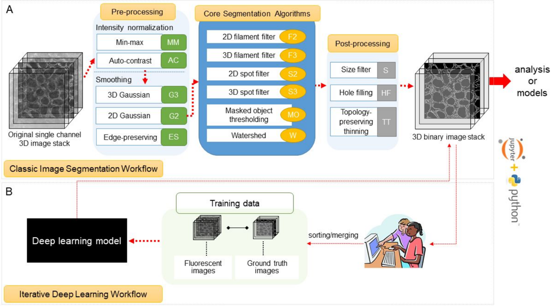
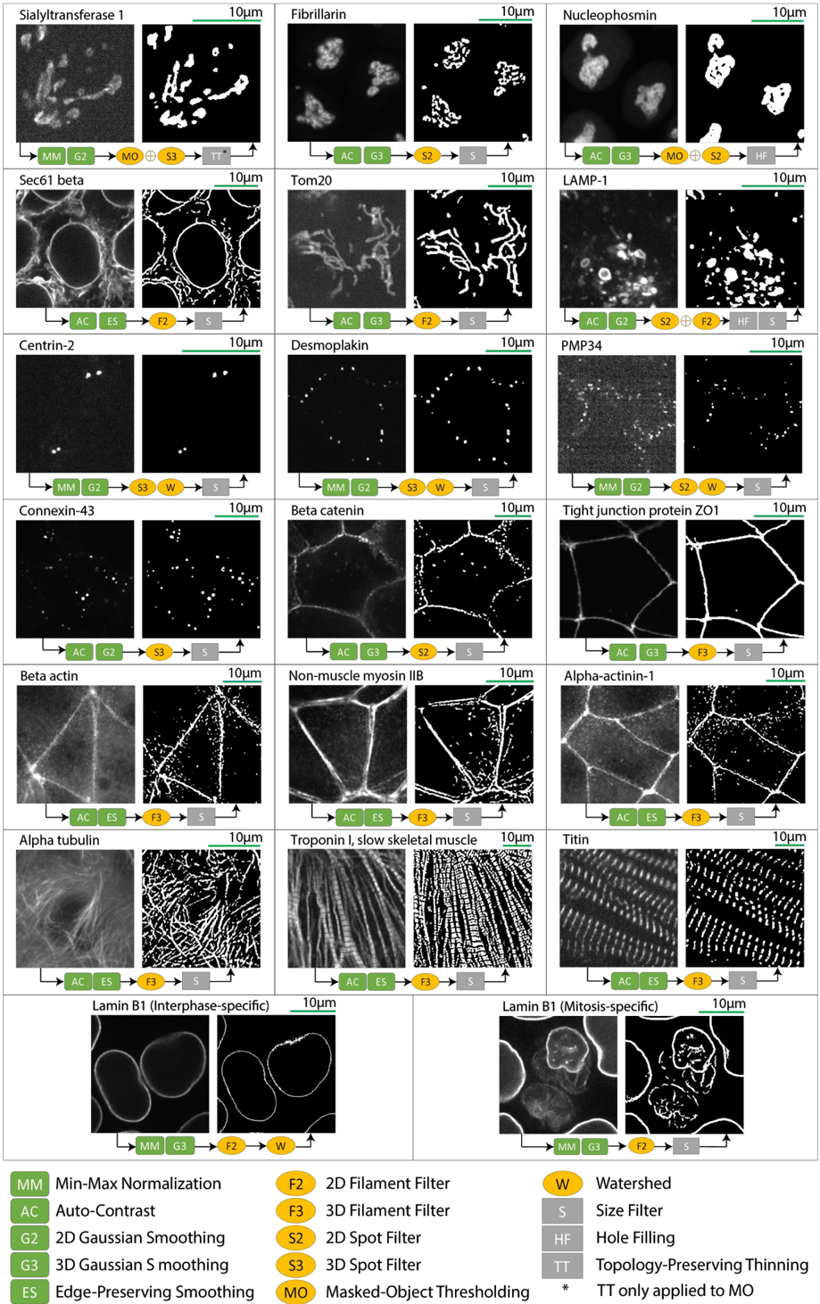
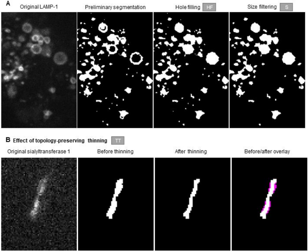
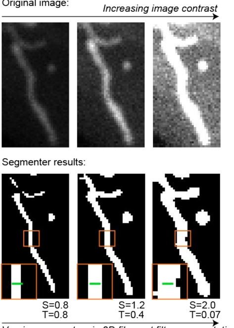
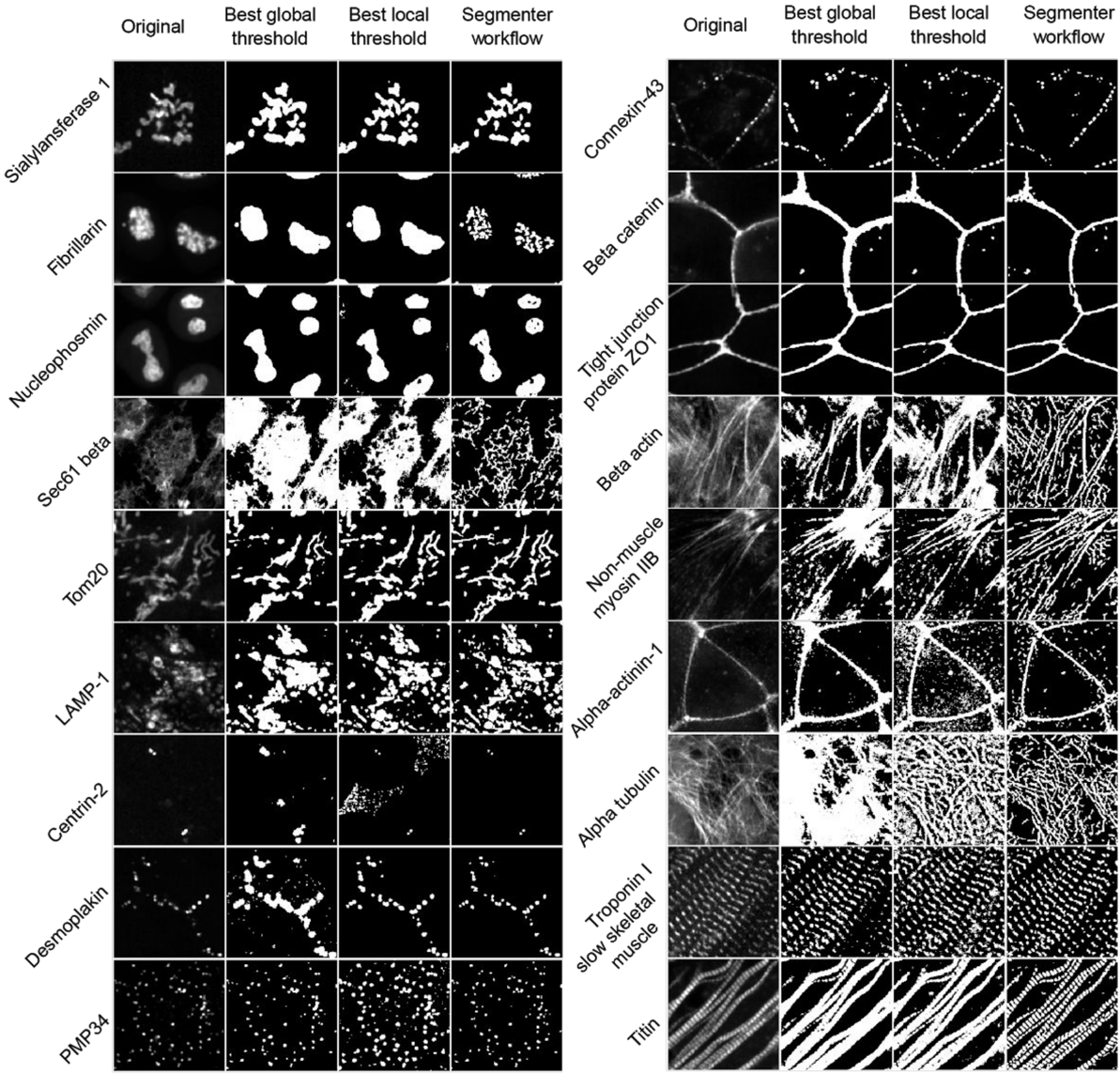
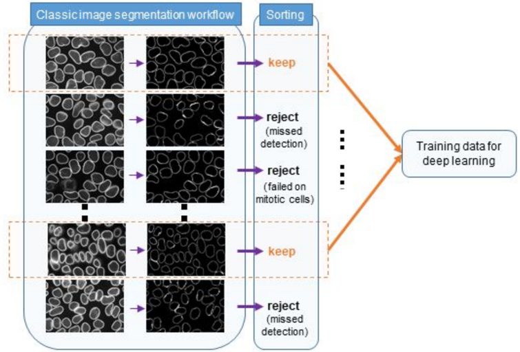
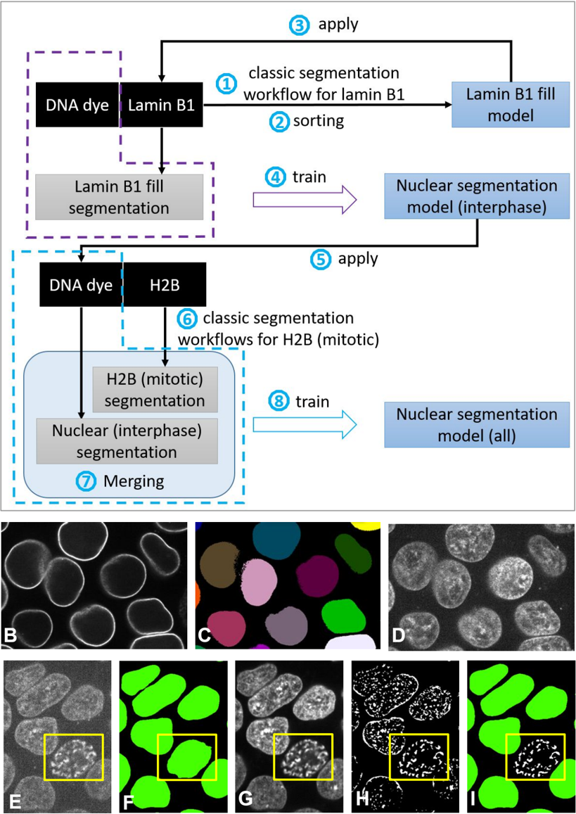
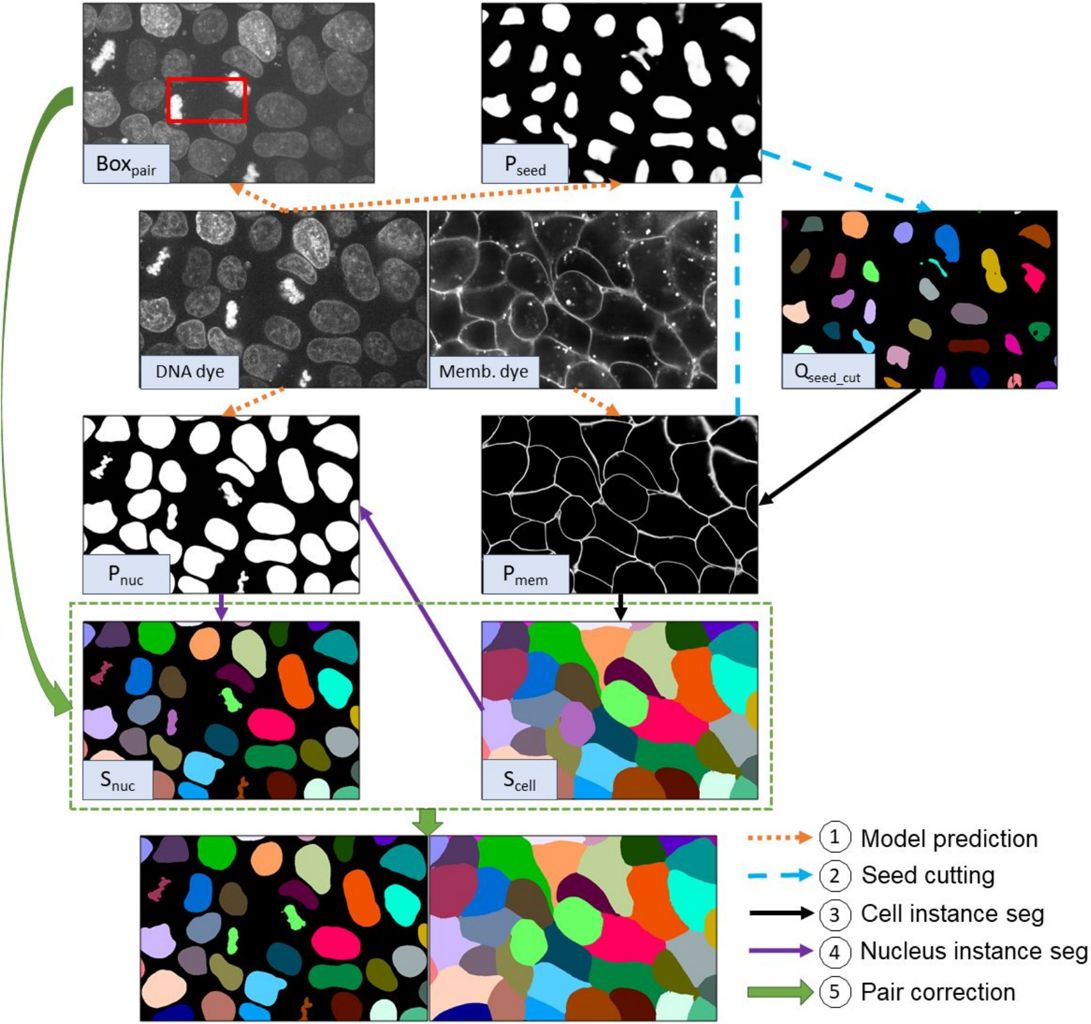
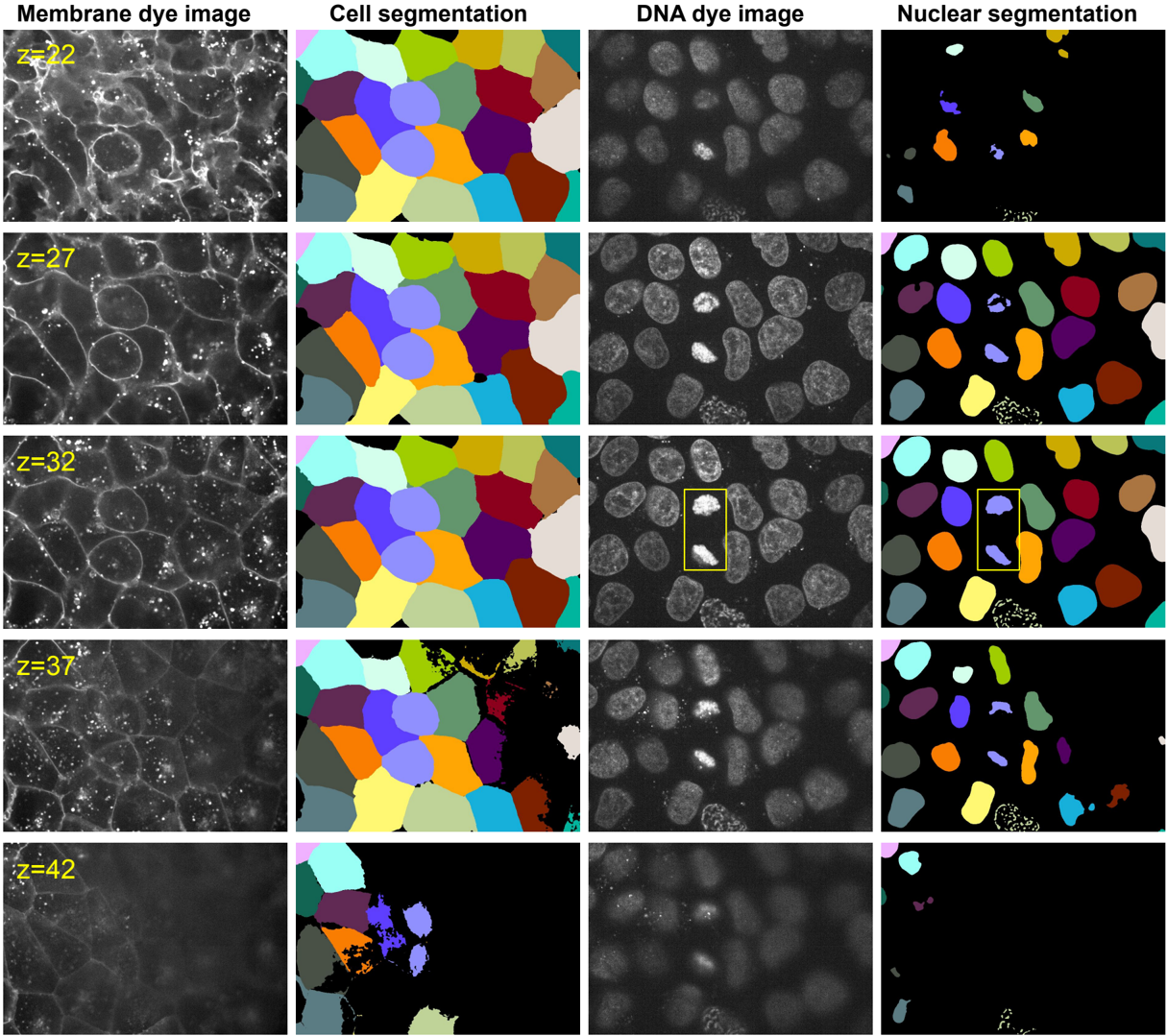
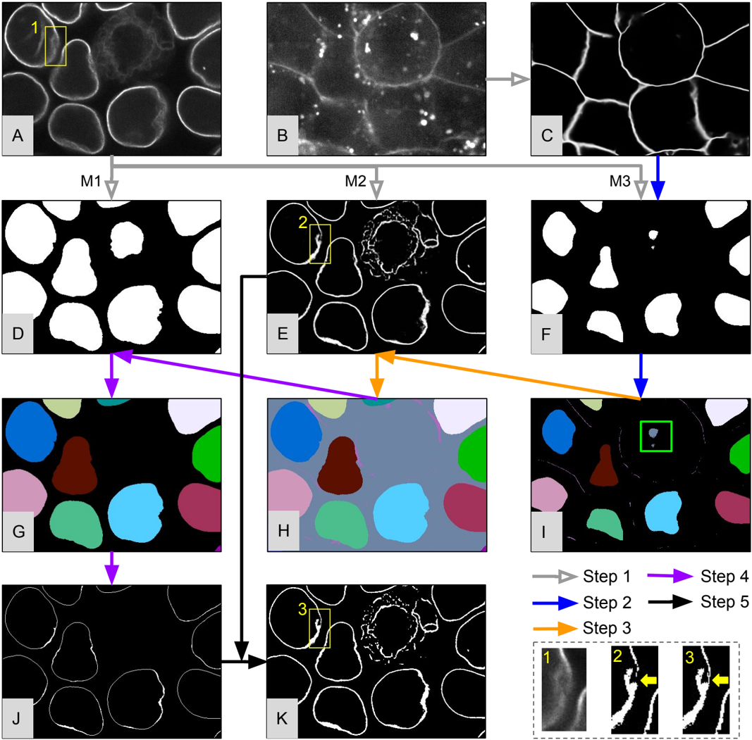
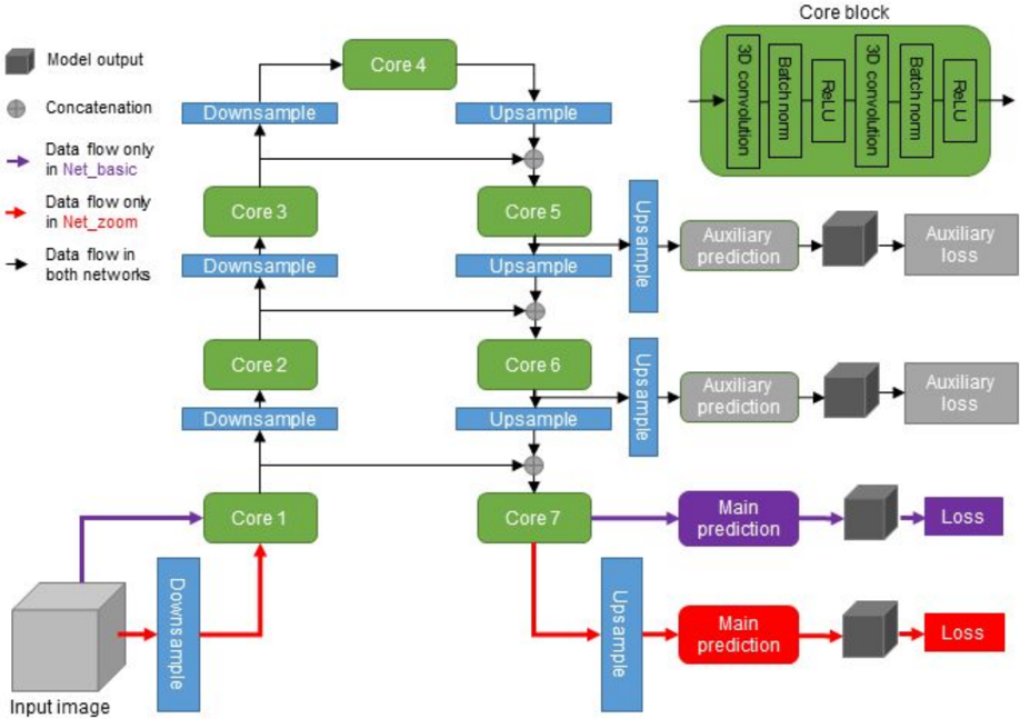
A continuing challenge in quantitative cell biology is the accurate and robust 3D segmentation of structures of interest from fluorescence microscopy images in an automated, reproducible, and widely accessible manner for subsequent interpretable data analysis. We describe the Allen Cell and Structure Segmenter (Segmenter), a Python-based open source toolkit developed for 3D segmentation of cells and intracellular structures in fluorescence microscope images. This toolkit brings together classic image segmentation and iterative deep learning workflows first to generate initial high-quality 3D intracellular structure segmentations and then to easily curate these results to generate the ground truths for building robust and accurate deep learning models. The toolkit takes advantage of the high-replicate 3D live cell image data collected at the Allen Institute for Cell Science of over 30 endogenous fluorescently tagged human induced pluripotent stem cell (hiPSC) lines. Each cell line represents a different intracellular structure with one or more distinct localization patterns within undifferentiated hiPS cells and hiPSC-derived cardiomyocytes. The Segmenter consists of two complementary elements, a classic image segmentation workflow with a restricted set of algorithms and parameters and an iterative deep learning segmentation workflow. We created a collection of 20 classic image segmentation workflows based on 20 distinct and representative intracellular structure localization patterns as a “lookup table” reference and starting point for users. The iterative deep learning workflow can take over when the classic segmentation workflow is insufficient. Two straightforward “human-in-the-loop” curation strategies convert a set of classic image segmentation workflow results into a set of 3D ground truth images for iterative model training without the need for manual painting in 3D. The deep learning model architectures used in this toolkit were designed and tested specifically for 3D fluorescence microscope images and implemented as readable scripts. The Segmenter thus leverages state of the art computer vision algorithms in an accessible way to facilitate their application by the experimental biology researcher. We include two useful applications to demonstrate how we used the classic image segmentation and iterative deep learning workflows to solve more challenging 3D segmentation tasks. First, we introduce the ‘Training Assay’ approach, a new experimental-computational co-design concept to generate more biologically accurate segmentation ground truths. We combined the iterative deep learning workflow with three Training Assays to develop a robust, scalable cell and nuclear instance segmentation algorithm, which could achieve accurate target segmentation for over 98% of individual cells and over 80% of entire fields of view. Second, we demonstrate how to extend the lamin B1 segmentation model built from the iterative deep learning workflow to obtain more biologically accurate lamin B1 segmentation by utilizing multi-channel inputs and combining multiple ML models. The steps and workflows used to develop these algorithms are generalizable to other similar segmentation challenges. More information, including tutorials and code repositories, are available at allencell.org/segmenter.
Overview
The claims and statistics of this paper haven’t been checked yet.
Free: it reads the open-access text and checks the first 5 claims that cite a source we can check. Create a free account to check every claim.
Integrity signals
Registry
Cites
Statistics
Figures




Verification
Claims the paper makes
The claims and statistics of this paper haven’t been checked yet.
Free: it reads the open-access text and checks the first 5 claims that cite a source we can check. Create a free account to check every claim.
Claims other papers make about this one
Each is checked against this paper’s own text: does it say what the citing paper says it does?
The cited source says what the claim attributes to it.
In the citing paper
“required deep-learning-based segmentation approaches to create a robust, scalable and highly accurate 3D cell and nuclear segmentation algorithm15 (Methods)”
From Integrated intracellular organization and its variations in human iPS cells (2023) · cites this paper as [15]
Self-check
Percentages, sums and reported differences re-computed from the paper’s own numbers.
The self-check needs the paper’s full text.
Citations
Cites
Adam: A Method for Stochastic Optimization
Kingma, D. P., & Ba, J. (2014) · arXiv · doi:10.48550/arxiv.1412.6980 · Cited by 98,946
Faster R-CNN: Towards Real-Time Object Detection with Region Proposal Networks
Ren, S., He, K., Girshick, R., & Sun, J. (2017) · IEEE Transactions on Pattern Analysis and Machine Intelligence · doi:10.1109/tpami.2016.2577031 · Cited by 73,472
Fiji: an open-source platform for biological-image analysis
Schindelin, J., Arganda-Carreras, I., Frise, E., Kaynig, V., Longair, M., Pietzsch, T., Preibisch, S., Rueden, C., Saalfeld, S., Schmid, B., et al. (2012) · Nature Methods · doi:10.1038/nmeth.2019 · Cited by 66,890
Fully convolutional networks for semantic segmentation
Long, J., Shelhamer, E., & Darrell, T. (2015) · 2015 IEEE Conference on Computer Vision and Pattern Recognition (CVPR) · doi:10.1109/cvpr.2015.7298965 · Cited by 38,834
SciPy 1.0: fundamental algorithms for scientific computing in Python
Virtanen, P., Gommers, R., Oliphant, T. E., Haberland, M., Reddy, T., Cournapeau, D., Burovski, E., Peterson, P., Weckesser, W., Bright, J., et al. (2020) · Nature Methods · doi:10.1038/s41592-019-0686-2 · Cited by 38,430
Harris, C. R., Millman, K. J., van der Walt, S. J., Gommers, R., Virtanen, P., Cournapeau, D., Wieser, E., Taylor, J., Berg, S., Smith, N. J., et al. (2020) · Nature · doi:10.1038/s41586-020-2649-2 · Cited by 23,826
Harris, C., Millman, K., van der Walt, S., Gommers, R., Virtanen, P., Cournapeau, D., Wieser, E., Taylor, J., Berg, S., Smith, N., et al. (2020) · RePEc: Research Papers in Economics · Cited by 17,844
Scale-space and edge detection using anisotropic diffusion
Perona, P., & Malik, J. (1990) · IEEE Transactions on Pattern Analysis and Machine Intelligence · doi:10.1109/34.56205 · Cited by 12,119
SciPy 1.0: fundamental algorithms for scientific computing in Python
Virtanen, P., Gommers, R., Oliphant, T. E., Haberland, M., Reddy, T., Cournapeau, D., Burovski, E., Peterson, P., Weckesser, W., Bright, J., et al. (2019) · Apollo - University of Cambridge Repository · doi:10.17863/cam.64134 · Cited by 10,460
Yushkevich, P. A., Piven, J., Hazlett, H. C., Smith, R. G., Ho, S., Gee, J. C., & Gerig, G. (2006) · NeuroImage · doi:10.1016/j.neuroimage.2006.01.015 · Cited by 8,868 · cited 1×
“Some algorithms are available as collections of basic functions in several open platforms, including the widely used ImageJ [Schindelin et al., 2012], CellProfiler [Carpenter et al., 2006, McQuin et al., 2018], Icy [De Chaumont et al., 2012], and ITK-SNAP [Yushkevich et al., 2006].”
Method
Cited by
Cellpose: a generalist algorithm for cellular segmentation
Stringer, C., Wang, T., Michaelos, M., & Pachitariu, M. (2021) · Nature Methods · doi:10.1038/s41592-020-01018-x · Cited by 4,369
Deep learning for cellular image analysis
Moen, E., Bannon, D., Kudo, T., Graf, W., Covert, M., & Van Valen, D. (2019) · Nature Methods · doi:10.1038/s41592-019-0403-1 · Cited by 1,225
Understanding metric-related pitfalls in image analysis validation
Reinke, A., Tizabi, M. D., Baumgartner, M., Eisenmann, M., Heckmann-Nötzel, D., Kavur, A. E., Rädsch, T., Sudre, C. H., Acion, L., Antonelli, M., et al. (2024) · Nature Methods · doi:10.1038/s41592-023-02150-0 · Cited by 241
Self-citationIntegrated intracellular organization and its variations in human iPS cells
Viana, M. P., Chen, J., Knijnenburg, T. A., Vasan, R., Yan, C., Arakaki, J. E., Bailey, M., Berry, B., Borensztejn, A., Brown, E. M., et al. (2023) · Nature · doi:10.1038/s41586-022-05563-7 · Cited by 132
Self-citationNucleus segmentation: towards automated solutions
Hollandi, R., Moshkov, N., Paavolainen, L., Tasnádi, E., Piccinini, F., & Horváth, P. (2022) · Trends in Cell Biology · doi:10.1016/j.tcb.2021.12.004 · Cited by 92
Metadata
- Authors
- Chen, J., Ding, L., Viana, M. P., Lee, H., Sluezwski, M. F., Morris, B., Hendershott, M. C., Yang, R., Mueller, I. A., & Rafelski, S. M.
- Affiliations
- Allen Institute for Cell Science (US)
- Corresponding author
- Rafelski, Susanne M.
- ORCID
- Venue
- bioRxiv (Cold Spring Harbor Laboratory)
- Publisher
- openRxiv
- Published
- 2018-12-08
- OpenAlex
- W2903709268
- Cited by
- 99
- References
- 41
- Citations by year
- 2026: 11 · 2025: 15 · 2024: 18 · 2023: 13 · 2022: 10 · 2021: 16 · 2020: 13 · 2019: 3
- Type
- posted content
- Language
- en
- Open access
- green
- doi.org
- Licence
- cc-by-nc
- Locations
- bioRxiv (Cold Spring Harbor Laboratory) (acceptedVersion, cc-by-nc)
- Topics
- Life Sciences › Biochemistry, Genetics and Molecular Biology › Biophysics › Cell Image Analysis Techniques
- Physical Sciences › Computer Science › Computer Vision and Pattern Recognition › Medical Image Segmentation Techniques
- Life Sciences › Biochemistry, Genetics and Molecular Biology › Biophysics › Advanced Fluorescence Microscopy Techniques
- Keywords
- 3D image segmentation, fluorescence microscopy, open source software, ground truth generation, cell instance segmentation, nuclei segmentation, lamin B1, hiPSC-derived cardiomyocytes, live-cell imaging
About these checks
Registry, retraction, Cites and statistics results are computed, not judged, with one exception: a printed reference that no index lists is read once by a model before it counts as a citation error. Claims are extracted by a model and kept only when their quote appears verbatim in the text; each cited source is then checked, with a model’s help, for whether it says what the claim relies on. Those verdicts can be wrong — read the claim’s quote and the cited source before relying on one.
Statistics and arithmetic are recomputed, not judged.
Confidence bands are shown only for fields calibrated on our annotated test set. None is calibrated yet, so no band is shown.
What counts as a claim ›How we measure confidence ›How we check this ›
Are you an author, or do you see a problem with this page? Request a review
How to cite
Cite CoChat for the checks on this page, and the paper for its own findings.
- This page
- CoChat. (2026). Citation check: The Allen Cell and Structure Segmenter: a new open source toolkit for segmenting 3D intracellular structures in fluorescence microscopy images. https://app.cochat.ai/papers/10.1101/491035
- The paper
- Chen, J., Ding, L., Viana, M. P., Lee, H., Sluezwski, M. F., Morris, B., Hendershott, M. C., Yang, R., Mueller, I. A., & Rafelski, S. M. (2018). The Allen Cell and Structure Segmenter: a new open source toolkit for segmenting 3D intracellular structures in fluorescence microscopy images. bioRxiv (Cold Spring Harbor Laboratory). https://doi.org/10.1101/491035
Data sources
Bibliographic data: Semantic Scholar, OpenAlex, Crossref.
Contains information from the Semantic Scholar Academic Graph Datasets, made available under the ODC Attribution License (ODC-BY 1.0).
Semantic Scholar’s data is described in: Kinney, R., et al. (2023). The Semantic Scholar Open Data Platform. arXiv:2301.10140.
