

The DOI matches its Crossref record; 28 of the 33 references it cites were found in a registry or an index.
Isolation of potent SARS-CoV-2 neutralizing antibodies and protection from disease in a small animal model
Open accessPublished version of a preprint
Abstract
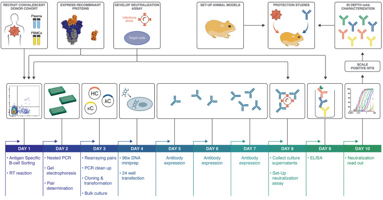
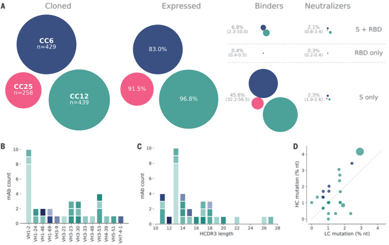
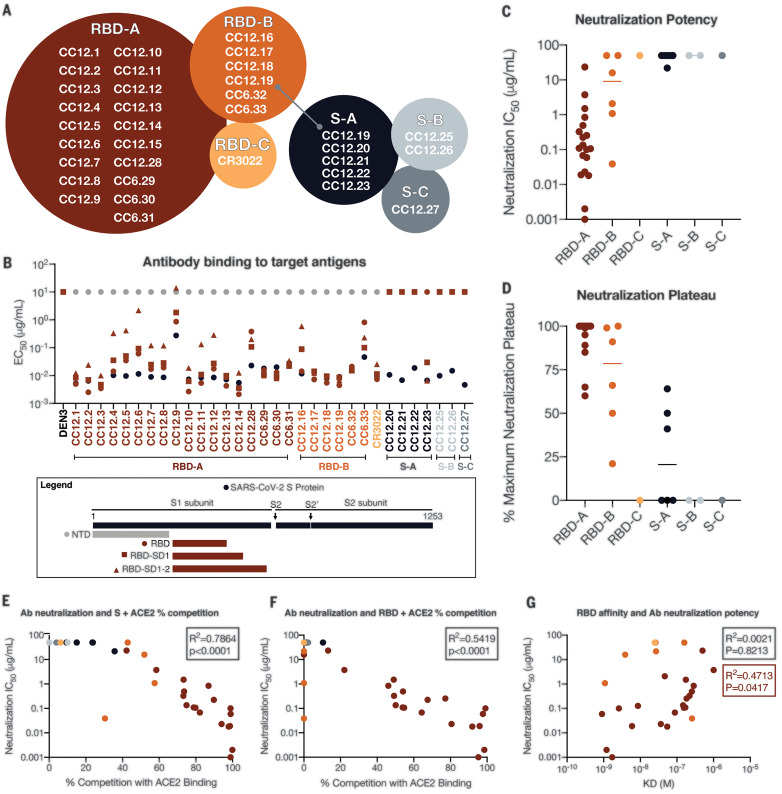
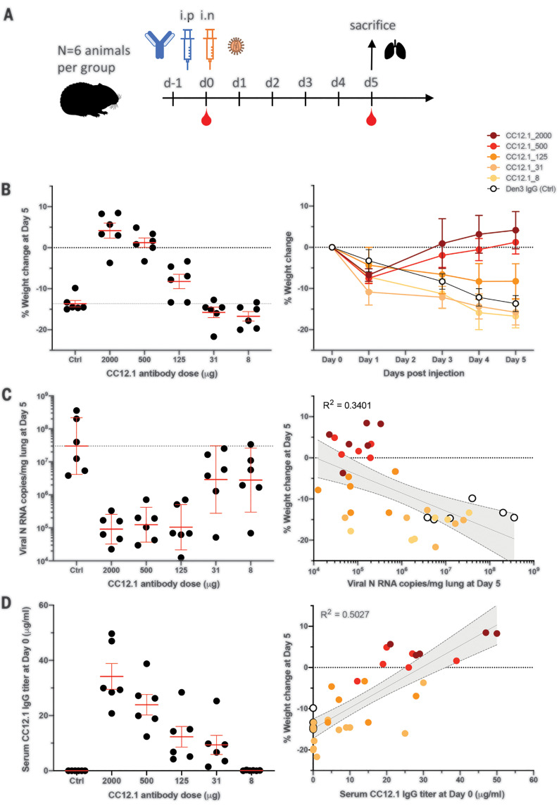
Protective neutralizing antibodies: Antibodies produced by survivors of coronavirus disease 2019 (COVID-19) may be leveraged to develop therapies. A first step is identifying neutralizing antibodies, which confer strong protection against severe acute respiratory syndrome coronavirus 2 (SARS-CoV-2). Rogers et al. used a high-throughput pipeline to isolate and characterize monoclonal antibodies from convalescent donors. Antibodies were selected for binding to the viral spike protein, which facilitates entry into host cells by binding to the angiotensin-converting enzyme 2 (ACE2) receptor. Most isolated antibodies bound to regions of the spike outside of the receptor binding domain (RBD); however, a larger proportion of the RBD-binding antibodies were neutralizing, with the most potent binding at a site that overlaps the ACE2 binding site. Two of the neutralizing antibodies were tested in Syrian hamsters and provided protection against SARS-CoV-2 infection. Science , this issue p. 956
Overview
The claims and statistics of this paper haven’t been checked yet.
Run the check — free, no account needed. It reads the open-access text and checks the sources it cites.
Integrity signals
Registry
Cites
Statistics
Figures
Verification
Claims the paper makes
The claims and statistics of this paper haven’t been checked yet.
Run the check — free, no account needed. It reads the open-access text and checks the sources it cites.
Claims other papers make about this one
Each is checked against this paper’s own text: does it say what the citing paper says it does?
In the citing paper
“A polyclonal antibody against multiple epitopes of the S protein beyond the RBD might, for example, inhibit viral attachment156–160, provide additional neutralizing activity161 and/or prevent post-attachment fusion162.”
From Immunological mechanisms of vaccine-induced protection against COVID-19 in humans (2021) · cites this paper as [159]
In the citing paper
“The vaccines elicit antibody responses against the RBD, the major target of neutralizing antibodies21–26, in a manner that resembles natural infection.”
From mRNA vaccine-elicited antibodies to SARS-CoV-2 and circulating variants (2021) · cites this paper as [26]
In the citing paper
“We focused on the RBD since it is the target of the majority of the more potent SARS-CoV-2 neutralizing antibodies discovered to date21–26.”
From mRNA vaccine-elicited antibodies to SARS-CoV-2 and circulating variants (2021) · cites this paper as [26]
In the citing paper
“Although antibodies to the N-terminal domain and other parts of S can also be neutralizing, we limited our analysis to memory B cells that produce anti-RBD antibodies because they are the most numerous and potent18,19.”
From Naturally enhanced neutralizing breadth against SARS-CoV-2 one year after infection (2021) · cites this paper as [18]
In the citing paper
“passive transfer of nAbs limits respiratory tract infection and protects against infection in animal models (16–20)”
From mRNA vaccination boosts cross-variant neutralizing antibodies elicited by SARS-CoV-2 infection (2021) · cites this paper as [16]
In the citing paper
“SARS-CoV-2 infection rapidly elicits nAbs (16, 21–24) that decline, but remain detectable, over several months (25–29).”
From mRNA vaccination boosts cross-variant neutralizing antibodies elicited by SARS-CoV-2 infection (2021) · cites this paper as [16]
In the citing paper
“Numerous neutralizing anti-RBD monoclonal antibodies (mAbs) have been characterized, the most potent of which block the RBD-ACE2 interaction (16, 17, 22–24, 32–37).”
From mRNA vaccination boosts cross-variant neutralizing antibodies elicited by SARS-CoV-2 infection (2021) · cites this paper as [16]
In the citing paper
“Several studies have now shown that authentic virus and pseudovirus neutralization correlate quite well (16, 86, 87).”
From mRNA vaccination boosts cross-variant neutralizing antibodies elicited by SARS-CoV-2 infection (2021) · cites this paper as [16]
In the citing paper
“The neutralizing antibodies are produced by B cells with a wide range of heavy chain and light chain V genes (Robbiani et al., 2020; Rogers et al., 2020). SARS-CoV-2 neutralizing antibodies also exhibit little to no somatic hypermutation (Gaebler et al., 2020; Robbiani et al., 2020; Rogers et al., 2020).”
From Adaptive immunity to SARS-CoV-2 and COVID-19 (2021) · cites this paper as Rogers et al., 2020
In the citing paper
“passive transfer of neutralizing antibodies in advance of SARS-CoV-2 infection (mimicking the conditions of 2° infection) can effectively prevent or limit URT infection, lung infection, and symptomatic disease in animal models”
From Adaptive immunity to SARS-CoV-2 and COVID-19 (2021) · cites this paper as Rogers et al., 2020
Self-check
Percentages, sums and reported differences re-computed from the paper’s own numbers.
The self-check needs the paper’s full text.
Citations
Cites
Cryo-EM structure of the 2019-nCoV spike in the prefusion conformation
Wrapp, D., Wang, N., Corbett, K. S., Goldsmith, J. A., Hsieh, C.-L., Abiona, O., Graham, B. S., & McLellan, J. S. (2020) · Science · doi:10.1126/science.abb2507 · Cited by 8,897
The IMpact-RSV Study Group (1998) · PEDIATRICS · doi:10.1542/peds.102.3.531 · Cited by 2,055
Broad and Potent Neutralizing Antibodies from an African Donor Reveal a New HIV-1 Vaccine Target
Walker, L., Phogat, S., Chan-hui, P., Wagner, D., Pham, P., Goss, J. L., Wrin, T., Simek, M., Fling, S., Mitcham, J., et al. (2009) · Science · doi:10.1126/science.1178746 · Cited by 1,849
Self-citationRational Design of Envelope Identifies Broadly Neutralizing Human Monoclonal Antibodies to HIV-1
Wu, X., Yang, Z., Li, Y., Högerkorp, C., Schief, W. R., Seaman, M. S., Zhou, T., Schmidt, S. D., Wu, L., Xu, L., et al. (2010) · Science · doi:10.1126/science.1187659 · Cited by 1,724
Tiller, T., Meffre, E., Yurasov, S., Tsuiji, M., Nussenzweig, M. C., & Wardemann, H. (2007) · Journal of Immunological Methods · doi:10.1016/j.jim.2007.09.017 · Cited by 1,078
Chan, J. F., Zhang, J., Yuan, S., Poon, V. K., Chan, C. C., Lee, A. C.-Y., Chan, W.-M., Fan, Z., Tsoi, H.-W., Wen, L., et al. (2020) · Clinical Infectious Diseases · doi:10.1093/cid/ciaa325 · Cited by 1,048
IMGT, the international ImMunoGeneTics information system®
Lefranc, M., Giudicelli, V., Kaas, Q., Duprat, E., Jabado-Michaloud, J., Scaviner, D., Ginestoux, C., Clément, O., Chaume, D., & Lefranc, G. (2004) · Nucleic Acids Res. · doi:10.1093/nar/gki065 · Cited by 879
Pre-fusion structure of a human coronavirus spike protein
Kirchdoerfer, R. N., Cottrell, C. A., Wang, N., Pallesen, J., Yassine, H. M., Turner, H. L., Corbett, K. S., Graham, B. S., McLellan, J. S., & Ward, A. B. (2016) · Nature · doi:10.1038/nature17200 · Cited by 765
Developmental pathway for potent V1V2-directed HIV-neutralizing antibodies
Doria‐Rose, N. A., Schramm, C. A., Gorman, J., Moore, P. L., Bhiman, J. N., DeKosky, B. J., Ernandes, M. J., Georgiev, I. S., Kim, H. J., Pancera, M., et al. (2014) · Nature · doi:10.1038/nature13036 · Cited by 713
Self-citationBiophysical properties of the clinical-stage antibody landscape
Jain, T., Sun, T., Durand, S., Hall, A., Houston, N. R., Nett, J. H., Sharkey, B., Bobrowicz, B., Caffry, I., Yu, Y., et al. (2017) · Proceedings of the National Academy of Sciences · doi:10.1073/pnas.1616408114 · Cited by 629
IMGT®, the international ImMunoGeneTics information system®
Lefranc, M., Giudicelli, V., Ginestoux, C., Jabado-Michaloud, J., Folch, G., Bellahcene, F., Wu, Y., Gemrot, E., Brochet, X., Lane, J., et al. (2004) · Nucleic Acids Res. · doi:10.1093/nar/gkn838 · Cited by 521
Sheridan, L., Blaauw, E., & Davies, G. (2003) · Trauma, Violence, & Abuse · doi:10.1177/1524838002250766 · Cited by 401 · cited 1×
“If ADE is found for SARS-CoV-2 and operates at sub-neutralizing concentrations of neutralizing antibodies as it can for dengue virus (13) then it would be important, from a vaccine standpoint, to carefully define the full range of nAb epitopes on the S protein as we have begun here.”
BackgroundProtective monotherapy against lethal Ebola virus infection by a potently neutralizing antibody
Corti, D., Misasi, J., Mulangu, S., Stanley, D. A., Kanekiyo, M., Wollen-Roberts, S. E., Ploquin, A., Doria‐Rose, N. A., Staupe, R. P., Bailey, M., et al. (2016) · Science · doi:10.1126/science.aad5224 · Cited by 398 · cited 1×
“Furthermore, nAbs prevent death from the emerging Ebola virus in macaques, even when given relatively late in infection, and thus have been proposed for use in humans in outbreaks (2, 3).”
BackgroundSok, D., van Gils, M. V., Pauthner, M., Julien, J., Saye-Francisco, K. L., Hsueh, J., Briney, B. S., Lee, J., Le, K., Lee, P. S., et al. (2014) · Proceedings of the National Academy of Sciences of the United States of America · doi:10.1073/pnas.1415789111 · Cited by 381
Self-citationDengue Antibody-Dependent Enhancement: Knowns and Unknowns
Halstead, S. (2014) · Microbiology spectrum · doi:10.1128/microbiolspec.aid-0022-2014 · Cited by 369
Severe Acute Respiratory Syndrome Coronavirus Infection of Golden Syrian Hamsters
Roberts, A., Vogel, L., Guarner, J., Hayes, N., Murphy, B. R., Zaki, S. R., & Subbarao, K. (2004) · Journal of Virology · doi:10.1128/jvi.79.1.503-511.2005 · Cited by 283 · cited 1×
“Syrian hamsters typically clear virus within one week after SARS-CoV-1 infection (10).”
BackgroundThe antiviral activity of antibodies in vitro and in vivo
Parren, P., & Burton, D. (2004) · Advances in Immunology · doi:10.1016/s0065-2776(01)77018-6 · Cited by 281 · cited 1×
“Sterilizing immunity at serum concentrations that represent a large multiplier of the in vitro neutralizing IC50 is observed for many viruses (11).”
BackgroundSelf-citationPassive immunotherapy of viral infections: 'super-antibodies' enter the fray
Walker, L. M., & Burton, D. R. (2018) · Nature reviews. Immunology · doi:10.1038/nri.2017.148 · Cited by 268
Self-citationStructural and molecular basis for Ebola virus neutralization by protective human antibodies
Misasi, J., Gilman, M. S. A., Kanekiyo, M., Gui, M., Cagigi, A., Mulangu, S., Corti, D., Ledgerwood, J. E., Lanzavecchia, A., Cunningham, J. M., et al. (2016) · Science · doi:10.1126/science.aad6117 · Cited by 207
McBride, C. E., Li, J., & Machamer, C. E. (2006) · Journal of Virology · doi:10.1128/jvi.02146-06 · Cited by 177 · cited 1×
“Unlike MLV-PSV, which buds at the plasma membrane, coronaviruses assemble in the ERGolgi intermediate compartment, so the C terminus of the SARS-CoV-1 Spike protein (S protein) contains an ER retrieval signal (6).”
BackgroundLongitudinal Analysis of the Human B Cell Response to Ebola Virus Infection
Davis, C. W., Jackson, K., McElroy, A. K., Halfmann, P., Huang, J. Y., Chennareddy, C., Piper, A. E., Leung, Y., Albariño, C. G., Crozier, I., et al. (2019) · Cell · doi:10.1016/j.cell.2019.04.036 · Cited by 161
Goodwin, E. C., Gilman, M. S. A., Wrapp, D., Chen, M., Ngwuta, J. O., Moin, S. M., Bai, P., Sivasubramanian, A., Connor, R. I., Wright, P. F., et al. (2018) · Immunity · doi:10.1016/j.immuni.2018.01.005 · Cited by 150
Quinlan, B. D., Mou, H., Zhang, L., Guo, Y., He, W., Ojha, A., Parcells, M. S., Luo, G., Li, W., Zhong, G., et al. (2020) · doi:10.1101/2020.04.10.036418 · Cited by 140
Self-citationMinimally Mutated HIV-1 Broadly Neutralizing Antibodies to Guide Reductionist Vaccine Design
Jardine, J., Sok, D., Julien, J., Briney, B. S., Sarkar, A., Liang, C.-H., Scherer, E., Dunand, C. J. H., Adachi, Y., Diwanji, D., et al. (2016) · PLoS Pathogens · doi:10.1371/journal.ppat.1005815 · Cited by 139
Self-citationRetroviral Vectors Pseudotyped with Severe Acute Respiratory Syndrome Coronavirus S Protein
Giroglou, T., Cinatl, J. J., Rabenau, H. F., Drosten, C., Schwalbe, H., Doerr, H. W., & von Laer, D. (2004) · Journal of Virology · doi:10.1128/jvi.78.17.9007-9015.2004 · Cited by 137 · cited 16×
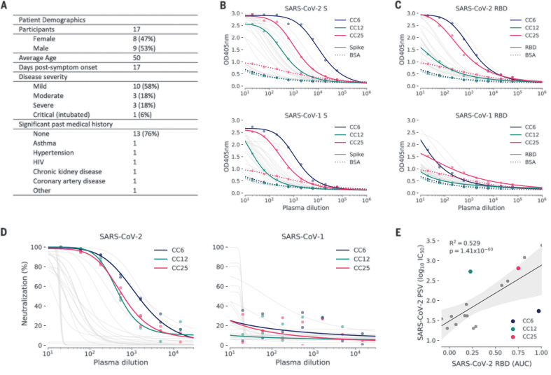
“OD405nm, optical density for wavelength of 405 nm. (D) Plasma was also tested for neutralization of pseudotyped (PSV) SARS-CoV-1 and SARS-CoV-2 virions.”
MethodInfluentialRational Vaccine Design in the Time of COVID-19
Burton, D., & Walker, L. (2020) · Cell Host and Microbe · doi:10.1016/j.chom.2020.04.022 · Cited by 112 · cited 1×
“The design of an immunogen that improves on the quality of nAbs elicited by natural infection may well emerge as an important goal of vaccine efforts (19).”
BackgroundSelf-citationYing, T., Prabakaran, P., Du, L., Shi, W., Feng, Y., Wang, Y., Wang, L., Li, W., Jiang, S., Dimitrov, D. S., et al. (2015) · Nature Communications · doi:10.1038/ncomms9223 · Cited by 110
Longitudinal dynamics of the human B cell response to the yellow fever 17D vaccine
Wec, A. Z., Haslwanter, D., Abdiche, Y. N., Shehata, L., Pedreño-López, N., Moyer, C. L., Bornholdt, Z. A., Lilov, A., Nett, J. H., Jangra, R. K., et al. (2020) · Proceedings of the National Academy of Sciences · doi:10.1073/pnas.1921388117 · Cited by 95
Cited by
Sorted by citations
What level of neutralising antibody protects from COVID-19?
Khoury, D. S., Cromer, D., Reynaldi, A., Schlub, T. E., Wheatley, A. K., Juno, J. A., Subbarao, K., Kent, S. J., Triccas, J. A., & Davenport, M. P. (2021) · doi:10.1101/2021.03.09.21252641 · Cited by 3,742
Khoury, D. S., Cromer, D., Reynaldi, A., Schlub, T. E., Wheatley, A. K., Juno, J. A., Subbarao, K., Kent, S. J., Triccas, J. A., & Davenport, M. P. (2021) · Nature Medicine · doi:10.1038/s41591-021-01377-8 · Cited by 3,742
Immunological memory to SARS-CoV-2 assessed for up to 8 months after infection.
Dan, J. M., Mateus, J., Kato, Y., Hastie, K. M., Yu, E. D., Faliti, C. E., Grifoni, A., Ramirez, S. I., Haupt, S., Frazier, A., et al. (2021) · Science · doi:10.1126/science.abf4063 · Cited by 2,656
“1, C and D), as RBD is the target of most neutralizing antibodies against SARS-CoV-2 (4, 27, 46, 47).”
BackgroundSelf-citationStarr, T. N., Greaney, A. J., Hilton, S. K., Ellis, D., Crawford, K. H. D., Dingens, A. S., Navarro, M. J., Bowen, J. E., Tortorici, M. A., Walls, A. C., et al. (2020) · Cell · doi:10.1016/j.cell.2020.08.012 · Cited by 1,956
Omicron escapes the majority of existing SARS-CoV-2 neutralizing antibodies
Cao, Y., Wang, J., Jian, F., Xiao, T., Song, W., Yisimayi, A., Huang, W., Li, Q., Wang, P., An, R., et al. (2022) · Nature · doi:10.1038/s41586-021-04385-3 · Cited by 2,006
Metadata
- Authors
- Rogers, T. F., Zhao, F., Huang, D., Beutler, N., Burns, A., He, W.-T., Limbo, O., Smith, C., Song, G., Woehl, J., Yang, L., Abbott, R. K., Callaghan, S., Garcia, E., Hurtado, J., Parren, M., Peng, L., Ramirez, S., Ricketts, J., Ricciardi, M. J., Rawlings, S. A., Wu, N. C., Yuan, M., Smith, D. M., Nemazee, D., Teijaro, J. R., Voss, J. E., Wilson, I. A., Andrabi, R., Briney, B., Landais, E., Sok, D., Jardine, J. G., & Burton, D. R.
- Affiliations
- Scripps Research Institute (US)
- Department of Immunology and Microbiology, The Scripps Research Institute, La Jolla, CA 92037, USA.
- La Jolla Institute for Immunology (US)
- George Washington University (US)
- University of California San Diego (US)
- Corresponding author
- Rogers, Thomas F.
- Zhao, Fangzhu
- Huang, Deli
- Beutler, Nathan
- Sok, Devin
- Jardine, Joseph G.
- Burton, Dennis R.
- ORCID
- Rogers, Thomas F.
- Zhao, Fangzhu
- Huang, Deli
- Beutler, Nathan
- Burns, Alison
- He, Wan-ting
- Limbo, Oliver
- Smith, Chloe
- Song, Ge
- Woehl, Jordan
- Yang, Linlin
- Abbott, Robert K.
- Callaghan, Sean
- Garcia, Elijah
- Hurtado, Jonathan
- Parren, Mara
- Peng, Linghang
- Ramirez, Sydney
- Ricketts, James
- Ricciardi, Michael J.
- Rawlings, Stephen A.
- Wu, Nicholas C.
- Yuan, Meng
- Smith, Davey M.
- Nemazee, David
- Teijaro, John R.
- Voss, James E.
- Wilson, Ian A.
- Andrabi, Raiees
- Briney, Bryan
- Landais, Elise
- Sok, Devin
- Jardine, Joseph G.
- Burton, Dennis R.
- Venue
- Science
- Published
- 2020-08-21
- Volume
- 369
- Issue
- 6506
- Pages
- 956-963
- ISSN
- 0036-8075 (ISSN-L)
- 1095-9203
- PMC
- PMC7299280
- OpenAlex
- W3035459690
- Cited by
- 1,230
- References
- 33
- Citations by year
- 2026: 23 · 2025: 47 · 2024: 68 · 2023: 126 · 2022: 242 · 2021: 504 · 2020: 217
- FWCI
- 35.77
- Type
- journal article
- Language
- en
- Open access
- hybrid
- doi.org
- Licence
- Locations
- Science (publishedVersion, cc-by)
- bioRxiv (Cold Spring Harbor Laboratory) (acceptedVersion, cc-by)
- PubMed Central (publishedVersion, cc-by)
- PubMed (publishedVersion)
- eScholarship (California Digital Library) (submittedVersion, cc-by)
- PubMed Central (submittedVersion, cc-by)
- eScholarship (California Digital Library) (submittedVersion)
- Funders
- National Institute of Allergy and Infectious Diseases (UM1 AI144462)
- Bill and Melinda Gates Foundation (OPP 1170236, OPP 1206647, OPP1196345)
- Topics
- Health Sciences › Medicine › Infectious Diseases › SARS-CoV-2 and COVID-19 Research
- Life Sciences › Agricultural and Biological Sciences › Animal Science and Zoology › Animal Virus Infections Studies
- Life Sciences › Immunology and Microbiology › Immunology › Monoclonal and Polyclonal Antibodies Research
- Keywords
- SARS-CoV-2, neutralizing antibodies, COVID-19 pandemic, receptor binding domain, spike protein epitopes, passive antibody transfer, Syrian hamster model, antibody isolation, high-throughput antibody screening, prophylaxis, vaccine design, lung viral titers
About these checks
Registry, retraction, Cites and statistics results are computed, not judged, with one exception: a printed reference that no index lists is read once by a model before it counts as a citation error. Claims are extracted by a model and kept only when their quote appears verbatim in the text; each cited source is then checked, with a model’s help, for whether it says what the claim relies on. Those verdicts can be wrong — read the claim’s quote and the cited source before relying on one.
Statistics and arithmetic are recomputed, not judged.
Confidence bands are shown only for fields calibrated on our annotated test set. None is calibrated yet, so no band is shown.
What counts as a claim ›How we measure confidence ›How we check this ›
Are you an author, or do you see a problem with this page? Request a review
