

The DOI matches its Crossref record; 47 of the 47 references it cites were found in a registry or an index.
Neuropilin-1 facilitates SARS-CoV-2 cell entry and infectivity
Open accessPublished version of a preprint
Abstract
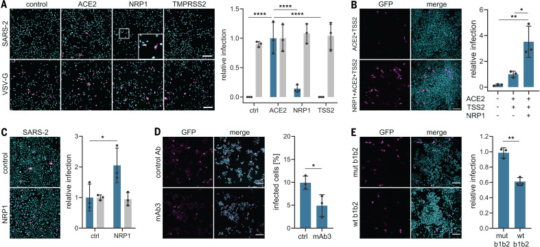
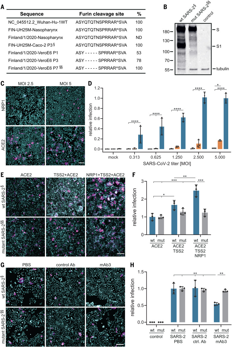
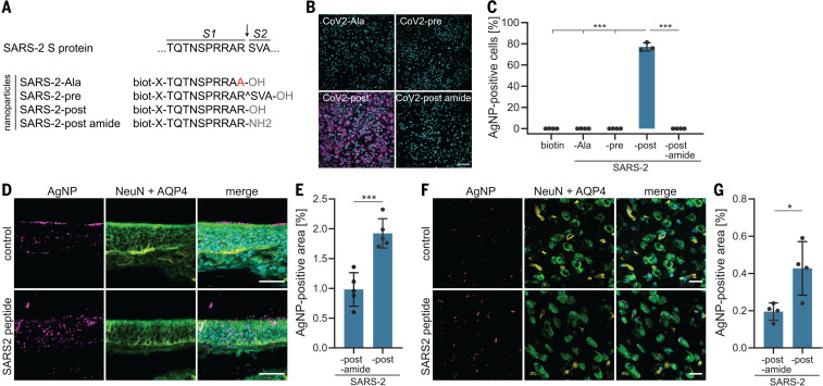
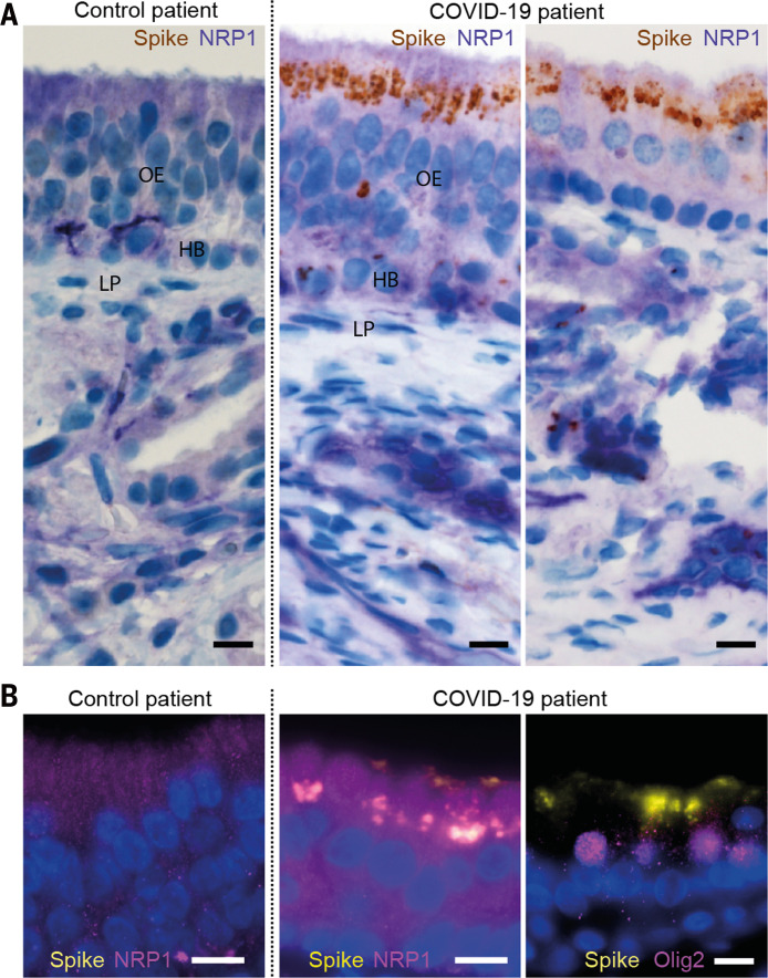
Another host factor for SARS-CoV-2: Virus-host interactions determine cellular entry and spreading in tissues. Severe acute respiratory syndrome coronavirus 2 (SARS-CoV-2) and the earlier SARS-CoV use angiotensin-converting enzyme 2 (ACE2) as a receptor; however, their tissue tropism differs, raising the possibility that additional host factors are involved. The spike protein of SARS-CoV-2 contains a cleavage site for the protease furin that is absent from SARS-CoV (see the Perspective by Kielian). Cantuti-Castelvetri et al. now show that neuropilin-1 (NRP1), which is known to bind furin-cleaved substrates, potentiates SARS-CoV-2 infectivity. NRP1 is abundantly expressed in the respiratory and olfactory epithelium, with highest expression in endothelial and epithelial cells. Daly et al. found that the furin-cleaved S1 fragment of the spike protein binds directly to cell surface NRP1 and blocking this interaction with a small-molecule inhibitor or monoclonal antibodies reduced viral infection in cell culture. Understanding the role of NRP1 in SARS-CoV-2 infection may suggest potential targets for future antiviral therapeutics. Science , this issue p. 856 , p. 861 ; see also p. 765
Overview
The claims and statistics of this paper haven’t been checked yet.
Run the check — free, no account needed. It reads the open-access text and checks the sources it cites.
Integrity signals
Registry
Cites
Statistics
Figures
Verification
Claims the paper makes
The claims and statistics of this paper haven’t been checked yet.
Run the check — free, no account needed. It reads the open-access text and checks the sources it cites.
Claims other papers make about this one
Each is checked against this paper’s own text: does it say what the citing paper says it does?
In the citing paper
“Most recently, Neuropilin1 (NRP1) was identified as an important cofactor for entry, particularly in cells with low level ACE2 expression (Cantuti-Castelvetri et al., 2020).”
From COVID-19 and the human innate immune system (2021) · cites this paper as Cantuti-Castelvetri et al., 2020
In the citing paper
“Mesenchymal cells across all ages and ventricles expressed Ace2 (Angiotensin I converting enzyme 2; Figure 4J,S4J), the SARS-CoV-2 receptor, and other known and putative accessory components, including cathepsins (Ctsl and Ctsb (Muus et al., 2020)) and Neuropilin-1 (NRP1 (Cantuti-Castelvetri et al., 2020)) (Figure 4J, S4J).”
From A cellular and spatial map of the choroid plexus across brain ventricles and ages (2021) · cites this paper as Cantuti-Castelvetri et al., 2020
In the citing paper
“We also validated the expression of NRP1 protein, an effector of SARS-CoV-2 cell entry and infectivity, in mesenchymal cells (Cantuti-Castelvetri et al., 2020) (Figure 4M).”
From A cellular and spatial map of the choroid plexus across brain ventricles and ages (2021) · cites this paper as Cantuti-Castelvetri et al., 2020
Self-check
Percentages, sums and reported differences re-computed from the paper’s own numbers.
The self-check needs the paper’s full text.
Citations
Cites
Trimmomatic: a flexible trimmer for Illumina sequence data
Bolger, A. M., Lohse, M., & Usadel, B. (2014) · Bioinformatics · doi:10.1093/bioinformatics/btu170 · Cited by 65,548
The Sequence Alignment/Map format and SAMtools
Li, H., Handsaker, B., Wysoker, A., Fennell, T., Ruan, J., Homer, N., Marth, G., Abecasis, G., Durbin, R., & 1000 Genome Project Data Processing Subgroup (2009) · Bioinformatics · doi:10.1093/bioinformatics/btp352 · Cited by 62,041
A novel coronavirus from patients with pneumonia in China, 2019
Zhu, N., Zhang, D., Wang, W., Li, X., Yang, B., Song, J., Zhao, X., Huang, B., Shi, W., Lu, R., et al. (2020) · New England Journal of Medicine · doi:10.1056/nejmoa2001017 · Cited by 28,244
A pneumonia outbreak associated with a new coronavirus of probable bat origin
Zhou, P., Yang, X.-L., Wang, X.-G., Hu, B., Zhang, L., Zhang, W., Si, H.-R., Zhu, Y., Li, B., Huang, C.-L., et al. (2020) · Nature · doi:10.1038/s41586-020-2012-7 · Cited by 21,969
Hoffmann, M., Kleine-Weber, H., Schroeder, S., Krüger, N., Herrler, T., Erichsen, S., Schiergens, T. S., Herrler, G., Wu, N.-H., Nitsche, A., et al. (2020) · Cell · doi:10.1016/j.cell.2020.02.052 · Cited by 20,286
Comprehensive Integration of Single-Cell Data
Stuart, T., Butler, A., Hoffman, P., Hafemeister, C., Papalexi, E., Mauck, W. M., Hao, Y., Stoeckius, M., Smibert, P., & Satija, R. (2019) · Cell · doi:10.1016/j.cell.2019.05.031 · Cited by 15,453
Cryo-EM structure of the 2019-nCoV spike in the prefusion conformation
Wrapp, D., Wang, N., Corbett, K. S., Goldsmith, J. A., Hsieh, C.-L., Abiona, O., Graham, B. S., & McLellan, J. S. (2020) · Science · doi:10.1126/science.abb2507 · Cited by 8,897
Structure, function and antigenicity of the SARS-CoV-2 spike glycoprotein
Walls, A. C., Park, Y.-J., Tortorici, M. A., Wall, A., McGuire, A. T., & Veesler, D. (2020) · doi:10.1101/2020.02.19.956581 · Cited by 8,586
Detection of 2019 novel coronavirus (2019-nCoV) by real-time RT-PCR
Corman, V. M., Landt, O., Kaiser, M., Molenkamp, R., Meijer, A., Chu, D. K. W., Bleicker, T., Brünink, S., Schneider, J. A., Schmidt, M. L., et al. (2020) · Eurosurveillance · doi:10.2807/1560-7917.es.2020.25.3.2000045 · Cited by 7,266
Neurologic manifestations of hospitalized patients with coronavirus disease 2019 in Wuhan, China
Mao, L., Jin, H., Wang, M., Hu, Y., Chen, S., He, Q., Chang, J., Hong, C., Zhou, Y., Wang, D., et al. (2020) · JAMA Neurology · doi:10.1001/jamaneurol.2020.1127 · Cited by 7,166
Virological assessment of hospitalized patients with COVID-2019
Wölfel, R., Corman, V. M., Guggemos, W., Seilmaier, M., Zange, S., Müller, M. A., Niemeyer, D., Jones, T. C., Vollmar, P., Rothe, C., et al. (2020) · Nature · doi:10.1038/s41586-020-2196-x · Cited by 6,661
ImageJ2: ImageJ for the next generation of scientific image data
Rueden, C., Schindelin, J., Hiner, M., DeZonia, B. E., Walter, A., Arena, E. T., & Eliceiri, K. W. (2017) · BMC Bioinformatics · doi:10.1186/s12859-017-1934-z · Cited by 6,087
Pulmonary Vascular Endothelialitis, Thrombosis, and Angiogenesis in Covid-19
Ackermann, M., Verleden, S. E., Kuehnel, M., Haverich, A., Welte, T., Laenger, F., Vanstapel, A., Werlein, C., Stark, H., Tzankov, A., et al. (2020) · New England Journal of Medicine · doi:10.1056/nejmoa2015432 · Cited by 5,613
Identification of a novel coronavirus in patients with severe acute respiratory syndrome
Drosten, C., Günther, S., Preiser, W., van der Werf, S., Brodt, H.-R., Becker, S., Rabenau, H., Panning, M., Kolesnikova, L., Fouchier, R. A. M., et al. (2003) · New England Journal of Medicine · doi:10.1056/nejmoa030747 · Cited by 4,574
Are patients with hypertension and diabetes mellitus at increased risk for COVID-19 infection?
Fang, L., Karakiulakis, G., & Roth, M. (2020) · The Lancet Respiratory Medicine · doi:10.1016/s2213-2600(20)30116-8 · Cited by 2,947
A subcellular map of the human proteome
Thul, P. J., Åkesson, L., Wiking, M., Mahdessian, D., Geladaki, A., Ait Blal, H., Alm, T., Asplund, A., Björk, L., Breckels, L. M., et al. (2017) · Science · doi:10.1126/science.aal3321 · Cited by 2,813
Single-cell landscape of bronchoalveolar immune cells in patients with COVID-19
Liao, M., Liu, Y., Yuan, J., Wen, Y., Xu, G., Zhao, J., Cheng, L., Li, J., Wang, X., Wang, F., et al. (2020) · Nature Medicine · doi:10.1038/s41591-020-0901-9 · Cited by 2,614
Hoffmann, M., Kleine-Weber, H., & Pöhlmann, S. (2020) · Molecular Cell · doi:10.1016/j.molcel.2020.04.022 · Cited by 1,852
Multiorgan and Renal Tropism of SARS-CoV-2
Puelles, V. G., Lütgehetmann, M., Lindenmeyer, M. T., Sperhake, J. P., Wong, M. N., Allweiss, L., Chilla, S., Heinemann, A., Wanner, N., Liu, S., et al. (2020) · New England Journal of Medicine · doi:10.1056/nejmc2011400 · Cited by 1,775
Coutard, B., Valle, C., de Lamballerie, X. N., Canard, B., Seidah, N. G., & Decroly, É. (2020) · Antiviral Research · doi:10.1016/j.antiviral.2020.104742 · Cited by 1,706
Wilm, A., Aw, P. P. K., Bertrand, D., Yeo, G. H. T., Ong, S. H., Wong, C. H., Khor, C. C., Petric, R., Hibberd, M. L., & Nagarajan, N. (2012) · Nucleic Acids Research · doi:10.1093/nar/gks918 · Cited by 1,370
Quick, J., Grubaugh, N. D., Pullan, S. T., Claro, I. M., Smith, A. D., Gangavarapu, K., Oliveira, G., Robles-Sikisaka, R., Rogers, T. F., Beutler, N. A., et al. (2017) · Nature Protocols · doi:10.1038/nprot.2017.066 · Cited by 1,190
Construction of a human cell landscape at single-cell level
Han, X., Zhou, Z., Fei, L., Sun, H., Wang, R., Chen, Y., Chen, H., Wang, J., Tang, H., Ge, W., et al. (2020) · Nature · doi:10.1038/s41586-020-2157-4 · Cited by 1,087
Mercer, J., Schelhaas, M., & Helenius, A. (2010) · Annual Review of Biochemistry · doi:10.1146/annurev-biochem-060208-104626 · Cited by 1,057
Self-citationThe protein expression profile of ACE2 in human tissues
Hikmet, F., Méar, L., Edvinsson, Å., Micke, P., Uhlén, M., & Lindskog, C. (2020) · Molecular Systems Biology · doi:10.15252/msb.20209610 · Cited by 1,016
C-end rule peptides mediate neuropilin-1-dependent cell, vascular, and tissue penetration
Teesalu, T., Sugahara, K. N., Kotamraju, V. R., & Ruoslahti, E. (2009) · Proceedings of the National Academy of Sciences · doi:10.1073/pnas.0908201106 · Cited by 812
Self-citationThe single-cell transcriptomic landscape of early human diabetic nephropathy
Wilson, P. C., Wu, H., Kirita, Y., Uchimura, K., Ledru, N., Rennke, H. G., Welling, P. A., Waikar, S. S., & Humphreys, B. D. (2019) · Proceedings of the National Academy of Sciences · doi:10.1073/pnas.1908706116 · Cited by 544
VEGF-A and the Induction of Pathological Angiogenesis
Nagy, J. A., Dvořàk, A. M., & Dvorak, H. F. (2007) · Annual Review of Pathology Mechanisms of Disease · doi:10.1146/annurev.pathol.2.010506.134925 · Cited by 483
Davidson, A. D., Williamson, M. K., Lewis, S., Shoemark, D., Carroll, M. W., Heesom, K. J., Zambon, M., Ellis, J., Lewis, P. A., Hiscox, J. A., et al. (2020) · Genome Medicine · doi:10.1186/s13073-020-00763-0 · Cited by 457
COVID-19 and the Chemical Senses: Supporting Players Take Center Stage
Cooper, K. W., Brann, D. H., Farruggia, M. C., Bhutani, S., Pellegrino, R., Tsukahara, T., Weinreb, C., Joseph, P. V., Larson, E. D., Parma, V., et al. (2020) · Neuron · doi:10.1016/j.neuron.2020.06.032 · Cited by 341
Goldberg, I., Allan, C., Burel, J., Creager, D. A., Falconi, A., Hochheiser, H., Johnston, J., Mellen, J., Sorger, P. K., & Swedlow, J. R. (2005) · Genome biology · doi:10.1186/gb-2005-6-5-r47 · Cited by 316
Single-cell analysis of olfactory neurogenesis and differentiation in adult humans
Durante, M. A., Kurtenbach, S., Sargi, Z. B., Harbour, J. W., Choi, R., Kurtenbach, S., Goss, G. M., Matsunami, H., & Goldstein, B. J. (2020) · Nature Neuroscience · doi:10.1038/s41593-020-0587-9 · Cited by 254
An Unbiased Screen for Human Cytomegalovirus Identifies Neuropilin-2 as a Central Viral Receptor
Martinez-Martín, N., Marcandalli, J., Huang, C., Arthur, C. P., Perotti, M., Foglierini, M., Ho, H., Dosey, A. M., Shriver, S. K., Payandeh, J., et al. (2018) · Cell · doi:10.1016/j.cell.2018.06.028 · Cited by 229
Neuropilin 1 is an entry factor that promotes EBV infection of nasopharyngeal epithelial cells
Wang, H., Zhang, H., Zhang, J., Li, Y., Zhao, B., Feng, G., Du, Y., Xiong, D., Zhong, Q., Liu, W.-L., et al. (2015) · Nature Communications · doi:10.1038/ncomms7240 · Cited by 217
Neuropilin-1 Is Involved in Human T-Cell Lymphotropic Virus Type 1 Entry
Ghez, D., Lepelletier, Y., Lambert, S., Fourneau, J., Blot, V., Janvier, S., Arnulf, B., Endert, P. V., Heveker, N., Pique, C., et al. (2006) · Journal of Virology · doi:10.1128/jvi.02719-05 · Cited by 211
How does SARS-CoV-2 cause COVID-19?
Matheson, N. J., & Lehner, P. J. (2020) · Science · doi:10.1126/science.abc6156 · Cited by 205
An endocytosis pathway initiated through neuropilin-1 and regulated by nutrient availability
Pang, H., Braun, G. B., Friman, T., Aza‐Blanc, P., Ruidíaz, M. E., Sugahara, K. N., Teesalu, T., & Ruoslahti, E. I. (2014) · Nature Communications · doi:10.1038/ncomms5904 · Cited by 196
Self-citationEtchable plasmonic nanoparticle probes to image and quantify cellular internalization
Braun, G. B., Friman, T., Pang, H. B., Pallaoro, A., de Mendoza, T. H., Willmore, A.-M. A., Kotamraju, V. R., Mann, A. P., She, Z. G., Sugahara, K. N., et al. (2014) · Nature Materials · doi:10.1038/nmat3982 · Cited by 174
Self-citationFunctional analysis of potential cleavage sites in the MERS-coronavirus spike protein
Kleine‐Weber, H., Elzayat, M. T., Hoffmann, M., & Pöhlmann, S. (2018) · Scientific Reports · doi:10.1038/s41598-018-34859-w · Cited by 164
Neuropilin Regulation of Angiogenesis, Arteriogenesis, and Vascular Permeability
Plein, A., Fantin, A., & Ruhrberg, C. (2014) · Microcirculation · doi:10.1111/micc.12124 · Cited by 130
NRP2 and CD63 Are Host Factors for Lujo Virus Cell Entry
Raaben, M., Jae, L. T., Herbert, A. S., Kuehne, A. I., Stubbs, S. H., Chou, Y., Blomen, V. A., Kirchhausen, T. L., Dye, J. M., Brummelkamp, T. R., et al. (2017) · Cell Host & Microbe · doi:10.1016/j.chom.2017.10.002 · Cited by 122
A Novel Activation Mechanism of Avian Influenza Virus H9N2 by Furin
Tse, L. V., Hamilton, A. M., Friling, T., & Whittaker, G. R. (2013) · Journal of Virology · doi:10.1128/jvi.02648-13 · Cited by 85
Single-cell longitudinal analysis of SARS-CoV-2 infection in human airway epitheliumPreprint
Ravindra, N. G., Alfajaro, M. M., Gasque, V., Habet, V., Wei, J., Filler, R. B., Huston, N. C., Wan, H., Szigeti-Buck, K., Wang, B., et al. (2020) · bioRxiv (Cold Spring Harbor Laboratory) · doi:10.1101/2020.05.06.081695 · Cited by 76
AL‐SHAMMA, H. A., & Vries, G. J. D. (1996) · Journal of Neurobiology · doi:10.1002/(sici)1097-4695(199601)29:1 · Cited by 45
Dorner, B. G., Steinbach, S., Hüser, M. B., Kroczek, R. A., & Scheffold, A. (2003) · Journal of Immunological Methods · doi:10.1016/s0022-1759(02)00498-2 · Cited by 32
Liao, K., Sikkema, D. J., Wang, C., & Lee, T. N. (2013) · Journal of Immunological Methods · doi:10.1016/j.jim.2012.12.010 · Cited by 22
Ullah, I., Chung, K., Beloor, J., Lee, S., & Kumar, P. (2019) · Journal of Visualized Experiments · doi:10.3791/59201 · Cited by 9
Research works printed but not linked: 1
- 10.1016/j.cell.2020.02.058 · DOI · Paper page
Cited by
Sorted by citations
Coronavirus biology and replication: implications for SARS-CoV-2
V’kovski, P., Kratzel, A., Steiner, S., Stalder, H., & Thiel, V. (2020) · Nature Reviews Microbiology · doi:10.1038/s41579-020-00468-6 · Cited by 3,371
Mechanisms of SARS-CoV-2 entry into cells
Jackson, C. B., Farzan, M., Chen, B., & Choe, H. (2022) · Nature Reviews Molecular Cell Biology · doi:10.1038/s41580-021-00418-x · Cited by 3,022
CD147-spike protein is a novel route for SARS-CoV-2 infection to host cells
Wang, K., Chen, W., Zhang, Z., Deng, Y., Lian, J., Du, P. F., Wei, D., Yang, Z., Sun, X., Gong, L., et al. (2020) · Signal Transduction and Targeted Therapy · doi:10.1038/s41392-020-00426-x · Cited by 1,630
Multiple SARS-CoV-2 variants escape neutralization by vaccine-induced humoral immunity
Garcia-Beltran, W. F., Lam, E. C., St. Denis, K., Nitido, A. D., Garcia, Z. H., Hauser, B. M., Feldman, J., Pavlovic, M. N., Gregory, D. J., Poznansky, M. C., et al. (2021) · Cell · doi:10.1016/j.cell.2021.03.013 · Cited by 1,353
Neuroinvasion of SARS-CoV-2 in human and mouse brain
Song, E., Zhang, C., Israelow, B., Lu-Culligan, A., Prado, A. V., Skriabine, S., Lu, P., Weizman, O.-E., Liu, F., Dai, Y., et al. (2020) · doi:10.1101/2020.06.25.169946 · Cited by 1,064
Metadata
- Authors
- Cantuti-Castelvetri, L., Ojha, R., Pedro, L. D., Djannatian, M., Franz, J., Kuivanen, S., van der Meer, F., Kallio, K., Kaya, T., Anastasina, M., Smura, T., Levanov, L., Szirovicza, L., Tobi, A., Kallio-Kokko, H., Österlund, P., Joensuu, M., Meunier, F. A., Butcher, S. J., Winkler, M. S., Mollenhauer, B., Helenius, A., Gokce, O., Teesalu, T., Hepojoki, J., Vapalahti, O., Stadelmann, C., Balistreri, G., & Simons, M.
- Affiliations
- Institute of Neuronal Cell Biology, Technical University Munich, Munich, Germany.
- University of Helsinki (FI)
- German Center for Neurodegenerative Diseases (DE)
- Universitätsmedizin Göttingen (DE)
- Department of Neuropathology, University Medical Center Göttingen, Göttingen, Germany.
- Department of Virology, Medicum, University of Helsinki, Helsinki, Finland.
- University of Tartu (EE)
- Department of Virology, University of Helsinki and Helsinki University Hospital, Helsinki, Finland.
- Finnish Institute for Health and Welfare (FI)
- The University of Queensland (AU)
- ETH Zurich (CH)
- Institute for Stroke and Dementia Research (ISD), University Hospital, LMU Munich, Munich, Germany.
- University of Zurich (CH)
- Corresponding author
- Ojha, Ravi
- Pedro, Liliana D.
- Djannatian, Minou
- Franz, Jonas
- Kuivanen, Suvi
- Balistreri, Giuseppe
- Simons, Mikael
- ORCID
- Cantuti-Castelvetri, Ludovico
- Ojha, Ravi
- Pedro, Liliana D.
- Djannatian, Minou
- Franz, Jonas
- Kuivanen, Suvi
- van der Meer, Franziska
- Kallio, Katri
- Kaya, Tuğberk
- Anastasina, Maria
- Smura, Teemu
- Levanov, Lev
- Szirovicza, Leonora
- Tobi, Allan
- Österlund, Pamela
- Joensuu, Merja
- Meunier, Frédéric A.
- Butcher, Sarah J.
- Winkler, Martin Sebastian
- Mollenhauer, Brit
- Gokce, Ozgun
- Teesalu, Tambet
- Hepojoki, Jussi
- Vapalahti, Olli
- Stadelmann, Christine
- Balistreri, Giuseppe
- Simons, Mikael
- Venue
- Science
- Published
- 2020-11-13
- Volume
- 370
- Issue
- 6518
- Pages
- 856-860
- ISSN
- 0036-8075 (ISSN-L)
- 1095-9203
- PMC
- PMC7857391
- OpenAlex
- W3093867667
- Cited by
- 1,925
- References
- 47
- Citations by year
- 2026: 56 · 2025: 120 · 2024: 211 · 2023: 277 · 2022: 461 · 2021: 669 · 2020: 128
- FWCI
- 50.07
- Type
- journal article
- Language
- en
- Open access
- hybrid
- doi.org
- Licence
- Locations
- Science (publishedVersion, cc-by)
- bioRxiv (Cold Spring Harbor Laboratory) (acceptedVersion)
- Europe PMC (PubMed Central) (publishedVersion, cc-by)
- PubMed Central (publishedVersion, cc-by)
- PubMed (publishedVersion)
- Open access LMU (Ludwid Maxmilian's Universitat Munchen) (submittedVersion, cc-by)
- Työväentutkimus Vuosikirja (submittedVersion)
- mediaTUM (Technical University of Munich) (submittedVersion)
- GoeScholar The Publication Server of the Georg-August-Universität Göttingen (Georg-August-Universität Göttingen) (submittedVersion, other-oa)
- Funders
- ERC CoV
- Topics
- Health Sciences › Medicine › Infectious Diseases › SARS-CoV-2 and COVID-19 Research
- Health Sciences › Medicine › Infectious Diseases › COVID-19 Clinical Research Studies
- Health Sciences › Medicine › Infectious Diseases › SARS-CoV-2 detection and testing
- Keywords
- neuropilin-1, SARS-CoV-2, viral entry, furin cleavage, spike protein, host factors, viral infectivity, monoclonal antibodies, small molecule inhibitors, antiviral agents, viral tropism, ACE2
- MeSH terms
- Protein Domains, Betacoronavirus, Host Microbial Interactions, Angiotensin-Converting Enzyme 2, COVID-19, SARS-CoV-2, Animals, Antibodies, Monoclonal, Female, Humans, Peptidyl-Dipeptidase A, Lung, Male, Mice, Inbred C57BL, Mutation, Olfactory Mucosa, Peptide Fragments, Pneumonia, Viral, Protein Binding, Serine Endopeptidases, Coronavirus Infections, Caco-2 Cells, Respiratory Mucosa, Neuropilin-1, Neuropilin-2, Mice, Virus Internalization, Metal Nanoparticles, HEK293 Cells, Pandemics, Spike Glycoprotein, Coronavirus
About these checks
Registry, retraction, Cites and statistics results are computed, not judged, with one exception: a printed reference that no index lists is read once by a model before it counts as a citation error. Claims are extracted by a model and kept only when their quote appears verbatim in the text; each cited source is then checked, with a model’s help, for whether it says what the claim relies on. Those verdicts can be wrong — read the claim’s quote and the cited source before relying on one.
Statistics and arithmetic are recomputed, not judged.
Confidence bands are shown only for fields calibrated on our annotated test set. None is calibrated yet, so no band is shown.
What counts as a claim ›How we measure confidence ›How we check this ›
Are you an author, or do you see a problem with this page? Request a review
