

The DOI matches its Crossref record; 124 of the 124 references it cites were found in a registry or an index.
Integrase-associated niche differentiation of endogenous large DNA viruses in crustaceans
Open accessPublished version of a preprint
Abstract
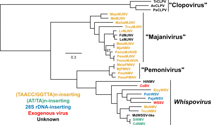
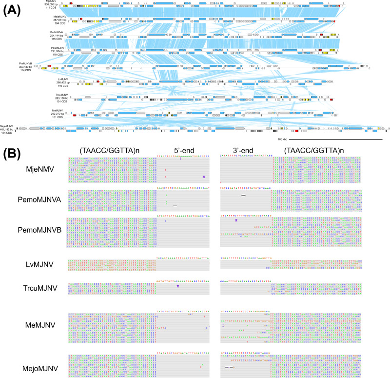
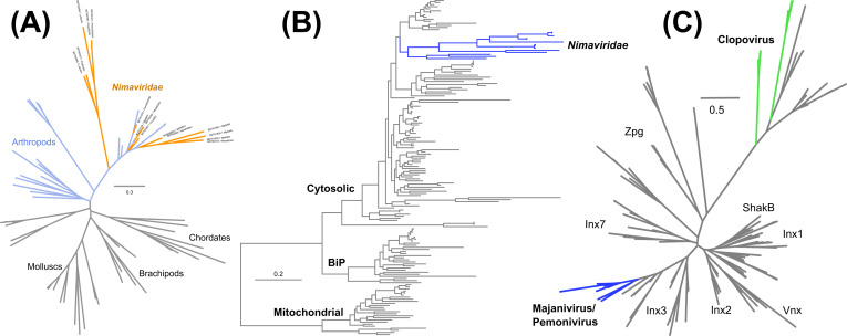
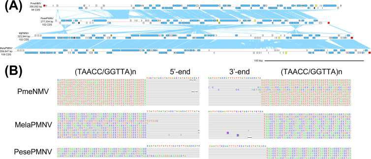
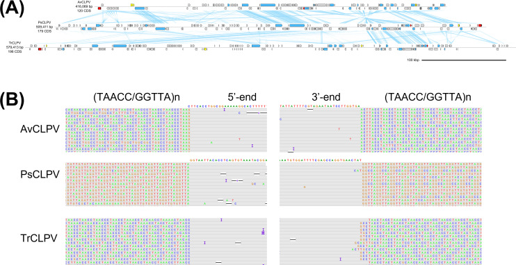
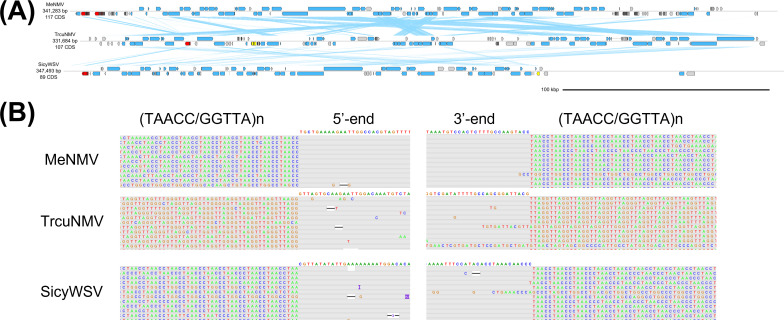
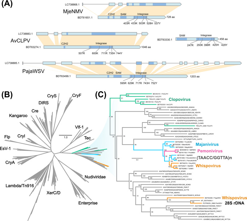
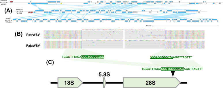
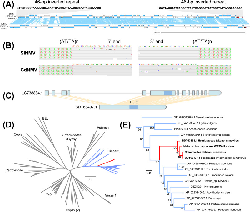
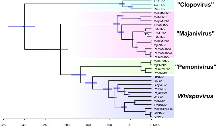
Crustacean genomes harbor sequences originating from nimaviruses, a family of large double-stranded DNA viruses infecting crustaceans. In this study, we recovered metagenome-assembled genomes of 27 endogenous nimaviruses from crustacean genome data. Phylogenetic analysis revealed four major lineages within Nimaviridae , and for three of these lineages, we propose novel genera of endogenous nimaviruses: “Majanivirus” and “Pemonivirus” identified from penaeid shrimp genomes, and “Clopovirus” identified from terrestrial isopods. Majanivirus genomes contain multiple eukaryotic-like genes such as baculoviral inhibitor of apoptosis repeat-containing genes, innexins, and heat shock protein 70-like genes, some of which contain introns. An alignment of long reads revealed that each endogenous nimavirus species specifically inserts into host microsatellites or within 28S rDNA. This insertion preference was associated with the type of virus-encoded DNA recombination enzymes, the integrases. Majaniviruses, pemoniviruses, some whispoviruses, and possibly clopoviruses specifically insert into the arthropod telomere repeat motif (TAACC/GGTTA)n and all possessed a specific tyrosine recombinase family. Pasiphaea japonica whispovirus and Portunus trituberculatus whispovirus, the closest relatives of white spot syndrome virus, integrate into the host 28S rDNA and are equipped with members of another family of tyrosine recombinases that are distantly related to telomere-specific tyrosine recombinases. Endogenous nimavirus genomes identified from sesarmid crabs, which lack tyrosine recombinases and are flanked by a 46-bp inverted terminal repeat, integrate into (AT/TA)n microsatellites through the acquisition of a Ginger2-like cut-and-paste DDE transposase. These results suggest that endogenous nimaviruses are giant transposable elements that occupy different sequence niches through the acquisition of different integrase families. IMPORTANCE: Crustacean genomes harbor sequences originating from a family of large DNA viruses called nimaviruses, but it is unclear why they are present. We show that endogenous nimaviruses selectively insert into repetitive sequences within the host genome, and this insertion specificity was correlated with different types of integrases, which are DNA recombination enzymes encoded by the nimaviruses themselves. This suggests that endogenous nimaviruses have colonized various genomic niches through the acquisition of integrases with different insertion specificities. Our results point to a novel survival strategy of endogenous large DNA viruses colonizing the host genomes. These findings may clarify the evolution and spread of nimaviruses in crustaceans and lead to measures to control and prevent the spread of pathogenic nimaviruses in aquaculture settings.
Overview
The claims and statistics of this paper haven’t been checked yet.
Run the check — free, no account needed. It reads the open-access text and checks the sources it cites.
Integrity signals
Registry
Cites
Statistics
Figures
Verification
Claims the paper makes
The claims and statistics of this paper haven’t been checked yet.
Run the check — free, no account needed. It reads the open-access text and checks the sources it cites.
Self-check
Percentages, sums and reported differences re-computed from the paper’s own numbers.
The self-check needs the paper’s full text.
Citations
Cites
The Sequence Alignment/Map format and SAMtools
Li, H., Handsaker, B., Wysoker, A., Fennell, T., Ruan, J., Homer, N., Marth, G., Abecasis, G., Durbin, R., & 1000 Genome Project Data Processing Subgroup (2009) · Bioinformatics · doi:10.1093/bioinformatics/btp352 · Cited by 62,041
Highly accurate protein structure prediction with AlphaFold
Jumper, J., Evans, R., Pritzel, A., Green, T., Figurnov, M., Ronneberger, O., Tunyasuvunakool, K., Bates, R., Žídek, A., Potapenko, A., et al. (2021) · Nature · doi:10.1038/s41586-021-03819-2 · Cited by 43,764
fastp: an ultra-fast all-in-one FASTQ preprocessor
Chen, S., Zhou, Y., Chen, Y., & Gu, J. (2018) · Bioinformatics · doi:10.1093/bioinformatics/bty560 · Cited by 29,721
BEDTools: a flexible suite of utilities for comparing genomic features
Quinlan, A. R., & Hall, I. M. (2010) · Bioinformatics · doi:10.1093/bioinformatics/btq033 · Cited by 27,437
IQ-TREE 2: New Models and Efficient Methods for Phylogenetic Inference in the Genomic Era
Minh, B. Q., Schmidt, H. A., Chernomor, O., Schrempf, D., Woodhams, M. D., von Haeseler, A., & Lanfear, R. (2020) · Molecular Biology and Evolution · doi:10.1093/molbev/msaa015 · Cited by 16,873
Robinson, J. T., Thorvaldsdóttir, H., Winckler, W., Guttman, M., Lander, E. S., Getz, G., & Mesirov, J. P. (2011) · Nature Biotechnology · doi:10.1038/nbt.1754 · Cited by 15,390
Graph-based genome alignment and genotyping with HISAT2 and HISAT-genotype
Kim, D., Paggi, J. M., Park, C., Bennett, C., & Salzberg, S. L. (2019) · Nature Biotechnology · doi:10.1038/s41587-019-0201-4 · Cited by 15,022
Minimap2: Pairwise alignment for nucleotide sequences
Li, H. (2018) · Bioinformatics · doi:10.1093/bioinformatics/bty191 · Cited by 14,807
trimAl: a tool for automated alignment trimming in large-scale phylogenetic analyses
Capella-Gutiérrez, S., Silla-Martínez, J. M., & Gabaldón, T. (2009) · Bioinformatics · doi:10.1093/bioinformatics/btp348 · Cited by 12,751
Prodigal: prokaryotic gene recognition and translation initiation site identification
Hyatt, D., Chen, G.-L., LoCascio, P. F., Land, M. L., Larimer, F. W., & Hauser, L. J. (2010) · BMC Bioinformatics · doi:10.1186/1471-2105-11-119 · Cited by 12,587
Cd-hit: a fast program for clustering and comparing large sets of protein or nucleotide sequences
Li, W., & Godzik, A. (2006) · Bioinformatics · doi:10.1093/bioinformatics/btl158 · Cited by 11,410
Posterior summarization in Bayesian phylogenetics using tracer 1.7
Rambaut, A., Drummond, A. J., Xie, D., Baele, G., & Suchard, M. A. (2018) · Systematic Biology · doi:10.1093/sysbio/syy032 · Cited by 11,159
UCSF ChimeraX: Structure visualization for researchers, educators, and developers
Pettersen, E. F., Goddard, T. D., Huang, C. C., Meng, E. C., Couch, G. S., Croll, T. I., Morris, J. H., & Ferrin, T. E. (2021) · Protein Science · doi:10.1002/pro.3943 · Cited by 9,514
ColabFold - Making protein folding accessible to all
Mirdita, M., Schütze, K., Moriwaki, Y., Heo, L., Ovchinnikov, S., & Steinegger, M. (2021) · doi:10.1101/2021.08.15.456425 · Cited by 9,391
Walker, B. J., Abeel, T., Shea, T., Priest, M., Abouelliel, A., Sakthikumar, S., Cuomo, C. A., Zeng, Q., Wortman, J., Young, S. K., et al. (2014) · PLoS ONE · doi:10.1371/journal.pone.0112963 · Cited by 9,197
MAFFT online service: multiple sequence alignment, interactive sequence choice and visualization
Katoh, K., Rozewicki, J., & Yamada, K. D. (2019) · Briefings in Bioinformatics · doi:10.1093/bib/bbx108 · Cited by 8,909
Canu: scalable and accurate long-read assembly via adaptive k -mer weighting and repeat separation
Koren, S., Walenz, B. P., Berlin, K., Miller, J. R., Bergman, N. H., & Phillippy, A. M. (2017) · Genome Research · doi:10.1101/gr.215087.116 · Cited by 7,304
BEAST 2: a software platform for Bayesian evolutionary analysis
Bouckaert, R., Heled, J., Kühnert, D., Vaughan, T., Wu, C.-H., Xie, D., Suchard, M. A., Rambaut, A., & Drummond, A. J. (2014) · PLoS Computational Biology · doi:10.1371/journal.pcbi.1003537 · Cited by 6,527
Assembly of long, error-prone reads using repeat graphs
Kolmogorov, M., Yuan, J., Lin, Y., & Pevzner, P. A. (2019) · Nature Biotechnology · doi:10.1038/s41587-019-0072-8 · Cited by 5,986
MITOS: Improved de novo metazoan mitochondrial genome annotation
Bernt, M., Donath, A., Jühling, F., Externbrink, F., Florentz, C., Fritzsch, G., Pütz, J., Middendorf, M., & Stadler, P. F. (2013) · Molecular Phylogenetics and Evolution · doi:10.1016/j.ympev.2012.08.023 · Cited by 5,354
DNA–DNA hybridization values and their relationship to whole-genome sequence similarities
Goris, J., Konstantinidis, K. T., Klappenbach, J. A., Coenye, T., Vandamme, P., & Tiedje, J. M. (2007) · International Journal of Systematic and Evolutionary Microbiology · doi:10.1099/ijs.0.64483-0 · Cited by 4,750
SeqKit: A Cross-Platform and Ultrafast Toolkit for FASTA/Q File Manipulation
Shen, W., Le, S., Li, Y., & Hu, F. (2016) · PLOS ONE · doi:10.1371/journal.pone.0163962 · Cited by 3,380
Bandage: interactive visualization of de novo genome assemblies
Wick, R. R., Schultz, M. B., Zobel, J., & Holt, K. E. (2015) · Bioinformatics · doi:10.1093/bioinformatics/btv383 · Cited by 3,029
AUGUSTUS: ab initio prediction of alternative transcripts
Stanke, M., Keller, O., Gunduz, I., Hayes, A., Waack, S., & Morgenstern, B. (2006) · Nucleic Acids Research · doi:10.1093/nar/gkl200 · Cited by 2,829
Selfish genes, the phenotype paradigm and genome evolution
Doolittle, W. F., & Sapienza, C. (1980) · Nature · doi:10.1038/284601a0 · Cited by 1,829
GFF Utilities: GffRead and GffCompare
Pertea, G., & Pertea, M. (2020) · F1000Research · doi:10.12688/f1000research.23297.2 · Cited by 1,152
HH-suite3 for fast remote homology detection and deep protein annotation
Steinegger, M., Meier, M., Mirdita, M., Vöhringer, H., Haunsberger, S. J., & Söding, J. (2019) · BMC Bioinformatics · doi:10.1186/s12859-019-3019-7 · Cited by 1,145
The enveomics collection: a toolbox for specialized analyses of microbial genomes and metagenomes
Rodriguez-R, L. M., & Konstantinidis, K. T. (2016) · doi:10.7287/peerj.preprints.1900v1 · Cited by 1,117
Formation of the Isthmus of Panama
O’Dea, A., Lessios, H. A., Coates, A. G., Eytan, R. I., Restrepo-Moreno, S. A., Cione, A. L., Collins, L. S., de Queiroz, A., Farris, D. W., Norris, R. D., et al. (2016) · Science Advances · doi:10.1126/sciadv.1600883 · Cited by 980
RefSeq: an update on mammalian reference sequences
Pruitt, K. D., Brown, G. R., Hiatt, S. M., Thibaud-Nissen, F., Astashyn, A., Ermolaeva, O., Farrell, C. M., Hart, J., Landrum, M. J., McGarvey, K. M., et al. (2014) · Nucleic Acids Research · doi:10.1093/nar/gkt1114 · Cited by 945
Rodriguez-R, L. M., & Konstantinidis, K. T. (2014) · Microbe Magazine · doi:10.1128/microbe.9.111.1 · Cited by 698
DALI and the persistence of protein shape
Holm, L. (2020) · Protein Science · doi:10.1002/pro.3749 · Cited by 692
Endogenous Viral Elements in Animal Genomes
Katzourakis, A., & Gifford, R. J. (2010) · PLoS Genetics · doi:10.1371/journal.pgen.1001191 · Cited by 661
Assembling Genomes and Mini-metagenomes from Highly Chimeric Reads
Nurk, S., Bankevich, A., Antipov, D., Gurevich, A., Korobeynikov, A., Lapidus, A., Prjibelsky, A., Pyshkin, A., Sirotkin, A., Sirotkin, Y., et al. (2013) · Lecture Notes in Computer Science · doi:10.1007/978-3-642-37195-0_13 · Cited by 661
Historic emergence, impact and current status of shrimp pathogens in Asia
Flegel, T. W. (2012) · Journal of Invertebrate Pathology · doi:10.1016/j.jip.2012.03.004 · Cited by 607
Penaeid shrimp genome provides insights into benthic adaptation and frequent molting
Zhang, X., Yuan, J., Sun, Y., Li, S., Gao, Y., Yu, Y., Liu, C., Wang, Q., Lv, X., Zhang, X., et al. (2019) · Nature Communications · doi:10.1038/s41467-018-08197-4 · Cited by 542
Disease will limit future food supply from the global crustacean fishery and aquaculture sectors
Stentiford, G. D., Neil, D. M., Peeler, E. J., Shields, J. D., Small, H. J., Flegel, T. W., Vlak, J. M., Jones, B., Morado, F., Moss, S., et al. (2012) · Journal of Invertebrate Pathology · doi:10.1016/j.jip.2012.03.013 · Cited by 496
Endogenous non-retroviral RNA virus elements in mammalian genomes
Horie, M., Honda, T., Suzuki, Y., Kobayashi, Y., Daito, T., Oshida, T., Ikuta, K., Jern, P., Gojobori, T., Coffin, J. M., et al. (2010) · Nature · doi:10.1038/nature08695 · Cited by 490
Rapid evolution to terrestrial life in Jamaican crabs
Schubart, C. D., Diesel, R., & Hedges, S. B. (1998) · Nature · doi:10.1038/30724 · Cited by 479
Complexities of Viral Mutation Rates
Peck, K. M., & Lauring, A. S. (2018) · Journal of Virology · doi:10.1128/jvi.01031-17 · Cited by 442
The enveomics collection: a toolbox for specialized analyses of microbial genomes and metagenomesPreprint
Rodriguez‐R, L. M., & Konstantinidis, K. T. (2016) · doi:10.7287/peerj.preprints.1900 · Cited by 410
Minh, B. Q., Schmidt, H. A., Chernomor, O., Schrempf, D., Woodhams, M. D., von Haeseler, A., & Lanfear, R. (2020) · Molecular Biology and Evolution · doi:10.1093/molbev/msaa131 · Cited by 408
The genome polishing tool POLCA makes fast and accurate corrections in genome assemblies
Zimin, A. V., & Salzberg, S. L. (2020) · PLOS Computational Biology · doi:10.1371/journal.pcbi.1007981 · Cited by 406
The integrase family of tyrosine recombinases: evolution of a conserved active site domain
Esposito, D., & Scocca, J. J. (1997) · Nucleic Acids Research · doi:10.1093/nar/25.18.3605 · Cited by 305
Historic emergence, impact and current status of shrimp pathogens in the Americas
Lightner, D. V., Redman, R. M., Pantoja, C. R., Tang, K. F. J., Noble, B. L., Schofield, P., Mohney, L. L., Nunan, L. M., & Navarro, S. A. (2012) · Journal of Invertebrate Pathology · doi:10.1016/j.jip.2012.03.006 · Cited by 296
The catalytic domain of all eukaryotic cut-and-paste transposase superfamilies
Yuan, Y.-W., & Wessler, S. R. (2011) · Proceedings of the National Academy of Sciences · doi:10.1073/pnas.1104208108 · Cited by 280
The ecology of the genome — mobile DNA elements and their hosts
Brookfield, J. F. Y. (2005) · Nature Reviews Genetics · doi:10.1038/nrg1524 · Cited by 228
Paleovirology—Modern Consequences of Ancient Viruses
Emerman, M., & Malik, H. S. (2010) · PLoS Biology · doi:10.1371/journal.pbio.1000301 · Cited by 193
Transposable elements in the mammalian germline: a comfortable niche or a deadly trap?
Zamudio, N., & Bourc’his, D. (2010) · Heredity · doi:10.1038/hdy.2010.53 · Cited by 192
The Double-Stranded DNA Virosphere as a Modular Hierarchical Network of Gene Sharing
Iranzo, J., Krupovìč, M., & Koonin, E. V. (2016) · mBio · doi:10.1128/mbio.00978-16 · Cited by 192
Paleozoic origin of insect large dsDNA viruses
Thézé, J., Bézier, A., Périquet, G., Drezen, J., & Herniou, E. A. (2011) · Proceedings of the National Academy of Sciences · doi:10.1073/pnas.1105580108 · Cited by 188
Dynamics of transposable elements: towards a community ecology of the genome
Venner, S., Feschotte, C., & Biémont, C. (2009) · Trends in Genetics · doi:10.1016/j.tig.2009.05.003 · Cited by 181
Molecular Phylogeny and Biogeography of the Marine ShrimpPenaeus
Baldwin, J. D., Bass, A. L., Bowen, B. W., & Clark, W. H. (1998) · Molecular Phylogenetics and Evolution · doi:10.1006/mpev.1998.0537 · Cited by 180
When parasitic wasps hijacked viruses: genomic and functional evolution of polydnaviruses
Herniou, E. A., Huguet, E., Thézé, J., Bézier, A., Périquet, G., & Drezen, J. (2013) · Philosophical Transactions of the Royal Society B Biological Sciences · doi:10.1098/rstb.2013.0051 · Cited by 175
The genomic ecosystem of transposable elements in maize
Stitzer, M. C., Anderson, S. N., Springer, N. M., & Ross‐Ibarra, J. (2021) · PLoS Genetics · doi:10.1371/journal.pgen.1009768 · Cited by 160
HyPo: Super Fast & Accurate Polisher for Long Read Genome AssembliesPreprint
Kundu, R., Casey, J., & Sung, W.-K. K. (2019) · bioRxiv (Cold Spring Harbor Laboratory) · doi:10.1101/2019.12.19.882506 · Cited by 136
Structural and sequence diversity of eukaryotic transposable elements
Kojima, K. K. (2018) · Genes & Genetic Systems · doi:10.1266/ggs.18-00024 · Cited by 132
Gupta, R. S., Sawnani, S., Adeolu, M., Alnajar, S., & Oren, A. (2018) · Antonie van Leeuwenhoek · doi:10.1007/s10482-018-1047-3 · Cited by 123
Cappello, J., Handelsman, K., & Lodish, H. F. (1985) · Cell · doi:10.1016/0092-8674(85)90016-9 · Cited by 121
Simple sequence repeats drive genome plasticity and promote adaptive evolution in penaeid shrimp
Yuan, J., Zhang, X., Wang, M., Sun, Y., Liu, C., Li, S., Yu, Y., Gao, Y., Liu, F., Zhang, X., et al. (2021) · Communications Biology · doi:10.1038/s42003-021-01716-y · Cited by 117
MaxAlign: maximizing usable data in an alignment
Gouveia‐Oliveira, R., Sackett, P. W., & Pedersen, A. G. (2007) · BMC Bioinformatics · doi:10.1186/1471-2105-8-312 · Cited by 109
The Apis mellifera Filamentous Virus Genome
Gauthier, L., Cornman, S., Hartmann, U., Cousserans, F., Evans, J. D., de Miranda, J. R., & Neumann, P. (2015) · Viruses · doi:10.3390/v7072798 · Cited by 106
Retroviral Integrase Structure and DNA Recombination Mechanism
Engelman, A., & Cherepanov, P. (2014) · Microbiology Spectrum · doi:10.1128/microbiolspec.mdna3-0024-2014 · Cited by 102
Cryptons: a group of tyrosine-recombinase-encoding DNA transposons from pathogenic fungi
Goodwin, T. J. D., Butler, M. I., & Poulter, R. T. M. (2003) · Microbiology · doi:10.1099/mic.0.26529-0 · Cited by 102
Long-Term Inheritance of the 28S rDNA-Specific Retrotransposon R2
Kojima, K. K., & Fujiwara, H. (2005) · Molecular Biology and Evolution · doi:10.1093/molbev/msi210 · Cited by 92
Wu, J., Namikoshi, A., Nishizawa, T., Mushiake, K., Teruya, K., & Muroga, K. (2001) · Diseases of Aquatic Organisms · doi:10.3354/dao047129 · Cited by 91
Polydnaviruses: From discovery to current insights
Strand, M. R., & Burke, G. R. (2015) · Virology · doi:10.1016/j.virol.2015.01.018 · Cited by 84
Polydnavirus hidden face: The genes producing virus particles of parasitic wasps
Bézier, A., Herbinière, J., Lanzrein, B., & Drezen, J. (2009) · Journal of Invertebrate Pathology · doi:10.1016/j.jip.2009.04.006 · Cited by 83
Crypton transposons: identification of new diverse families and ancient domestication events
Kojima, K. K., & Jurka, J. (2011) · Mobile DNA · doi:10.1186/1759-8753-2-12 · Cited by 82
Requirements for catalysis in the Cre recombinase active site
Gibb, B., Gupta, K., Ghosh, K., Sharp, R. B., Chen, J., & Duyne, G. D. V. (2010) · Nucleic Acids Research · doi:10.1093/nar/gkq384 · Cited by 80
ICTV Virus Taxonomy Profile: Nimaviridae
Wang, H., Hırono, I., Maningas, M. B. B., Somboonwiwat, K., & Stentiford, G. D. (2019) · Journal of General Virology · doi:10.1099/jgv.0.001248 · Cited by 78
Tang, B., Zhang, D.-Z., Li, H., Jiang, S., Zhang, H., Xuan, F., Ge, B., Wang, Z., Liu, Y., Sha, Z., et al. (2020) · GigaScience · doi:10.1093/gigascience/giz161 · Cited by 73
The Enterprise , a massive transposon carrying Spok meiotic drive genesPreprint
Vogan, A. A., Ament‐Velásquez, S. L., Bastiaans, E., Wallerman, O., Saupe, S. J., Suh, A., & Johannesson, H. (2021) · Genome Research · doi:10.1101/gr.267609.120 · Cited by 72
Crustacean Genome Exploration Reveals the Evolutionary Origin of White Spot Syndrome Virus
Kawato, S., Shitara, A., Wang, Y., Nozaki, R., Kondo, H., & Hırono, I. (2018) · Journal of Virology · doi:10.1128/jvi.01144-18 · Cited by 71
Self-citationPhylogeny of penaeoid shrimps (Decapoda: Penaeoidea) inferred from nuclear protein-coding genes
Ma, K. Y., Chan, T., & Chu, K. H. (2009) · Molecular Phylogenetics and Evolution · doi:10.1016/j.ympev.2009.05.019 · Cited by 70
Automated cell tracking using StarDist and TrackMate
Fazeli, E., Roy, N. H., Follain, G., Laine, R., von Chamier, L., Hänninen, P., Eriksson, J., Tinevez, J., & Jacquemet, G. (2020) · bioRxiv · doi:10.12688/f1000research.27019.1 · Cited by 69
Evolution of Target Specificity in R1 Clade Non-LTR Retrotransposons
Kojima, K. K., & Fujiwara, H. (2003) · Molecular Biology and Evolution · doi:10.1093/molbev/msg031 · Cited by 66
Bao, W., Kapitonov, V. V., & Jurka, J. (2010) · Mobile DNA · doi:10.1186/1759-8753-1-3 · Cited by 66
Chebbi, M. A., Becking, T., Moumen, B., Giraud, I., Gilbert, C., Peccoud, J., & Cordaux, R. (2019) · Molecular Biology and Evolution · doi:10.1093/molbev/msz010 · Cited by 65
Remarkable Diversity of Endogenous Viruses in a Crustacean Genome
Thézé, J., Leclercq, S., Moumen, B., Cordaux, R., & Gilbert, C. (2014) · Genome Biology and Evolution · doi:10.1093/gbe/evu163 · Cited by 62
Tuyen, N. X., Verreth, J. A. J., Vlak, J. M., & de Jong, M. C. M. (2014) · Preventive Veterinary Medicine · doi:10.1016/j.prevetmed.2014.08.007 · Cited by 58
Delaroque, N., & Boland, W. (2008) · BMC Evolutionary Biology · doi:10.1186/1471-2148-8-110 · Cited by 57
Complete fusion of a transposon and herpesvirus created the Teratorn mobile element in medaka fish
Inoue, Y., Saga, T., Aikawa, T., Kumagai, M., Shimada, A., Kawaguchi, Y., Naruse, K., Morishita, S., Koga, A., & Takeda, H. (2017) · Nature Communications · doi:10.1038/s41467-017-00527-2 · Cited by 55
Smyshlyaev, G., Barábas, O., & Bateman, A. (2021) · Molecular Systems Biology · doi:10.15252/msb.20209880 · Cited by 51
The Oldest Shrimp (Devonian: Famennian) and Remarkable Preservation of Soft Tissue
Feldmann, R. M., & Schweitzer, C. E. (2010) · Journal of Crustacean Biology · doi:10.1651/09-3268.1 · Cited by 49
Brown Planthopper Nudivirus DNA Integrated in Its Host Genome
Cheng, R., Xi, Y., Lou, Y.-H., Wang, Z., Xu, J.-Y., Xu, H., & Zhang, C. (2014) · Journal of Virology · doi:10.1128/jvi.03166-13 · Cited by 48
Wallace, M. A., Coffman, K. A., Gilbert, C., Ravindran, S., Albery, G. F., Abbott, J. K., Argyridou, E., Bellosta, P., Betancourt, A. J., Colinet, H., et al. (2021) · Virus Evolution · doi:10.1093/ve/veab031 · Cited by 47
Analysis of the Genome of the Sexually Transmitted Insect Virus Helicoverpa zea Nudivirus 2
Burand, J. P., Kim, W., Afonso, C. L., Tulman, E. R., Kutish, G. F., Lu, Z., & Rock, D. L. (2012) · Viruses · doi:10.3390/v4010028 · Cited by 43
Evolutionary History of Endogenous Human Herpesvirus 6 Reflects Human Migration out of Africa
Aswad, A., Aimola, G., Wight, D. J., Roychoudhury, P., Zimmermann, C., Hill, J. A., Laßner, D., Xie, H. J., Huang, M.-L. W., Parrish, N. F., et al. (2020) · Molecular Biology and Evolution · doi:10.1093/molbev/msaa190 · Cited by 41
Genome and transcriptome assemblies of the kuruma shrimp,Marsupenaeus japonicus
Kawato, S., Nishitsuji, K., Arimoto, A., Hisata, K., Kawamitsu, M., Nozaki, R., Kondo, H., Shinzato, C., Ohira, T., Satoh, N., et al. (2021) · G3 Genes Genomes Genetics · doi:10.1093/g3journal/jkab268 · Cited by 41
Self-citationPetersen, J. M., Bézier, A., Drezen, J., & van Oers, M. M. (2022) · Journal of Invertebrate Pathology · doi:10.1016/j.jip.2022.107718 · Cited by 41
Huang, S.-W., Lin, Y. Y., You, E.-M., Liu, T. T., Shu, H., Wu, K. M., Tsai, S., Lo, C., Kou, G. H., Chin, G., et al. (2011) · BMC Genomics · doi:10.1186/1471-2164-12-242 · Cited by 39
Alcivar‐Warren, A., Meehan-Meola, D., Wang, Y., Guo, X., Zhou, L., Xiang, J., Moss, S. M., Arce, S. M., Warren, W., Xu, Z., et al. (2006) · Marine Biotechnology · doi:10.1007/s10126-005-6031-z · Cited by 38
Hyper-expansion of large DNA segments in the genome of kuruma shrimp, Marsupenaeus japonicus
Koyama, T., Asakawa, S., Katagiri, T., Shimizu, A., Fagutao, F. F., Mavichak, R., Santos, M. D., Fuji, K., Sakamoto, T., Kitakado, T., et al. (2010) · BMC Genomics · doi:10.1186/1471-2164-11-141 · Cited by 38
Self-citationEndogenization and excision of human herpesvirus 6 in human genomes
Liu, X., Kosugi, S., Koide, R., Kawamura, Y., Ito, J., Miura, H., Matoba, N., Matsuzaki, M., Fujita, M., Kamada, A. J., et al. (2020) · PLoS Genetics · doi:10.1371/journal.pgen.1008915 · Cited by 36
Utari, H. B., Soowannayan, C., Flegel, T. W., Whityachumnarnkul, B., & Kruatrachue, M. (2017) · Developmental & Comparative Immunology · doi:10.1016/j.dci.2017.07.011 · Cited by 35
Yuan, J., Zhang, X., Li, F., & Xiang, J. (2021) · Frontiers in Genetics · doi:10.3389/fgene.2021.658619 · Cited by 32
Maisey, J. G., & Carvalho, M. D. G. P. D. (1995) · American Museum Novitates · Cited by 30
Cheng, J., Chan, T., Zhang, N., Sun, S., & Sha, Z. (2018) · Zoologica Scripta · doi:10.1111/zsc.12298 · Cited by 29
Rod-shaped nuclear viruses of crustaceans hemocyte-infecting species
Johnson, P. (1988) · Diseases of Aquatic Organisms · doi:10.3354/dao005111 · Cited by 28
Paleovirology of the DNA viruses of eukaryotes
Barreat, J. G. N., & Katzourakis, A. (2021) · Trends in Microbiology · doi:10.1016/j.tim.2021.07.004 · Cited by 28
Nudivirus Remnants in the Genomes of Arthropods
Cheng, R., Li, X., & Zhang, C. (2020) · Genome Biology and Evolution · doi:10.1093/gbe/evaa074 · Cited by 27
Description of the first haemocytic rod-shaped virus from a penaeid prawn
Owens, L. (1993) · Diseases of Aquatic Organisms · doi:10.3354/dao016217 · Cited by 26
Convergent evolution of tRNA gene targeting preferences in compact genomes
Spaller, T., Kling, E., Glöckner, G., Hillmann, F., & Winckler, T. (2016) · Mobile DNA · doi:10.1186/s13100-016-0073-9 · Cited by 26
Paleovirology of bornaviruses: What can be learned from molecular fossils of bornaviruses
Horie, M., & Tomonaga, K. (2018) · Virus Research · doi:10.1016/j.virusres.2018.04.006 · Cited by 26
The Origin of Large-Bodied Shrimp that Dominate Modern Global Aquaculture
Robalino, J., Wilkins, B., Bracken‐Grissom, H. D., Chan, T., & O’Leary, M. A. (2016) · PLoS ONE · doi:10.1371/journal.pone.0158840 · Cited by 25
Genome size estimation of brackishwater fishes and penaeid shrimps by flow cytometry
Swathi, A., Shekhar, M. S., Katneni, V. K., & Vijayan, K. K. (2018) · Molecular Biology Reports · doi:10.1007/s11033-018-4243-3 · Cited by 24
A novel viral lineage distantly related to herpesviruses discovered within fish genome sequence data
Aswad, A., & Katzourakis, A. (2017) · Virus Evolution · doi:10.1093/ve/vex016 · Cited by 23
Bao, W., Tang, K. F. J., & Alcivar‐Warren, A. (2020) · Genes · doi:10.3390/genes11010094 · Cited by 23
The oldest peracarid crustacean reveals a Late Devonian freshwater colonization by isopod relatives
Robin, N., Guériau, P., Luque, J., Jarvis, D., Daley, A. C., & Vonk, R. (2021) · Biology Letters · doi:10.1098/rsbl.2021.0226 · Cited by 23
Huerlimann, R., Cowley, J. A., Wade, N. M., Wang, Y., Kasinadhuni, N., Chan, C. K., Jabbari, J. S., Siemering, K. R., Gordon, L., Tinning, M., et al. (2022) · G3 Genes Genomes Genetics · doi:10.1093/g3journal/jkac034 · Cited by 20
Diseases of Cultured Kuruma Shrimp in Japan: a Review
Momoyama, K., & Muroga, K. (2005) · Fish Pathology · doi:10.3147/jsfp.40.1 · Cited by 19
Ma, C.-H., Kwiatek, A., Bolusani, S., Voziyanov, Y., & Jayaram, M. (2007) · Journal of Molecular Biology · doi:10.1016/j.jmb.2007.02.022 · Cited by 18
Genomics and proteomics of Apis mellifera filamentous virus isolated from honeybees in China
Yang, D., Wang, J., Wang, X., Dèng, F., Diao, Q., Wang, M., Hú, Z., & Hou, C. (2022) · Virologica Sinica · doi:10.1016/j.virs.2022.02.007 · Cited by 18
Functional Peroral Infectivity Complex of White Spot Syndrome Virus of Shrimp
Wang, X., Chen, C., Zhang, N., et al. (2022) · Journal of Virology · doi:10.1128/jvi.01173-22 · Cited by 18
Taengchaiyaphum, S., Srisala, J., Bunphimpapha, P., Supungul, P., Tassanakajon, A., Chaiyapechara, S., Borwornpinyo, S., Sritunyalucksana, K., & Flegel, T. W. (2019) · Developmental & Comparative Immunology · doi:10.1016/j.dci.2019.03.004 · Cited by 16
Rozenberg, A., Brand, P., Rivera, N. T., Leese, F., & Schubart, C. D. (2015) · BMC Evolutionary Biology · doi:10.1186/s12862-015-0380-7 · Cited by 15
Zhao, C., Zhang, X., Liu, C., Huan, P., Li, F., Xiang, J., & Huang, C. (2011) · Chinese Journal of Oceanology and Limnology · doi:10.1007/s00343-012-1159-y · Cited by 14
Taengchaiyaphum, S., Wongkhaluang, P., Sittikankaew, K., Karoonuthaisiri, N., Flegel, T. W., & Sritunyalucksana, K. (2022) · BMC Genomics · doi:10.1186/s12864-022-08802-3 · Cited by 13
Vega, E. D. L. R. (2006) · The University of Queensland · doi:10.14264/293358 · Cited by 12
Russell, A., Borrelli, S., Fontana, R., Laricchiuta, J., Pascar, J., Becking, T., Giraud, I., Cordaux, R., & Chandler, C. H. (2021) · Heredity · doi:10.1038/s41437-021-00457-2 · Cited by 9
Bead-free long fragment LSK109 library preparation v1Preprint
Tyson, J. D. (2019) · doi:10.17504/protocols.io.7eshjee · Cited by 7
The Paris Biota decapod (Arthropoda) fauna and the diversity of Triassic decapods
Smith, C. P. A., Charbonnier, S., Jenks, J. F., Bylund, K. G., Escarguel, G., Olivier, N., Fara, E., & Brayard, A. (2022) · Journal of Paleontology · doi:10.1017/jpa.2022.34 · Cited by 7
Shitara, A., Kondo, H., & Hırono, I. (2013) · Fisheries Science · doi:10.1007/s12562-013-0608-7 · Cited by 2
Self-citation
Research works printed but not linked: 1
- 10.1038/s41592-022-01488-1 · DOI · Paper page
Metadata
- Authors
- Kawato, S., Nozaki, R., Kondo, H., & Hirono, I.
- Affiliations
- Tokyo University of Marine Science and Technology (JP)
- Laboratory of Genome Science, Tokyo University of Marine Science and Technology , Tokyo, Japan
- Venue
- Microbiology Spectrum
- Publisher
- American Society for Microbiology
- Published
- 2024-01-11
- Volume
- 12
- Issue
- 1
- Pages
- e0055923-e0055923
- ISSN
- 2165-0497 (ISSN-L)
- PMC
- PMC10871703
- OpenAlex
- W4389488615
- Cited by
- 9
- References
- 124
- Citations by year
- 2026: 3 · 2025: 3 · 2024: 3
- FWCI
- 0.98
- Type
- journal article
- Language
- en
- Open access
- gold
- doi.org
- Licence
- cc-by
- creativecommons.org/licenses/by/4.0/ (vor)
- journals.asm.org/non-commercial-tdm-license (tdm)
- Locations
- Microbiology Spectrum (publishedVersion, cc-by)
- bioRxiv (Cold Spring Harbor Laboratory) (acceptedVersion)
- PubMed Central (publishedVersion, cc-by)
- PubMed (publishedVersion)
- DOAJ (DOAJ: Directory of Open Access Journals) (submittedVersion, cc-by-sa)
- Funders
- MEXT | Japan Science and Technology Agency (JPMJSA1806)
- MEXT | Japan Society for the Promotion of Science (19J21518, JP22H00379)
- Topics
- Life Sciences › Immunology and Microbiology › Immunology › Aquaculture disease management and microbiota
- Life Sciences › Immunology and Microbiology › Virology › Bacteriophages and microbial interactions
- Life Sciences › Biochemistry, Genetics and Molecular Biology › Genetics › Chromosomal and Genetic Variations
- Keywords
- usage, crustacean viruses, integrase, tyrosine recombinases, metagenome-assembled genomes, white spot syndrome virus, transposable elements, host-virus coevolution, terrestrial isopods, swimming crab, inverted terminal repeats
- MeSH terms
- DNA Viruses, Repetitive Sequences, Nucleic Acid, Genome, Integrases
About these checks
Registry, retraction, Cites and statistics results are computed, not judged, with one exception: a printed reference that no index lists is read once by a model before it counts as a citation error. Claims are extracted by a model and kept only when their quote appears verbatim in the text; each cited source is then checked, with a model’s help, for whether it says what the claim relies on. Those verdicts can be wrong — read the claim’s quote and the cited source before relying on one.
Statistics and arithmetic are recomputed, not judged.
Confidence bands are shown only for fields calibrated on our annotated test set. None is calibrated yet, so no band is shown.
What counts as a claim ›How we measure confidence ›How we check this ›
Are you an author, or do you see a problem with this page? Request a review
Data sources
Bibliographic data: Semantic Scholar, OpenAlex, Crossref.
Contains information from the Semantic Scholar Academic Graph Datasets, made available under the ODC Attribution License (ODC-BY 1.0).
Semantic Scholar’s data is described in: Kinney, R., et al. (2023). The Semantic Scholar Open Data Platform. arXiv:2301.10140.
