

The DOI matches its Crossref record; 1 of the 1 references it cites were found in a registry or an index.
Antioxidant and Anticancer Efficacies of Anethum graveolens against Human Breast Carcinoma Cells through Oxidative Stress and Caspase Dependency
Abstract
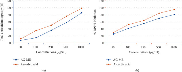
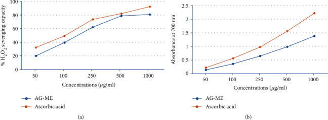
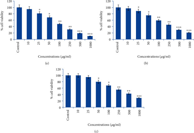
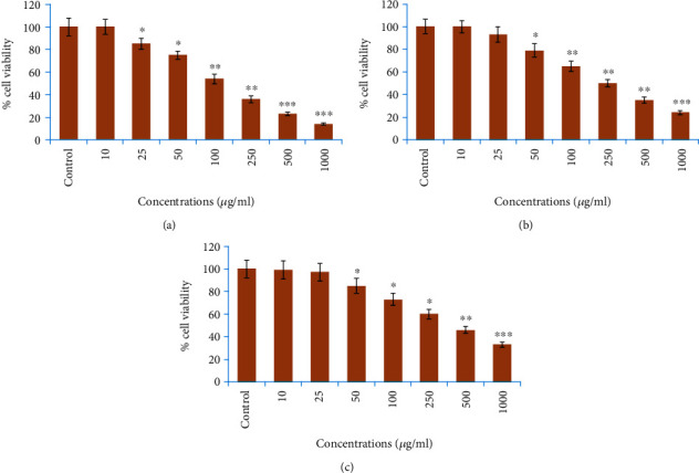
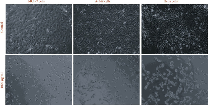
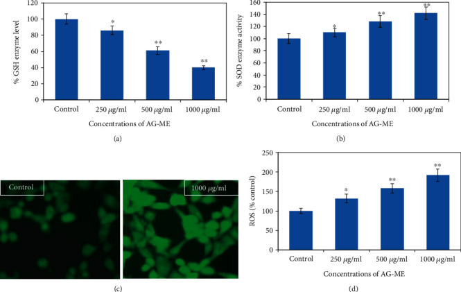
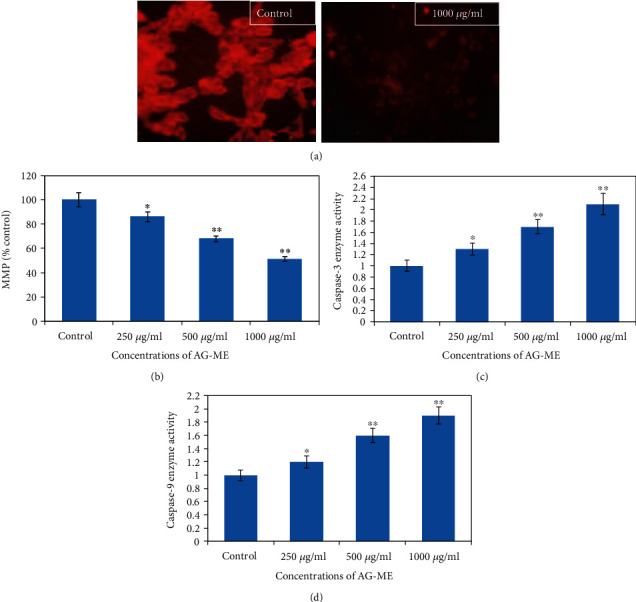
Anethum graveolens, belonging to the family Apiaceae, has been extensively used for medicinal and therapeutic purposes since long. Plants encompass rich number of effective constituents with less toxicity. Thus, nowadays, the attempts are being made to search plant constituents that can prevent and reverse the chronic diseases, such as cancer. In this study, an in vitro antioxidant and anticancer efficacies of Anethum graveolens (AG‐ME) were studied on human breast (MCF‐7), lung (A‐549), and cervical (HeLa) carcinoma cell lines. The antioxidant efficacies of AG‐ME were evaluated by total antioxidant, DPPH radical scavenging, H 2 O 2 scavenging, and ferrous reducing antioxidant assays. Further, the anticancer potential of AG‐ME was also determined against different cancer cell lines. The AG‐ME exhibited strong antioxidant activities as observed by antioxidant assays. AG‐ME also showed a dose‐dependent anticancer/cytotoxic potential against MCF‐7, A‐549, and HeLa cell lines. The AG‐ME‐induced reduction in GSH and increase in SOD activities indicates the role of oxidative stress in AG‐ME‐induced MCF‐7 cell death. The results also exhibited that AG‐ME triggered ROS production and significantly reduced MMP level. Moreover, a dose‐dependent increase in caspase‐3 and caspase‐9 activities suggests that the AG‐ME‐induced MCF‐7 cell death is caspase‐dependent. Together, the present study provides reasoning and reassurance for the uses of A. graveleons for medical purposes as an antioxidant and anticancer agent. Additional investigations are required to examine biological and anticancer activities under an in vivo system to discover a possible beneficial use of AG‐ME against diseases.
Overview
The claims and statistics of this paper haven’t been checked yet.
Run the check — free, no account needed. It reads the open-access text and checks the sources it cites.
Integrity signals
Registry
Cites
Statistics
Figures
Verification
Claims the paper makes
The claims and statistics of this paper haven’t been checked yet.
Run the check — free, no account needed. It reads the open-access text and checks the sources it cites.
Claims other papers make about this one
Each is checked against this paper’s own text: does it say what the citing paper says it does?
In the citing paper
“However, although the cytotoxic effects of A. graveolens products have been previously reported [20,22,23,24], these effects were usually described without the investigation of the underlying mechanisms of action.”
From Maceration, Soxhlet Extraction, and Steam Hydrodistillation of Anethum graveolens Seeds: Phytochemical Profiles and Cytotoxic Effects (2026) · cites this paper as [20]
In the citing paper
“Similarly, mechanistic studies in MCF-7 cells indicated that the A. graveolens methanolic extract increased ROS levels, altered the mitochondrial membrane potential and activated caspases-3 and -9 [20].”
From Maceration, Soxhlet Extraction, and Steam Hydrodistillation of Anethum graveolens Seeds: Phytochemical Profiles and Cytotoxic Effects (2026) · cites this paper as [20]
In the citing paper
“For example, a methanolic dill extract was reported to exert dose-dependent cytotoxic effects in MCF-7, A549 and HeLa cells [20]; similarly, Mohammed et al. revealed that an ethyl acetate extract fraction of dill seeds was able to inhibit HepG2 cell proliferation [47].”
From Maceration, Soxhlet Extraction, and Steam Hydrodistillation of Anethum graveolens Seeds: Phytochemical Profiles and Cytotoxic Effects (2026) · cites this paper as [20]
In the citing paper
“Mohammed et al. calculated an IC50 of 0.393 mg/mL for HepG2 cells in the case of ethyl acetate extract fraction of dill [47], while Al-Oqail et al. reported three different values of IC50 for specific cell lines, given by A. graveolens methanolic extract: IC50 of 104 μg/mL for MCF-7, 122 μg/mL for A-549, and 156 μg/mL for HeLa cells [20].”
From Maceration, Soxhlet Extraction, and Steam Hydrodistillation of Anethum graveolens Seeds: Phytochemical Profiles and Cytotoxic Effects (2026) · cites this paper as [20]
In the citing paper
“As mentioned previously, both AGEO and A. graveolens L. extracts have shown promising cytotoxic effects in multiple cell lines [20,22,23,24].”
From Maceration, Soxhlet Extraction, and Steam Hydrodistillation of Anethum graveolens Seeds: Phytochemical Profiles and Cytotoxic Effects (2026) · cites this paper as [20]
1 more claim about this paper
Create a free account to see every claim other papers make about this one.
Self-check
Percentages, sums and reported differences re-computed from the paper’s own numbers.
The self-check needs the paper’s full text.
Citations
Cites
Anethum graveolens: An Indian traditional medicinal herb and spice
Shekhawat, G., & Jana, S. (2010) · Pharmacognosy Reviews · doi:10.4103/0973-7847.70915 · Cited by 269
Research works printed but not linked: 64
- 10.4103/0973-7847.70902 · DOI · Paper page
- 10.1007/s12291-014-0446-0 · DOI · Paper page
- 10.1016/0748-5514(85)90140-0 · DOI
- 10.1186/s12937-016-0186-5 · DOI · Paper page
- 10.1007/978-3-540-74603-4 · DOI
- 10.7324/japs.2015.50110 · DOI
- 10.1021/jf0207530 · DOI
- 10.1093/ajcn/78.3.517s · DOI
- 10.4103/1687-4293.123791 · DOI
- 10.4103/0973-1296.62901 · DOI
- 10.1007/s11694-020-00571-5 · DOI
- 10.3390/molecules201219753 · DOI · Paper page
- 10.1007/s11694-018-9952-8 · DOI
- 10.1007/s11694-019-00227-z · DOI
- 10.1111/jfbc.12782 · DOI · Paper page
- Naseri M., 2012, The study of anti-inflammatory activity of oil-based dill (Anethum graveolens L.) extract used topically in formalin-induced inflammation male rat paw, Iran. J. Pharm. Res, 11, 1169
- 10.1155/2016/1098916 · DOI · Paper page
- 10.1080/03235408.2016.1243999 · DOI · Paper page
- 10.1016/j.jsps.2019.09.001 · DOI · Paper page
- 10.1016/j.foodchem.2008.12.039 · DOI
- 10.1080/10942910903474401 · DOI · Paper page
- 10.1016/S0098-2997(00)00007-8 · DOI
- 10.4314/tjpr.v14i11.6 · DOI
- 10.1016/j.anres.2016.07.007 · DOI
- 10.2478/s11756-020-00535-6 · DOI
- Devasagayam T. P., 2002, Immune system and antioxidants, especially those derived from Indian medicinal plants, Indian Journal of Experimental Biology, 40, 639
- 10.4103/0973-1296.185708 · DOI
- 10.1007/s10616-016-0048-1 · DOI
- 10.1186/1756-0500-6-24 · DOI
- 10.1038/1811199a0 · DOI
- 10.1016/j.foodchem.2008.04.071 · DOI
- 10.1155/2016/6392656 · DOI · Paper page
- 10.2478/s11756-019-00222-1 · DOI
- 10.4103/pm.pm_163_18 · DOI
- 10.1016/j.mam.2011.10.006 · DOI
- 10.1155/2014/761264 · DOI
- 10.3390/molecules15107313 · DOI
- 10.1080/10715760290006411 · DOI
- 10.1016/j.foodchem.2005.07.058 · DOI
- 10.1007/s11694-019-00114-7 · DOI
- Chekuri S., 2018, Radical scavenging activity (2, 2-diphenyl-1-picrylhydrazyl) of acalypha indica linn. (Euphorbeace family), International Journal of Pharmaceutical Sciences and Research, 9, 313
- 10.1021/jf0502698 · DOI
- 10.1111/1541-4337.12204 · DOI
- Aliyu A. B., 2013, Free radical scavenging and total antioxidant capacity of root extracts of Anchomanes difformis Engl.(Araceae), Acta Poloniae Pharmaceutica, 70, 115
- 10.1016/j.bjbas.2016.12.003 · DOI
- Oshaghi E. A., 2016, Aqueous extract of Anethum Graveolens L. has potential antioxidant and antiglycation effects, Iran. J. Med. Sci, 41, 328
- 10.1021/bc200141h · DOI
- 10.1021/ja303372u · DOI
- 10.3390/plants8040096 · DOI
- 10.1016/S0278-6915(99)00096-4 · DOI
- Salmanian S., 2014, Phenolic content, antiradical, antioxidant, and antibacterial properties of hawthorn (Crataegus elbursensis) seed and pulp extract, Journal of Agricultural Science and Technology, 16, 343
- 10.5772/intechopen.71923 · DOI
- 10.1007/BF01404408 · DOI
- 10.1007/BF01666038 · DOI
- Sharopov F. S., 2013, Composition and bioactivity of the essential oil of Anethum graveolens L. from Tajikistan, Int. J. Med. Arom. Plants, 3, 125
- 10.1016/S0378-8741(02)00116-2 · DOI
- 10.3390/ijms20133238 · DOI
- 10.1186/1472-6882-12-220 · DOI
- 10.1074/jbc.M203842200 · DOI
- 10.1016/j.bbamcr.2016.09.012 · DOI · Paper page
- 10.1038/74994 · DOI · Paper page
- 10.1155/2015/756482 · DOI
- 10.1186/s12906-020-2858-0 · DOI
- 10.1248/cpb.50.501 · DOI
Cited by
Hadi, N., Drioiche, A., Bouchra, E. M., Baammi, S., Abdelaziz Shahat, A., Tagnaout, I., Radi, M., Remok, F., Bouzoubaa, A., & Zair, T. (2024) · Pharmaceuticals · doi:10.3390/ph17070862 · Cited by 30
Goldiș, C., Racoviceanu, R., Negrea-Ghiulai, R., Mioc, A., Prodea, A., Atyim, E., Maksimovic, T., Lukinich-Gruia, A. T., Pricop, M.-A., & Șoica, C. (2026) · Molecules · doi:10.3390/molecules31183337
Metadata
- Authors
- Al-Oqail, M. M., & Farshori, N. N.
- ORCID
- Farshori, Nida N.
- Venue
- BioMed Research International
- Publisher
- Wiley
- Published
- 2021-01
- Volume
- 2021
- Issue
- 1
- ISSN
- 2314-6133
- 2314-6141
- PMC
- PMC8112917
- Cited by
- 18
- References
- 1
- Language
- en
- Funders
- King Saud University
About these checks
Registry, retraction, Cites and statistics results are computed, not judged, with one exception: a printed reference that no index lists is read once by a model before it counts as a citation error. Claims are extracted by a model and kept only when their quote appears verbatim in the text; each cited source is then checked, with a model’s help, for whether it says what the claim relies on. Those verdicts can be wrong — read the claim’s quote and the cited source before relying on one.
Statistics and arithmetic are recomputed, not judged.
Confidence bands are shown only for fields calibrated on our annotated test set. None is calibrated yet, so no band is shown.
What counts as a claim ›How we measure confidence ›How we check this ›
Are you an author, or do you see a problem with this page? Request a review
Data sources
Bibliographic data: Semantic Scholar, OpenAlex, Crossref.
Contains information from the Semantic Scholar Academic Graph Datasets, made available under the ODC Attribution License (ODC-BY 1.0).
Semantic Scholar’s data is described in: Kinney, R., et al. (2023). The Semantic Scholar Open Data Platform. arXiv:2301.10140.
