

The DOI matches its Crossref record; 58 of the 60 references it cites were found in a registry or an index.
Towards the creation of decellularized organ constructs using irreversible electroporation and active mechanical perfusion
Open accessAbstract
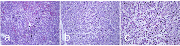
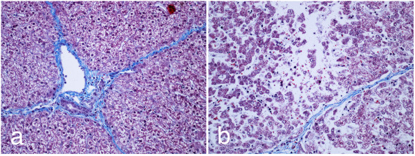
Background: Despite advances in transplant surgery and general medicine, the number of patients awaiting transplant organs continues to grow, while the supply of organs does not. This work outlines a method of organ decellularization using non-thermal irreversible electroporation (N-TIRE) which, in combination with reseeding, may help supplement the supply of organs for transplant. Methods: In our study, brief but intense electric pulses were applied to porcine livers while under active low temperature cardio-emulation perfusion. Histological analysis and lesion measurements were used to determine the effects of the pulses in decellularizing the livers as a first step towards the development of extracellular scaffolds that may be used with stem cell reseeding. A dynamic conductivity numerical model was developed to simulate the treatment parameters used and determine an irreversible electroporation threshold. Results: Ninety-nine individual 1000 V/cm 100-μs square pulses with repetition rates between 0.25 and 4 Hz were found to produce a lesion within 24 hours post-treatment. The livers maintained intact bile ducts and vascular structures while demonstrating hepatocytic cord disruption and cell delamination from cord basal laminae after 24 hours of perfusion. A numerical model found an electric field threshold of 423 V/cm under specific experimental conditions, which may be used in the future to plan treatments for the decellularization of entire organs. Analysis of the pulse repetition rate shows that the largest treated area and the lowest interstitial density score was achieved for a pulse frequency of 1 Hz. After 24 hours of perfusion, a maximum density score reduction of 58.5 percent had been achieved. Conclusions: This method is the first effort towards creating decellularized tissue scaffolds that could be used for organ transplantation using N-TIRE. In addition, it provides a versatile platform to study the effects of pulse parameters such as pulse length, repetition rate, and field strength on whole organ structures.
Overview
The claims and statistics of this paper haven’t been checked yet.
Run the check — free, no account needed. It reads the open-access text and checks the sources it cites.
Integrity signals
Registry
Cites
Statistics
Figures
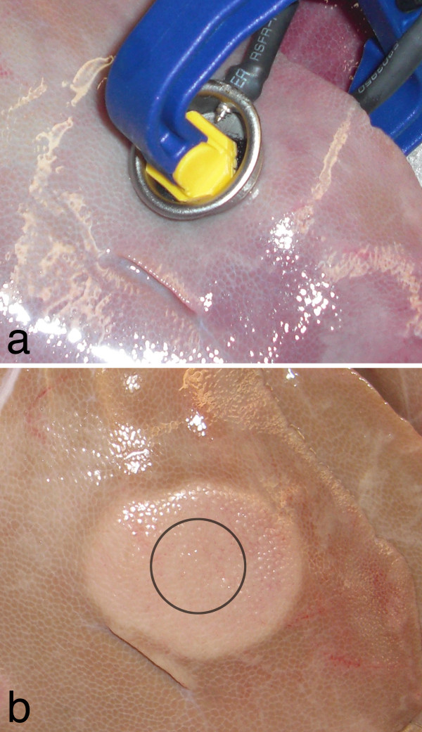
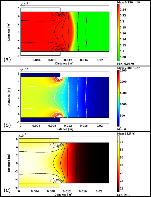
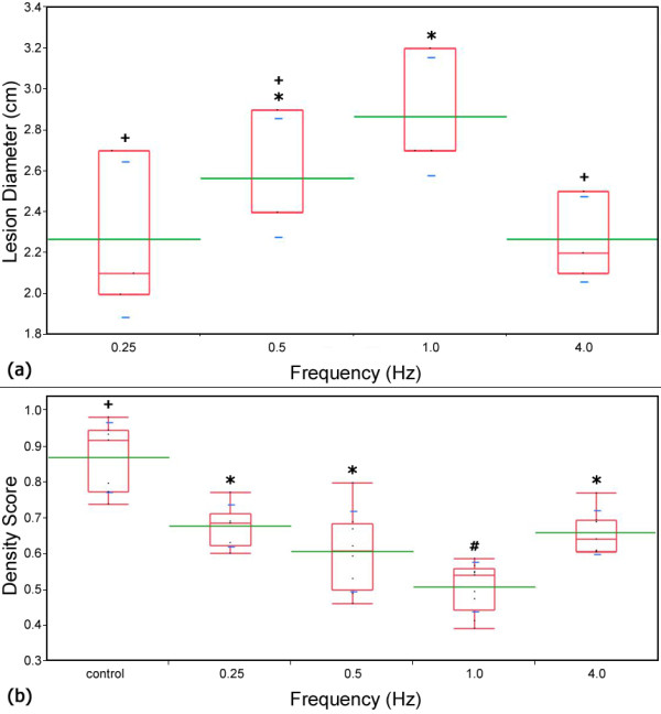
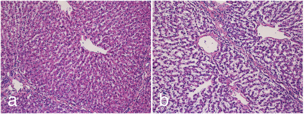
Verification
Claims the paper makes
The claims and statistics of this paper haven’t been checked yet.
Run the check — free, no account needed. It reads the open-access text and checks the sources it cites.
Claims other papers make about this one
Each is checked against this paper’s own text: does it say what the citing paper says it does?
The cited source says what the claim attributes to it.
In the citing paper
“Sano et al. utilized N-TIRE accompanied by lactated Ringer's solution perfusion through the portal vein and hepatic artery for porcine liver decellularization, facilitating the removal of cellular debris resulting from electroporation [139].”
From Liver tissue engineering using decellularized scaffolds: Current progress, challenges, and opportunities (2024) · cites this paper as [139]
Self-check
Percentages, sums and reported differences re-computed from the paper’s own numbers.
The self-check needs the paper’s full text.
Citations
Cites
Perfusion-decellularized matrix: using nature's platform to engineer a bioartificial heart
Ott, H. C., Matthiesen, T. S., Goh, S.-K., Black, L. D., Kren, S. M., Netoff, T. I., & Taylor, D. A. (2008) · Nature Medicine · doi:10.1038/nm1684 · Cited by 2,650
Soft Lithography in Biology and Biochemistry
Whitesides, G. M., Ostuni, E., Takayama, S., Jiang, X., & Ingber, D. E. (2001) · Annual Review of Biomedical Engineering · doi:10.1146/annurev.bioeng.3.1.335 · Cited by 2,603
Physical properties of tissue : a comprehensive reference book
Duck, F. A. (1990) · Cited by 1,675
Microscale technologies for tissue engineering and biology
Khademhosseini, A., Langer, R., Borenstein, J. T., & Vacanti, J. P. (2006) · Proceedings of the National Academy of Sciences · doi:10.1073/pnas.0507681102 · Cited by 1,553
Uygun, B. E., Soto-Gutierrez, A., Yagi, H., Izamis, M.-L., Guzzardi, M. A., Shulman, C., Milwid, J., Kobayashi, N., Tilles, A., Berthiaume, F., et al. (2010) · Nature Medicine · doi:10.1038/nm.2170 · Cited by 1,370
Tissue Ablation with Irreversible Electroporation
Davalos, R. V., Mir, L. M., & Rubinsky, B. (2005) · Annals of Biomedical Engineering · doi:10.1007/s10439-005-8981-8 · Cited by 1,296
Self-citationHematopoietic colony-forming cells derived from human embryonic stem cells
Kaufman, D. S., Hanson, E. T., Lewis, R. L., Auerbach, R., & Thomson, J. A. (2001) · Proceedings of the National Academy of Sciences · doi:10.1073/pnas.191362598 · Cited by 925
SELF-REGULATION OF GROWTH IN THREE DIMENSIONS
Folkman, J., & Hochberg, M. (1973) · The Journal of Experimental Medicine · doi:10.1084/jem.138.4.745 · Cited by 829
The use of whole organ decellularization for the generation of a vascularized liver organoid
Baptista, P. M., Siddiqui, M. M., Lozier, G., Rodriguez, S. R., Atala, A., & Soker, S. (2011) · Hepatology · doi:10.1002/hep.24067 · Cited by 681
Yamada, K., Yazawa, K., Shimizu, A., Iwanaga, T., Hisashi, Y., Nuhn, M. G., O'Malley, P., Nobori, S., Vagefi, P. A., Patience, C., et al. (2004) · Nature Medicine · doi:10.1038/nm1172 · Cited by 609
Research works printed but not linked: 4
- Fabre: Report of the British Transplantation Society Working Party on Organ Donation. 1995.
- Davalos RV: Irreversible Electroporation to Create Tissue Scaffolds. United States patent 2009.
- Davalos RV, Mir LM, Rubinsky B: Tissue Ablation with Irreversible Electroporation. Ann Biomed Eng 2005, 33: 223–231. 10.1007/s10439-005-8981-8 · DOI
- Edd JF, Horowitz L, Davalos RV, Mir LM, Rubinsky B: In Vivo Results of a New Focal Tissue Ablation Technique: Irreversible Electroporation. Ieee Transactions on Biomedical Engineering 2006, 53: 1409–1415. 10.1109/TBME.2006.873745 · DOI
Cited by
Sorted by citations
An overview of tissue and whole organ decellularization processes
Crapo, P. M., Gilbert, T. W., & Badylak, S. F. (2011) · Biomaterials · doi:10.1016/j.biomaterials.2011.01.057 · Cited by 3,591
Electroporation-based technologies for medicine: principles, applications, and challenges.
Yarmush, M., Golberg, A., Serša, G., Kotnik, T., & Miklavčič, D. (2014) · Annual Review of Biomedical Engineering · doi:10.1146/annurev-bioeng-071813-104622 · Cited by 813
Extracellular matrix-based materials for regenerative medicine
Hussey, G. S., Dziki, J. L., & Badylak, S. F. (2018) · Nature Reviews Materials · doi:10.1038/s41578-018-0023-x · Cited by 844
A Review of Basic to Clinical Studies of Irreversible Electroporation Therapy
Jiang, C., Davalos, R. V., & Bischof, J. C. (2014) · IEEE Transactions on Biomedical Engineering · doi:10.1109/tbme.2014.2367543 · Cited by 334
Self-citationBiocompatibility evaluation of tissue-engineered decellularized scaffolds for biomedical application
Hussein, K. H., Park, K.-M., Kang, K.-S., & Woo, H.-M. (2016) · Materials Science and Engineering: C · doi:10.1016/j.msec.2016.05.068 · Cited by 218
Aycock, K. N., & Davalos, R. V. (2019) · Bioelectricity · doi:10.1089/bioe.2019.0029 · Cited by 189
Self-citationDecellularization and Recellularization Technologies in Tissue Engineering
Fu, R., Wang, Y.-C., Liu, S., Shih, T.-R., Lin, H.-L., Chen, Y.-M., Sung, J.-H., Lu, C.-H., Wei, J.-R., Wang, Z.-W., et al. (2014) · Cell Transplantation · doi:10.3727/096368914x678382 · Cited by 165
Sabetkish, S., Kajbafzadeh, A.-M., Sabetkish, N., Khorramirouz, R., Akbarzadeh, A., Seyedian, S. L., Pasalar, P., Orangian, S., Beigi, R. S. H., Aryan, Z., et al. (2015) · Journal of Biomedical Materials Research Part A · doi:10.1002/jbm.a.35291 · Cited by 152
Decellularized extracellular matrix mediates tissue construction and regeneration
Liu, C., Pei, M., Li, Q., & Zhang, Y. (2021) · Frontiers of Medicine · doi:10.1007/s11684-021-0900-3 · Cited by 151
Preparation and Use of Decellularized Extracellular Matrix for Tissue Engineering
McInnes, A. D., Moser, M., & Chen, D. (2022) · Journal of Functional Biomaterials · doi:10.3390/jfb13040240 · Cited by 146
Liao, J., Xu, B., Zhang, R., Fan, Y., Xie, H., & Li, X. (2020) · Journal of Materials Chemistry B · doi:10.1039/d0tb01534b · Cited by 133
Sano, M. B., Arena, C. B., DeWitt, M. R., Saur, D., & Davalos, R. V. (2014) · Bioelectrochemistry · doi:10.1016/j.bioelechem.2014.07.010 · Cited by 125
Self-citationA Three-Dimensional In Vitro Tumor Platform for Modeling Therapeutic Irreversible Electroporation
Arena, C. B., Szot, C. S., Garcia, P. A., Rylander, M. N., & Davalos, R. V. (2012) · Biophysical Journal · doi:10.1016/j.bpj.2012.09.017 · Cited by 112
Self-citationEmerging Technologies for Pulmonary Vein Isolation
Ramirez, F. D., Reddy, V. Y., Viswanathan, R., Hocini, M., & Jaı̈s, P. (2020) · Circulation Research · doi:10.1161/circresaha.120.316402 · Cited by 113
Yao, C., Dong, S., Zhao, Y., Lv, Y., Liu, H., Gong, L., Ma, J., Wang, H., & Sun, Y. (2017) · IEEE Transactions on Biomedical Engineering · doi:10.1109/tbme.2017.2690624 · Cited by 94
Contribution of Physical Methods in Decellularization of Animal Tissues
Rabbani, M., Zakian, N., & Alimoradi, N. (2021) · Journal of Medical Signals & Sensors · doi:10.4103/jmss.jmss_2_20 · Cited by 87
Barbulescu, G. I., Bojin, F. M., Ordodi, V. L., Goje, I.-D., Barbulescu, A. S., & Păunescu, V. (2022) · International Journal of Molecular Sciences · doi:10.3390/ijms232113040 · Cited by 89
Graybill, P. M., & Davalos, R. V. (2020) · Cancers · doi:10.3390/cancers12051132 · Cited by 84
Self-citationStrategies for development of decellularized heart valve scaffolds for tissue engineering
Snyder, Y., & Jana, S. (2022) · Biomaterials · doi:10.1016/j.biomaterials.2022.121675 · Cited by 74
In Vitro Cell Selectivity of Reversible and Irreversible
Hunter, D. W., Kostecki, G., Fish, J. M., Jensen, J. A., & Tandri, H. S. (2021) · Circulation Arrhythmia and Electrophysiology · doi:10.1161/circep.120.008817 · Cited by 69
Sano, M. B., Fan, R. E., & Xing, L. (2017) · Scientific Reports · doi:10.1038/srep40747 · Cited by 69
Self-citationAgnass, P., van Veldhuisen, E., van Gemert, M. J. C., van der Geld, C. W. M., van Lienden, K. P., van Gulik, T. M., Meijerink, M. R., Besselink, M. G., Kok, H. P., & Crezee, J. (2020) · International Journal of Hyperthermia · doi:10.1080/02656736.2020.1753828 · Cited by 66
Khan, A. A., Vishwakarma, S. K., Bardia, A., & Venkateshwarulu, J. (2014) · Journal of Artificial Organs · doi:10.1007/s10047-014-0780-2 · Cited by 59
Decellularized blood vessel development: Current state-of-the-art and future directions
Wang, X., Chan, V., & Corridon, P. R. (2022) · Frontiers in Bioengineering and Biotechnology · doi:10.3389/fbioe.2022.951644 · Cited by 64
Zhang, Z., Li, W., Procissi, D., Tyler, P. D., Omary, R. A., & Larson, A. C. (2013) · Nanomedicine · doi:10.2217/nnm.13.72 · Cited by 59
Tan, J., Zhang, Q.-Y., Huang, L.-P., Huang, K., & Xie, H.-Q. (2021) · Biomaterials Science · doi:10.1039/d1bm00470k · Cited by 57
Irreversible Electroporation: An In Vivo Study with Dorsal Skin Fold Chamber
Qin, Z., Jiang, J., Long, G., Lindgren, B. R., & Bischof, J. C. (2012) · Annals of Biomedical Engineering · doi:10.1007/s10439-012-0686-1 · Cited by 55
Miklovic, T., Latouche, E. L., DeWitt, M. R., Davalos, R. V., & Sano, M. B. (2017) · Annals of Biomedical Engineering · doi:10.1007/s10439-017-1889-2 · Cited by 54
Self-citationMethods to generate tissue-derived constructs for regenerative medicine applications
Paulo Zambon, J., Atala, A., & Yoo, J. J. (2020) · Methods · doi:10.1016/j.ymeth.2019.09.016 · Cited by 51
Wendler, J. J., Pech, M., Blaschke, S., Porsch, M., Janitzky, A., Ülrich, M., Dudeck, O., Ricke, J., & Liehr, U. B. (2011) · CardioVascular and Interventional Radiology · doi:10.1007/s00270-011-0187-x · Cited by 44
Nerve regeneration using decellularized tissues: challenges and opportunities
Mahdian, M., Tabatabai, T. S., Abpeikar, Z., Rezakhani, L., & Khazaei, M. (2023) · Frontiers in Neuroscience · doi:10.3389/fnins.2023.1295563 · Cited by 43
Rat liver regeneration following ablation with irreversible electroporation
Golberg, A., Bruinsma, B. G., Jaramillo, M., Yarmush, M. L., & Uygun, B. E. (2016) · PeerJ · doi:10.7717/peerj.1571 · Cited by 38
Preparation of a three-dimensional extracellular matrix by decellularization of rabbit livers
Nari, G., Cid, M. P., Comín, R., Reyna, L., Juri, G., Taborda, R., & Salvatierra, N. A. (2013) · Revista Española de Enfermedades Digestivas · doi:10.4321/s1130-01082013000300004 · Cited by 36
Membrane-Targeting Approaches for Enhanced Cancer Cell Destruction with Irreversible Electroporation
Jiang, C., Qin, Z., & Bischof, J. C. (2013) · Annals of Biomedical Engineering · doi:10.1007/s10439-013-0882-7 · Cited by 33
Decellularized Cell‐Secreted Extracellular Matrices as Biomaterials for Tissue Engineering
Ramos‐Rodriguez, D. H., & Leach, J. K. (2024) · Small Science · doi:10.1002/smsc.202400335 · Cited by 35
Sano, M. B., DeWitt, M. R., Teeter, S. D., & Xing, L. (2018) · Computers in Biology and Medicine · doi:10.1016/j.compbiomed.2018.02.009 · Cited by 33
Self-citationCharacterization of Irreversible Electroporation Ablation with a Validated Perfused Organ Model
Bhonsle, S. P., Bonakdar, M., Neal, R. E., Aardema, C. H., Robertson, J. L., Howarth, J., Kavnoudias, H., Thomson, K. R., Goldberg, S. N., & Davalos, R. V. (2016) · Journal of Vascular and Interventional Radiology · doi:10.1016/j.jvir.2016.07.012 · Cited by 32
Self-citationJiang, C., Shao, Q., & Bischof, J. C. (2014) · Annals of Biomedical Engineering · doi:10.1007/s10439-014-1133-2 · Cited by 32
Skin regeneration with all accessory organs following ablation with irreversible electroporation
Golberg, A., Villiger, M., Broelsch, G. F., Quinn, K. P., Albadawi, H., Khan, S. I., Watkins, M. T., Georgakoudi, I., Austen, W. G., Bei, M., et al. (2016) · Journal of Tissue Engineering and Regenerative Medicine · doi:10.1002/term.2374 · Cited by 31
Yao, C., Lv, Y., Zhao, Y., Dong, S., Liu, H., & Ma, J. (2017) · Scientific Reports · doi:10.1038/s41598-017-15494-3 · Cited by 31
Sano, M. B., Fan, R. E., Hwang, G. L., Sonn, G. A., & Xing, L. (2016) · Journal of Vascular and Interventional Radiology · doi:10.1016/j.jvir.2016.05.032 · Cited by 31
Self-citationChang, T. T., Zhou, V. X., & Rubinsky, B. (2017) · BioTechniques · doi:10.2144/000114547 · Cited by 30
Hussein, K. H., Ahmadzada, B., Correa, J. C., Sultan, A., Wilken, S., Amiot, B., & Nyberg, S. L. (2024) · Bioactive Materials · doi:10.1016/j.bioactmat.2024.06.001 · Cited by 28
“…the electric field surpasses a relatively high threshold (e.g., 700 V/cm for eight 100 μ s pulses at 1 Hz in the liver), the cell membrane undergoes permanent changes in permeability, ultimately leading to cellular death, a process known as non-thermal irreversible electroporation (N-TIRE) [139].”
BackgroundMethodCasciola, M., Keck, D. M., Feaster, T. K., & Blinova, K. (2022) · Physiological Reports · doi:10.14814/phy2.15493 · Cited by 27
Kakabadze, A., & Kakabadze, Z. (2015) · Transplantation Proceedings · doi:10.1016/j.transproceed.2014.09.181 · Cited by 27
Campelo, S. N., Massimo Valerio, A., Ahmed, H. U., Hu, Y., Arena, S. L., Neal, R. E., Emberton, M., & Arena, C. B. (2017) · APL Bioengineering · doi:10.1063/1.5005828 · Cited by 27
Self-citationLiu, H., Xing, F., Yu, P., Lu, R., Ma, S., Shakya, S., Xiang, Z., Peng, K., Zhang, D., & Liu, M. (2024) · Materials & Design · doi:10.1016/j.matdes.2024.113072 · Cited by 31
Dermol‐Černe, J., & Damijan Miklavčič, A. (2017) · IEEE Transactions on Biomedical Engineering · doi:10.1109/tbme.2017.2773126 · Cited by 26
High-Performance Acellular Tissue Scaffold Combined with Hydrogel Polymers for Regenerative Medicine
Lee, E., Kim, H. J., Shaker, M. R., Ryu, J. R., Ham, M. S., Seo, S. H., Kim, D. H., Lee, K., Jung, N., Choe, Y., et al. (2019) · ACS Biomaterials Science & Engineering · doi:10.1021/acsbiomaterials.9b00219 · Cited by 24
Liver scaffolds obtained by decellularization: A transplant perspective in liver bioengineering
Dias, M. L., Paranhos, B. A., & Goldenberg, R. C. D. S. (2022) · Journal of Tissue Engineering · doi:10.1177/20417314221105305 · Cited by 23
Irreversible Electroporation in Liver Cancers and Whole Organ Engineering
Saini, A., Breen, I. D., Alzubaidi, S. J., Pershad, Y., Sheth, R. A., Naidu, S., Knuttinen, M. G., Albadawi, H., & Öklü, R. (2018) · Journal of Clinical Medicine · doi:10.3390/jcm8010022 · Cited by 26
Kidney bioengineering in regenerative medicine: An emerging therapy for kidney disease
Lin, Y.-Q., Wang, L., Pan, L., Wang, H., Zhu, G., Liu, W., Wang, J., Braddock, M., & Zheng, M. (2015) · Cytotherapy · doi:10.1016/j.jcyt.2015.10.004 · Cited by 22
Mahnič-Kalamiza, S., Kotnik, T., & Damijan Miklavčič, A. (2012) · BMC Medical Education · doi:10.1186/1472-6920-12-102 · Cited by 20
Rolong, A., Davalos, R. V., & Rubinsky, B. (2017) · Springer eBooks · doi:10.1007/978-3-319-55113-5_2 · Cited by 19
Self-citationMußbach, F., Settmacher, U., Dirsch, O., Xie, C., & Dahmen, U. (2016) · European Surgical Research · doi:10.1159/000446211 · Cited by 18
Shevchuk, O. I., Korcheva, V., Moskalenko, N. S., Kyryk, V. M., Kot, K., & Krasnienkov, D. S. (2025) · Frontiers in Bioengineering and Biotechnology · doi:10.3389/fbioe.2025.1621641 · Cited by 20
Comprehensive review on thermal aspects of nonthermal irreversible electroporation
Wijayanta, A. T., & Kurata, K. (2023) · Heat Transfer · doi:10.1002/htj.22880 · Cited by 17
Assessment of ex vivo perfused liver health by Raman spectroscopy
Zu, T. N. K., Athamneh, A. I. M., Collakova, E., Robertson, J. L., Hawken, T. N., Aardema, C. H., & Senger, R. S. (2015) · Journal of Raman Spectroscopy · doi:10.1002/jrs.4688 · Cited by 16
Self-citationSoltanmohammadi, F., Gharehbaba, A. M., Alizadeh, E., & Javadzadeh, Y. (2024) · International Journal of Biological Macromolecules · doi:10.1016/j.ijbiomac.2024.138893 · Cited by 15
Computational and experimental model of electroporation for human aorta
Filipović, N., Saveljic, I. B., Jovicic, N. U., Tanasković, I., & Zdravkovic, N. D. (2016) · PubMed · doi:10.5277/abb-00444-2015-02 · Cited by 15
Sano, M. B., Petrella, R. A., Kaufman, J. D., Fesmire, C. C., Xing, L., Gerber, D. A., & FOGLE, C. A. (2020) · Computers in Biology and Medicine · doi:10.1016/j.compbiomed.2020.103807 · Cited by 15
Self-citationHan, Y., Zhang, B., Li, J., Cen, L., Zhao, L., & Xi, Z. (2023) · Bioresources and Bioprocessing · doi:10.1186/s40643-022-00621-4 · Cited by 16
Histological and Mathematical Analysis of the Irreversibly Electroporated Liver Tissue
Sung, C. K., Kim, H. B., Jung, J. H., Baik, K. Y., Moon, K.-W., Kim, H.-S., Yi, J., & Chung, J. H. (2016) · Technology in Cancer Research & Treatment · doi:10.1177/1533034616640642 · Cited by 14
Zhang, B., Liu, F., Fang, Z. G., Ding, L., Moser, M., & Zhang, W. (2021) · International Journal of Hyperthermia · doi:10.1080/02656736.2021.1912417 · Cited by 13
Decellularized Green and Brown Macroalgae as Cellulose Matrices for Tissue Engineering
Berry-Kilgour, C., Oey, I., Cabral, J. D., Dowd, G. C., & Wise, L. M. (2024) · Journal of Functional Biomaterials · doi:10.3390/jfb15120390 · Cited by 12
Soltanmohammadi, F., Gharehbaba, A. M., & Javadzadeh, Y. (2025) · Biomedicine & Pharmacotherapy · doi:10.1016/j.biopha.2025.118200 · Cited by 14
Khorasani, A. (2020) · Polish Journal of Medical Physics And Engineering · doi:10.2478/pjmpe-2020-0008 · Cited by 12
Numerical mesoscale tissue model of electrochemotherapy in liver based on histological findings
Cindrič, H., Gašljević, G., Edhemović, I., Brecelj, E., Žmuc, J., Čemažar, M., Seliškar, A., Damijan Miklavčič, A., & Kos, B. (2022) · Scientific Reports · doi:10.1038/s41598-022-10426-2 · Cited by 12
Lopez-Ichikawa, M. A., Vu, N. K., Nijagal, A., Rubinsky, B., & Chang, T. T. (2021) · Scientific Reports · doi:10.1038/s41598-021-94016-8 · Cited by 11
Vishwakarma, S. K., Lakkireddy, C., Bardia, A., Paspala, S. A. B., Tripura, C., Habeeb, M. A., & Khan, A. A. (2018) · World Journal of Hepatology · doi:10.4254/wjh.v10.i11.822 · Cited by 9
The Effect of Small Intestine Heterogeneity on Irreversible Electroporation Treatment Planning
Phillips, M. (2014) · Journal of Biomechanical Engineering · doi:10.1115/1.4027815 · Cited by 8
Impact of Pulsed Electric Field Ablation on His Bundle Conduction: A Preclinical Canine Study
Zhai, Z., Wang, Y., Shi, L., & Liu, X. (2024) · Medical Science Monitor · doi:10.12659/msm.945007 · Cited by 7
RetractedKhorasani, A., Firoozabadi, S. M., & Shankayi, Z. (2019) · Polish Journal of Medical Physics And Engineering · doi:10.2478/pjmpe-2019-0031 · Cited by 6
The Effect of Conductivity Changes on Temperature Rise During Irreversible Electroporation
Khorasani, A. (2020) · Frontiers in biomedical technologies · doi:10.18502/fbt.v7i3.4620 · Cited by 6
Application of pulsed electric field technology to skin engineering
Berry-Kilgour, C., Wise, L., King, J., & Oey, I. (2024) · Frontiers in Bioengineering and Biotechnology · doi:10.3389/fbioe.2024.1386725 · Cited by 6
Zhai, Z., Ling, Y., Wang, Y., Shi, L., & Liu, X. (2024) · Frontiers in Cardiovascular Medicine · doi:10.3389/fcvm.2024.1426920 · Cited by 5
„Liver engineering“ als neue Quelle von Spenderorganen
Mußbach, F., Dahmen, U., Dirsch, O., & Settmacher, U. (2015) · Der Chirurg · doi:10.1007/s00104-015-0015-y · Cited by 5
Decellularized dermal tissue substitutes
Chaves, C., Alshomer, F., Alhujayri, A. K., & Kalaskar, D. M. (2019) · Elsevier eBooks · doi:10.1016/b978-0-08-102546-8.00004-2 · Cited by 5
Arena, C. B., Neal, R. E., & Davalos, R. V. (2014) · Elsevier eBooks · doi:10.1016/b978-0-444-53632-7.01016-9 · Cited by 5
Self-citationCindrič, H., Kos, B., & Damijan Miklavčič, A. (2021) · Slovenian Medical Journal · doi:10.6016/zdravvestn.2954 · Cited by 5
Sano, M. B., & DeWitt, M. R. (2020) · Bioelectricity · doi:10.1089/bioe.2020.0023 · Cited by 4
Self-citationOverview of Decellularized Materials for Tissue Repair and Organ Replacement
Liao, J., Guo, Q., Xu, B., & Li, X. (2021) · Springer eBooks · doi:10.1007/978-981-33-6962-7_1 · Cited by 4
Vera-Tizatl, A. L., van der Hee, R., Cornelissen, J. J. L. M., Vera-Tizatl, C. E., Abayazid, M., & Fütterer, J. J. (2023) · Bioengineering & Translational Medicine · doi:10.1002/btm2.10607 · Cited by 4
Wang, Y., Ma, R., Huang, Z., Zhou, Y., Wang, K., Xiao, Z., Guo, Q., Yang, D., Han, M., Shen, S., et al. (2023) · Computers in Biology and Medicine · doi:10.1016/j.compbiomed.2023.107824 · Cited by 4
Thermal Considerations with Tissue Electroporation
O’Brien, T. J., Arena, C. B., & Davalos, R. V. (2017) · Springer eBooks · doi:10.1007/978-3-319-32003-8_68-1 · Cited by 4
Self-citationStrategies for decellularization, re-cellularization and crosslinking in liver bioengineering
Wang, J., & Jin, X. (2024) · The International Journal of Artificial Organs · doi:10.1177/03913988231218566 · Cited by 3
The Decellularization of Tissues
Cao, G., & Li, X. (2021) · Springer eBooks · doi:10.1007/978-981-33-6962-7_2 · Cited by 3
Lee, J. G., Bak, S. Y., Nahm, J. H., Lee, S. W., Min, S. O., & Kim, K. S. (2015) · Korean Journal of Hepato-Biliary-Pancreatic Surgery · doi:10.14701/kjhbps.2015.19.2.47 · Cited by 3
Khorasani, A. (2022) · Electromagnetic Biology and Medicine · doi:10.1080/15368378.2022.2114493 · Cited by 3
Liver tissue remodeling following ablation with irreversible electroporation in a porcine model
Monleón, E., Lucía, Ó., Güemes, A., López-Alonso, B., del Amo, M. D. A., Sarnago, H., Hernaez, A., Burdio, J. M., & Junquera, C. (2022) · Frontiers in Veterinary Science · doi:10.3389/fvets.2022.1014648 · Cited by 3
Mechanical Properties of the Extracellular Matrix
Sanz-Fraile, H., & Otero, J. (2024) · Springer eBooks · doi:10.1007/978-3-031-56363-8_5 · Cited by 3
Mechanical Properties of the Extracellular Matrix
Sanz-Fraile, H., & Otero, J. (2023) · Springer eBooks · doi:10.1007/978-3-030-92090-6_5-1 · Cited by 2
Conductivity Variation during Irreversible Electroporation
Xu, C., & Yang, L. (2021) · E3S Web of Conferences · doi:10.1051/e3sconf/202127101032 · Cited by 2
Wang, Z., Li, Y., Liang, M., Sun, J., Zhang, J., Xu, L., & Han, Y. (2025) · Frontiers in Physiology · doi:10.3389/fphys.2025.1632680 · Cited by 2
Tissue Engineering with Electroporation
Ho, M. P. (2016) · Springer eBooks · doi:10.1007/978-3-319-26779-1_49-1 · Cited by 2
Gong, L., Yao, C., Dong, S., & Zhao, Y. (2017) · IEEE International Pulsed Power Conference (PPC) · doi:10.1109/ppc.2017.8291223 · Cited by 2
ON TISSUE ENGINEERING OF PIG, HUMAN, AND NON-HUMAN PRIMATE TISSUES
Nayakawde, N. B. (2020) · Gothenburg University Publications Electronic Archive (Gothenburg University) · doi:10.13140/rg.2.2.31294.72008 · Cited by 2
Research Progress on the Application of Irreversible Electroporation Ablation in Cancers
Zhang, R., Xie, L., Jin, Q., Zhang, C., Guo, T., Zhao, Q., & Jiang, T. (2025) · Advanced ultrasound in diagnosis and therapy · doi:10.26599/audt.2025.240052 · Cited by 1
Thermal Considerations with Tissue Electroporation
O’Brien, T. J., Arena, C. B., & Davalos, R. V. (2018) · Springer eBooks · doi:10.1007/978-3-319-26695-4_68 · Cited by 1
Self-citationExtracellular Matrix Specification of Regenerative Cells in the Adult Lung
Burgess, J. K., & Tjin, G. (2015) · Stem cell biology and regenerative medicine · doi:10.1007/978-3-319-21082-7_11 · Cited by 1
Metadata
- Authors
- Sano, M. B., Neal, R. E., Garcia, P. A., Gerber, D., Robertson, J., & Davalos, R. V.
- Affiliations
- Virginia Tech - Wake Forest University School of Biomedical Engineering & Sciences (US)
- University of North Carolina at Chapel Hill (US)
- Virginia–Maryland College of Veterinary Medicine (US)
- Corresponding author
- Davalos, Rafael V
- Venue
- BioMedical Engineering OnLine
- Published
- 2010-12
- Volume
- 9
- Issue
- 1
- Pages
- 83-83
- ISSN
- 1475-925X (ISSN-L)
- PMC
- PMC3018380
- OpenAlex
- W2024731442
- Cited by
- 117
- References
- 60
- Citations by year
- 2026: 1 · 2025: 4 · 2024: 11 · 2023: 8 · 2022: 9 · 2021: 10 · 2020: 9 · 2019: 6 · 2018: 5 · 2017: 11 · 2016: 15 · 2015: 6 · 2014: 9 · 2013: 5 · 2012: 4
- FWCI
- 4.39
- Type
- journal article
- Language
- en
- Open access
- gold
- doi.org
- Locations
- BioMedical Engineering OnLine (publishedVersion, cc-by)
- PubMed Central (publishedVersion, cc-by)
- PubMed (publishedVersion)
- DOAJ (DOAJ: Directory of Open Access Journals) (submittedVersion)
- VTechWorks (Virginia Tech) (submittedVersion, cc-by)
- Topics
- Health Sciences › Medicine › Surgery › Tissue Engineering and Regenerative Medicine
- Life Sciences › Biochemistry, Genetics and Molecular Biology › Biotechnology › Microbial Inactivation Methods
- Physical Sciences › Engineering › Biomedical Engineering › 3D Printing in Biomedical Research
- Keywords
- irreversible electroporation, porcine liver, extracellular matrix scaffolds, threshold electric field, pulse repetition rate, organ transplantation
- MeSH terms
- Animals, Humans, Liver, Perfusion, Swine, Electroporation, Tissue Engineering, Tissue Scaffolds, Mechanical Phenomena
About these checks
Registry, retraction, Cites and statistics results are computed, not judged, with one exception: a printed reference that no index lists is read once by a model before it counts as a citation error. Claims are extracted by a model and kept only when their quote appears verbatim in the text; each cited source is then checked, with a model’s help, for whether it says what the claim relies on. Those verdicts can be wrong — read the claim’s quote and the cited source before relying on one.
Statistics and arithmetic are recomputed, not judged.
Confidence bands are shown only for fields calibrated on our annotated test set. None is calibrated yet, so no band is shown.
What counts as a claim ›How we measure confidence ›How we check this ›
Are you an author, or do you see a problem with this page? Request a review
