

The DOI matches its Crossref record; 44 of the 55 references it cites were found in a registry or an index.
Mesothelin confers pancreatic cancer cell resistance to TNF-α-induced apoptosis through Akt/PI3K/NF-κB activation and IL-6/Mcl-1 overexpression
Open accessAbstract
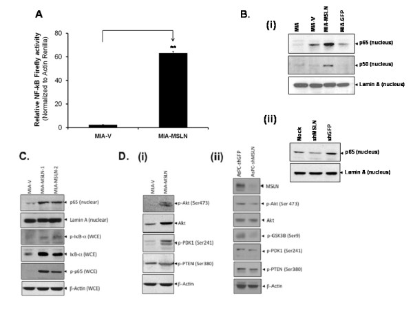
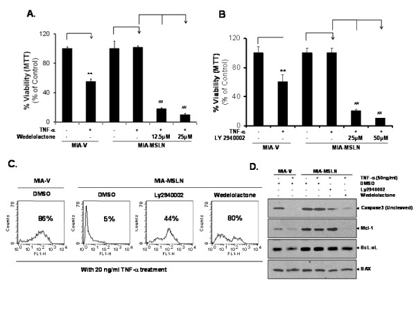
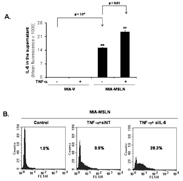
Background: Previous studies showed that mesothelin (MSLN) plays important roles in survival of pancreatic cancer (PC) cells under anchorage dependent/independent conditions as well as resistance to chemotherapy. The recent success of intratumorally-injected adeno-encoded, chemo/radiation-inducible-promoter driven hTNF-α, (TNFerade) + gemcitabine in pre-clinical models of PC have renewed interest in use of TNF-α as a therapeutic component. To help find additional factors which might affect the therapy, we examined the resistance of MSLN-overexpressing pancreatic cancer cell lines to TNF-α-induced growth inhibition/apoptosis. Methods: Stable MSLN overexpressing MIA PaCa-2 cells (MIA-MSLN), stable MSLN-silenced AsPC-1 cells (AsPC-shMSLN) and other pancreatic cells (MIA-PaCa2, Panc 28, Capan-1, BxPC3, PL 45, Hs 766T, AsPC-1, Capan-2, Panc 48) were used. NF-κB activation was examined by western blots and luciferase reporter assay. TNF-α induced growth inhibition/apoptosis was measured by MTT, TUNEL assay and caspase activation. IL-6 was measured using luminex based assay. Results: Compared to low endogenous MSLN-expressing MIA PaCa-2 and Panc 28 cells, high endogenous MSLN-expressing Capan-1, BxPC3, PL 45, Hs 766T, AsPC-1, Capan-2, Panc 48 cells were resistant to TNF-α induced growth inhibition. Stable MSLN overexpressing MIA-PaCa2 cells (MIA-MSLN) were resistant to TNF-α-induced apoptosis while stable MSLN-silenced AsPC1 cells (AsPC-shMSLN) were sensitive. Interestingly, TNF-α-treated MIA-MSLN cells showed increased cell cycle progression and cyclin A induction, both of which were reversed by caspase inhibition. We further found that MIA-MSLN cells showed increased expression of anti-apoptotic Bcl-XL and Mcl-1; deactivated (p-Ser 75 ) BAD, and activated (p-Ser 70 ) Bcl-2. Constitutively activated NF-κB and Akt were evident in MIA-MSLN cells that could be suppressed by MSLN siRNA with a resultant increase in sensitivity of TNF-α induced apoptosis. Blocking NF-κB using IKK inhibitor wedelolactone also increased sensitivity to TNF-α-mediated cytotoxicity with concomitant decrease in Mcl-1. Blocking Akt using PI3K inhibitor also had a likewise effect presumably affecting cell cycle. MIA-MSLN cells produced increased IL-6 and were increased furthermore by TNF-α treatment. SiRNA-silencing of IL-6 increased TNF-α sensitivity of MIA-MSLN cells. Conclusions: Our study delineates a MSLN-Akt-NF-κB-IL-6-Mcl-1 survival axis that may be operative in PC cells, and might help cancer cells' survival in the highly inflammatory milieu evident in PC. Further, for the success of TNFerade + gemcitabine to be successful, we feel the simultaneous inhibition of components of this axis is also essential.
Overview
The claims and statistics of this paper haven’t been checked yet.
Run the check — free, no account needed. It reads the open-access text and checks the sources it cites.
Integrity signals
Registry
Cites
Statistics
Figures
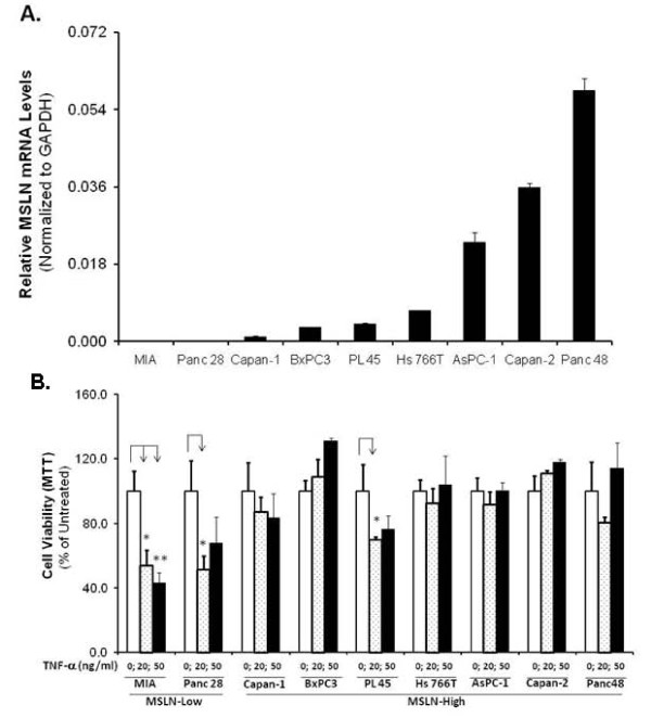
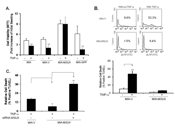
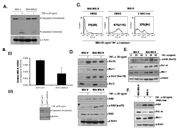
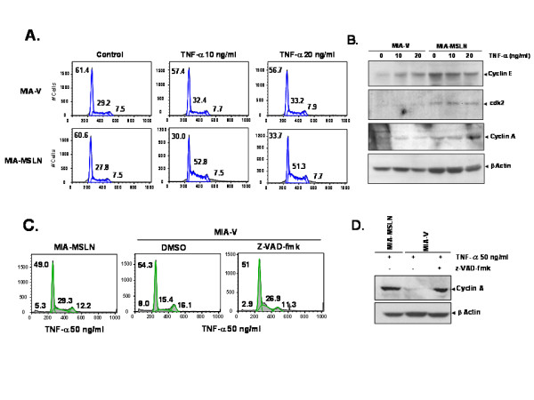
Verification
Claims the paper makes
The claims and statistics of this paper haven’t been checked yet.
Run the check — free, no account needed. It reads the open-access text and checks the sources it cites.
Claims other papers make about this one
Each is checked against this paper’s own text: does it say what the citing paper says it does?
The cited source says what the claim attributes to it.
In the citing paper
“It confers pancreatic cancer cell resistance to TNFα-induced apoptosis through NF-κB activation and the enhanced expression of IL-6 (319).”
From IL-6 in inflammation, autoimmunity and cancer (2021) · cites this paper as [319]
Self-check
Percentages, sums and reported differences re-computed from the paper’s own numbers.
The self-check needs the paper’s full text.
Citations
Cites
An endotoxin-induced serum factor that causes necrosis of tumors
Carswell, E. A., Old, L. J., Kassel, R. L., Green, S., Fiore, N., & Williamson, B. (1975) · Proceedings of the National Academy of Sciences · doi:10.1073/pnas.72.9.3666 · Cited by 4,532 · cited 1×
“TNF-a was identified as a cytokine that induces tumor necrosis/regression in animals [9].”
BackgroundThe Bcl2 family: regulators of the cellular life-or-death switch
Cory, S., & Adams, J. M. (2002) · Nature Reviews Cancer · doi:10.1038/nrc883 · Cited by 4,032 · cited 1×
“Dephosphorylated BAD promotes apoptosis by binding and sequestering Bcl-2 and/or Bcl-XL away from proapoptotic Bax/Bak proteins [22,23].”
BackgroundSignalling pathways of the TNF superfamily: a double-edged sword
Aggarwal, B. B. (2003) · Nature Reviews Immunology · doi:10.1038/nri1184 · Cited by 2,793 · cited 2×
“TNF-a induces NF- B activation in most cells [17,18] but this is not always sufficient to avoid the pro-apoptotic effects of TNF-a; rather, a constitutive activation of NF- B seems to be necessary [20].”
BackgroundInflammation and cancer: how hot is the link?
Aggarwal, B. B., Shishodia, S., Sandur, S. K., Pandey, M. K., & Sethi, G. (2006) · Biochemical Pharmacology · doi:10.1016/j.bcp.2006.06.029 · Cited by 1,312 · cited 8×
“Another interesting observation is that TNF-a induces MIA-MSLN cells to go into cell cycle rather than stalling the process, similar to previous reports of auto/paracrine growth stimulation by TNF-a [17].”
BackgroundResultInfluentialWang, C.-Y., Cusack, J. C., Liu, R., & Baldwin, A. S. (1999) · Nature Medicine · doi:10.1038/7410 · Cited by 986 · cited 1×
“However, a large percentage of patients and/or cell lines are resistant to TNF-a treatment [14,15].”
BackgroundTNF-α in promotion and progression of cancer
Balkwill, F. R. (2006) · Cancer and Metastasis Reviews · doi:10.1007/s10555-006-9005-3 · Cited by 919
Bcl-2 family proteins and cancer
Yip, K. W., & Reed, J. C. (2008) · Oncogene · doi:10.1038/onc.2008.307 · Cited by 913 · cited 1×
“Bcl-2 regulates apoptosis by heterodimerization with its pro-apoptotic partner, Bax, to maintain mitochondrial integrity [40].”
BackgroundMammalian cell-cycle regulation: several Cdks, numerous cyclins and diverse compensatory mechanisms
Satyanarayana, A., & Kaldis, P. (2009) · Oncogene · doi:10.1038/onc.2009.170 · Cited by 800 · cited 1×
“We further examined S-phase promoting cyclins A/E [28] and cdk2.”
BackgroundTumour necrosis factor-α as a tumour promoter
Szlosarek, P. W., Charles, K. A., & Balkwill, F. R. (2006) · European Journal of Cancer · doi:10.1016/j.ejca.2006.01.012 · Cited by 367
Mesothelin targeted cancer immunotherapy.
Hassan, R., & Ho, M. (2008) · European Journal of Cancer · doi:10.1016/j.ejca.2007.08.028 · Cited by 337 · cited 1×
“The importance of mesothelin (MSLN) as a biomarker and preferred immunotherapeutic target is steadily growing for many cancers, including pancreatic cancer (PC) [1-3].”
Background
Research works printed but not linked: 6
- Li M, Bharadwaj U, Zhang R, Zhang S, Mu H, Fisher WE, Brunicardi FC, Chen C, Yao Q: Mesothelin is a malignant factor and therapeutic vaccine target for pancreatic cancer. Mol Cancer Ther. 2008, 7: 286-296. 10.1158/1535-7163.MCT-07-0483 · DOI
- Fernandez A, Marin MC, McDonnell T, Ananthaswamy HN: Differential sensitivity of normal and Ha-ras-transformed C3H mouse embryo fibroblasts to tumor necrosis factor: induction of bcl-2, c-myc, and manganese superoxide dismutase in resistant cells. Oncogene. 1994, 9: 2009-2017.
- Komori A, Yatsunami J, Suganuma M, Okabe S, Abe S, Sakai A, Sasaki K, Fujiki H: Tumor necrosis factor acts as a tumor promoter in BALB/3T3 cell transformation. Cancer Res. 1993, 53: 1982-1985.
- Eisenmann KM, VanBrocklin MW, Staffend NA, Kitchen SM, Koo HM: Mitogen-activated protein kinase pathway-dependent tumor-specific survival signaling in melanoma cells through inactivation of the proapoptotic protein bad. Cancer Res. 2003, 63: 8330-8337.
- Argani P, Iacobuzio-Donahue C, Ryu B, Rosty C, Goggins M, Wilentz RE, Murugesan SR, Leach SD, Jaffee E, Yeo CJ: Mesothelin is overexpressed in the vast majority of ductal adenocarcinomas of the pancreas: identification of a new pancreatic cancer marker by serial analysis of gene expression (SAGE). Clin Cancer Res. 2001, 7: 3862-3868.
- Kalthoff H, Roeder C, Humburg I, Thiele HG, Greten H, Schmiegel W: Modulation of platelet-derived growth factor A- and B-chain/c-sis mRNA by tumor necrosis factor and other agents in adenocarcinoma cells. Oncogene. 1991, 6: 1015-1021.
Cited by
Sorted by citations
IL-6 in inflammation, autoimmunity and cancer
Hirano, T. (2021) · International Immunology · doi:10.1093/intimm/dxaa078 · Cited by 1,453
The Journal of Clinical Investigation
Witkin, E. M. (2016) · Journal of the American Dietetic Association · doi:10.1016/0002-8223(94)92394-9 · Cited by 920
Mesothelin-Targeted CARs: Driving T Cells to Solid Tumors
Morello, A., Sadelain, M., & Adusumilli, P. S. (2015) · Cancer Discovery · doi:10.1158/2159-8290.cd-15-0583 · Cited by 491
“Overexpression of MSLN alone is sufficient to constitutively activate the NFκB, MAPK, and PI3K intracellular pathways promoting cell proliferation and resistance to apoptosis (25).”
BackgroundMesothelin Immunotherapy for Cancer: Ready for Prime Time?
Hassan, R., Thomas, A., Alewine, C., Le, D., Jaffee, E., & Pastan, I. (2016) · Journal of Clinical Oncology · doi:10.1200/jco.2016.68.3672 · Cited by 301
Targeting Apoptosis Pathways in Cancer and Perspectives with Natural Compounds from Mother Nature
Millimouno, F. M., Dong, J., Liu, Y., Li, J., & Li, X. (2014) · Cancer Prevention Research · doi:10.1158/1940-6207.capr-14-0136 · Cited by 276
Golfier, S., Kopitz, C., Kahnert, A., Heisler, I., Schatz, C., Stelte-Ludwig, B., Mayer-Bartschmid, A., Unterschemmann, K., Bruder, S., Linden, L., et al. (2014) · Molecular Cancer Therapeutics · doi:10.1158/1535-7163.mct-13-0926 · Cited by 232
The role of mesothelin in tumor progression and targeted therapy
Tang, Z.-W., Qian, M., & Ho, M. (2013) · Anti-Cancer Agents in Medicinal Chemistry · doi:10.2174/1871520611313020014 · Cited by 136
“Cyclin A was also increased in mesothelin overexpressing MIA PaCa-2 cells by TNF-α treatment [18].”
BackgroundInfluentialMesothelin-targeted CAR-T cell therapy for solid tumors
Klampatsa, A., Dimou, V., & Albelda, S. (2020) · Expert Opinion on Biological Therapy · doi:10.1080/14712598.2021.1843628 · Cited by 130
“overexpression in pancreatic cancer cell lines has been associated with resistance to tumor necrosis factor-alpha (TNF-α)mediated apoptosis [48], anoikis-induced apoptosis [49] and resistance to paclitaxel treatment [50].”
BackgroundMesothelin as a biomarker for targeted therapy
Lv, J., & Li, P. (2019) · Biomarker Research · doi:10.1186/s40364-019-0169-8 · Cited by 133
“The overexpression of MSLN could activate the NFκB, MAPK, and PI3K pathways and subsequently induce resistance to apoptosis [10] or promote cell proliferation, migration, and metastasis by inducing the activation and expression of MMP7 [9] and MMP9 [5].”
BackgroundHarnessing Tumor Necrosis Factor Alpha to Achieve Effective Cancer Immunotherapy
Mercogliano, M. F., Bruni, S., Mauro, F. L., Elizalde, P. V., & Schillaci, R. (2021) · Cancers · doi:10.3390/cancers13030564 · Cited by 145
CAR-T Cell Therapy for Breast Cancer: From Basic Research to Clinical Application
Yang, Y.-H., Liu, J., Lu, C., & Wei, J. (2022) · International Journal of Biological Sciences · doi:10.7150/ijbs.70120 · Cited by 135
“Overexpression of MSLN constitutively activates the intracellular signaling pathways (such as MAPK, PI3K, and NF-kB) to promote tumor development and progression[99, 101].”
BackgroundHe, X., Wang, L., Riedel, H., Wang, K., Yang, Y. S., Dinu, C. Z., & Rojanasakul, Y. (2017) · Molecular Cancer · doi:10.1186/s12943-017-0633-8 · Cited by 132
“Preclinical studies showed that MSLN is involved in cell proliferation, anoikis resistant and survival [10–12], and its downregulation promotes drug-induced apoptosis and chemosensitivity [13, 14].”
BackgroundRegulatory mechanisms and clinical perspectives of miRNA in tumor radiosensitivity
Zhao, L., Bode, A. M., Cao, Y., & Dong, Z. (2012) · Carcinogenesis · doi:10.1093/carcin/bgs235 · Cited by 114
Mesothelin/mucin 16 signaling in activated portal fibroblasts regulates cholestatic liver fibrosis
Koyama, Y., Wang, P., Liang, S., Iwaisako, K., Liu, X., Xu, J., Zhang, M., Sun, M., Cong, M., Karin, D., et al. (2017) · Journal of Clinical Investigation · doi:10.1172/jci88845 · Cited by 115
Tozbikian, G., Brogi, E., Kadota, K., Catalano, J. P., Akram, M. M., Patil, S., Ho, A., Reis-Filho, J., Weigelt, B., Norton, L., et al. (2014) · PLoS ONE · doi:10.1371/journal.pone.0114900 · Cited by 97
“Mesothelin expression increases resistance to TNFa-induced apoptosis through Akt/PI3K/NF-kB activation and IL-6/Mcl-1 expression in pancreatic carcinoma cell lines [12].”
BackgroundMesothelin Expression in Human Tumors: A Tissue Microarray Study on 12,679 Tumors
Weidemann, S., Gagelmann, P., Gorbokon, N., Lennartz, M., Menz, A., Luebke, A., Kluth, M., Hube-Magg, C., Blessin, N., Fraune, C., et al. (2021) · Biomedicines · doi:10.3390/biomedicines9040397 · Cited by 95
Hagemann, U. B., Ellingsen, C., Schuhmacher, J., Kristian, A., Mobergslien, A., Cruciani, V., Wickstroem, K., Schatz, C. A., Kneip, C., Golfier, S., et al. (2019) · Clinical Cancer Research · doi:10.1158/1078-0432.ccr-18-3476 · Cited by 117
Marin-Muller, C., Li, D., Bharadwaj, U., Li, M., Chen, C., Hodges, S., Fisher, W., Mo, Q., Hung, M., & Yao, Q. (2013) · Clinical Cancer Research · doi:10.1158/1078-0432.ccr-12-3776 · Cited by 85
Self-citationIL-6 roles – Molecular pathway and clinical implication in pancreatic cancer – A systemic review
Pop, V.-V., Seicean, A., Lupan, I., Samaşcă, G., & Burz, C. C. (2016) · Immunology Letters · doi:10.1016/j.imlet.2016.11.010 · Cited by 82
Luminal membrane expression of mesothelin is a prominent poor prognostic factor for gastric cancer
Einama, T., Homma, S., Kamachi, H., Kawamata, F., Takahashi, K., Takahashi, N., Taniguchi, M., Kamiyama, T., Furukawa, H., Matsuno, Y., et al. (2012) · British Journal of Cancer · doi:10.1038/bjc.2012.235 · Cited by 74
“In addition, mesothelin-activated nuclear factor-kappaB (NF-kB) induces elevated interleukin (IL)-6 expression, which acts as a growth factor to support pancreatic cancer cell survival/proliferation through a novel auto/paracrine IL-6/soluble IL-6R trans-signalling (Bharadwaj et al, 2011a, b).”
BackgroundWang, Y., Wang, X., Zhao, H., Liang, B., & Du, Q. (2012) · Journal of Chemotherapy · doi:10.1179/1973947812y.0000000049 · Cited by 74
The Impact of Mesothelin in the Ovarian Cancer Tumor Microenvironment
Hilliard, T. S. (2018) · Cancers · doi:10.3390/cancers10090277 · Cited by 73
Bassagañas, S., Allende, H., Cobler, L., Ortiz, M. R., Llop, E., de Bolós, C., & Peracaula, R. (2015) · Cytokine · doi:10.1016/j.cyto.2015.04.006 · Cited by 62
Aliabadi, H. M., Maranchuk, R. A., Kucharski, C., Mahdipoor, P., Hugh, J. C., & Uludağ, H. (2013) · Journal of Controlled Release · doi:10.1016/j.jconrel.2013.08.012 · Cited by 56
Regulation of invasion and peritoneal dissemination of ovarian cancer by mesothelin manipulation
Coelho, R., Ricardo, S., Amaral, A. L., Huang, Y.-L., Nunes, M., Neves, J. P., Mendes, N., López, M. N., Bartosch, C., Ferreira, V., et al. (2020) · Oncogenesis · doi:10.1038/s41389-020-00246-2 · Cited by 55
Chen, H.-Y., Lin, L.-T., Wang, M.-L., Lee, S.-H., Tsai, M., Tsai, C., Liu, W.-H., Chen, T.-C., Yang, Y.-P., Lee, Y.-Y., et al. (2016) · OncoTarget · doi:10.18632/oncotarget.9890 · Cited by 55
“For instance, IL-6-induced PI3K/AKT activation was essential for anti-apoptotic signaling cascade, which has long be linked to therapeutic resistance [19].”
BackgroundPlant coumestans: recent advances and future perspectives in cancer therapy.
Nehybová, T., Šmarda, J., & Beneš, P. (2014) · Anti-Cancer Agents in Medicinal Chemistry · doi:10.2174/1871520614666140713172949 · Cited by 54
“Wedelolactone senzitizes pancreatic cancer cells to proapoptotic effects of TNFα by inhibiting the NFκB activity and decreasing expression of Mcl-1, a member of the Bcl-2 protein family [134].”
BackgroundInhibition of Mesothelin as a Novel Strategy for Targeting Cancer Cells
Wang, K., Bodempudi, V., Liu, Z., Borrego-Diaz, E., Yamoutpoor, F., Meyer, A. C., Woo, R. A., Pan, W., Dudek, A., Olyaee, M., et al. (2012) · PLoS ONE · doi:10.1371/journal.pone.0033214 · Cited by 53
Zheng, C.-N., Jia, W., Tang, Y., Zhao, H., Jiang, Y., & Sun, S.-C. (2012) · Journal of experimental & clinical cancer research : CR · doi:10.1186/1756-9966-31-84 · Cited by 50
“Otherwise, knockdown of mesothelin sensitized pancreatic cancer cells to radiation and TNF-a-induced apoptosis [17,18].”
BackgroundAl‐Lamki, R. S., Lu, W., Manalo, P. B., Wang, J., Warren, A. Y., Tolkovsky, A. M., Pober, J. S., & Bradley, J. R. (2016) · Cell Death and Disease · doi:10.1038/cddis.2016.184 · Cited by 48
Intracellular localization of mesothelin predicts patient prognosis of extrahepatic bile duct cancer
Kawamata, F., Kamachi, H., Einama, T., Homma, S., Tahara, M., MIYAZAKI, M., Tanaka, S., Kamiyama, T., Nishihara, H., Taketomi, A., et al. (2012) · International Journal of Oncology · doi:10.3892/ijo.2012.1662 · Cited by 61
Lu, Y., Chen, Y., Wang, Z., Shen, H.-Y., Xu, L., Huang, C., Tong, Y., Shao, Y., Zhang, H.-Q., & Fu, Z. (2025) · Journal of Translational Medicine · doi:10.1186/s12967-025-06236-5 · Cited by 45
Pandey, M., Gowda, K., Doi, K., Sharma, A. K., Wang, H.-G., & Amin, S. (2013) · PLoS ONE · doi:10.1371/journal.pone.0078570 · Cited by 43
Huo, Q.-Y., Xu, C., Shao, Y., Yu, Q., Huang, L.-H., Liu, Y.-D., & Bao, H. (2021) · International Journal on Biological Sciences · doi:10.7150/ijbs.52097 · Cited by 42
“In addition, MSLN protects cells from drug-induced apoptosis and mediates drug resistance in cancer [58,59,60].”
Ouladan, S., & Orouji, E. (2025) · Journal of Clinical Oncology · doi:10.1200/jco-24-02081 · Cited by 48
Ye, L., Lou, Y., Lu, L., & Fan, X. (2018) · Experimental and Therapeutic Medicine · doi:10.3892/etm.2018.7015 · Cited by 42
“It can activate nuclear factor-κB, mitogen-activated protein kinase and PI3K intracellular pathways that contribute to cell proliferation and resistance to apoptosis (60-62).”
BackgroundCAR T-cells for colorectal cancer immunotherapy: Ready to go?
Ghazi, B., Ghanmi, A. E., Kandoussi, S., Ghouzlani, A., & Badou, A. (2022) · Frontiers in Immunology · doi:10.3389/fimmu.2022.978195 · Cited by 47
PD-1 silencing improves anti-tumor activities of human mesothelin-targeted CAR T cells.
Liu, G.-D., Zhang, Q., Li, D., Zhang, L.-S., Gu, Z., Liu, J., Liu, G.-P., Yang, M., Gu, J., Cui, X., et al. (2020) · Human Immunology · doi:10.1016/j.humimm.2020.12.002 · Cited by 40
Montemagno, C., Cassim, S., Pouysségur, J., Broisat, A., & Pagès, G. (2020) · International Journal of Molecular Sciences · doi:10.3390/ijms21114067 · Cited by 39
“MSLN signaling is also involved in the resistance to TNF-α-induced apoptosis through the activation of the Akt/PI3K/NFκB axis in PDAC [48].”
BackgroundEffectiveness of mesothelin family proteins and osteopontin for malignant mesothelioma
Pantazopoulos, I., Boura, P., Xanthos, T., & Syrigos, K. (2012) · European Respiratory Journal · doi:10.1183/09031936.00226111 · Cited by 38
Metformin Antagonizes Ovarian Cancer Cells Malignancy Through MSLN Mediated IL-6/STAT3 Signaling
Yang, X., Huang, M., Zhang, Q., Chen, J., Li, J., Han, Q., Zhang, L., Li, J.-Q., Liu, S., Ma, Y., et al. (2021) · Cell Transplantation · doi:10.1177/09636897211027819 · Cited by 37
Amatuximab and novel agents targeting mesothelin for solid tumors
Baldo, P., & Cecco, S. (2017) · OncoTargets and Therapy · doi:10.2147/ott.s145105 · Cited by 37
“14,15 Importantly, in recent years, the activation and stimulation of immune reactions using engineered T-cells with chimeric antigen receptors (CARs) allowed the characterization of antigens, including MSLN, overexpressed in solid tumors and in several B-cell malignancies, including acute…”
Kobayashi, K., Sasaki, T., Takenaka, F., Yakushiji, H., Fujii, Y., Kishi, Y., Kita, S., Shen, L.-H., Kumon, H., & Matsuura, E. (2015) · Journal of Immunological Research · doi:10.1155/2015/268172 · Cited by 36
“against Mesothelin Commonly Expressed on Cancer Cells Kazuko Kobayashi,1,2 Takanori Sasaki,3 Fumiaki Takenaka,3 Hiromasa Yakushiji,1 Yoshihiro Fujii,4 Yoshiro Kishi,4 Shoichi Kita,4 Lianhua Shen,1,5 Hiromi Kumon,2 and Eiji Matsuura1,3 1Department of Cell Chemistry, Okayama University Graduate School of Medicine, Denti…”
BackgroundC-ERC/mesothelin provokes lymphatic invasion of colorectal adenocarcinoma
Kawamata, F., Homma, S., Kamachi, H., Einama, T., Kato, Y., Tsuda, M., Tanaka, S., Maeda, M., Kajino, K., Hino, O., et al. (2013) · Journal of gastroenterology · doi:10.1007/s00535-013-0773-6 · Cited by 35
“Many researchers have investigated the role of the mesothelin expression in tumor biology, and demonstrated the importance of mesothelin expression for tumor progression in vitro [17-20] and in vivo [20, 21], and we recently explored that the luminal membrane expression of mesothelin was related to an unfavorable pati…”
BackgroundTargeting solid tumor antigens with chimeric receptors: cancer biology meets synthetic immunology
Kembuan, G. J., Kim, J. Y., Maus, M. V., & Jan, M. (2024) · Trends in cancer · doi:10.1016/j.trecan.2024.01.003 · Cited by 37
Yang, Y., Vedvyas, Y., Alcaina, Y., Trumper, S. J., Babu, D. S., Min, I. M., Tremblay, J. M., Shoemaker, C. B., & Jin, M. M. (2024) · JCI Insight · doi:10.1172/jci.insight.186268 · Cited by 37
“Additionally, aberrant expression of MSLN has been associated with tumor aggressiveness, malignant transformation, increased invasion and metastasis, and resistance to apoptosis (3–6).”
The dark side of immunotherapy: pancreatic cancer
Mucciolo, G., Roux, C., Scagliotti, A., Brugiapaglia, S., Novelli, F., & Cappello, P. (2020) · Cancer Drug Resistance · doi:10.20517/cdr.2020.13 · Cited by 38
“MSLN contributes to cell proliferation and resistance to apoptosis([105]), and by activating matrix metalloproteases 9 (MMP-9)([104]) and MMP-7([106]), it can induce tumor cell migration and invasion.”
BackgroundLi, Y., Tian, W., Zhang, H., Zhang, Z., Zhao, Q., Chang, L., Lei, N., & Zhang, W. (2022) · Frontiers in Oncology · doi:10.3389/fonc.2022.830570 · Cited by 31
“Studies have shown that overexpression of MSLN can activate the PI3K pathway and induce drug resistance in pancreatic cancer cells (19).”
BackgroundNucleic acid combinations: A new frontier for cancer treatment
Remant Bahadur, K. C., Thapa, B., Valencia‐Serna, J., Aliabadi, H. M., & Uludağ, H. (2017) · Journal of Controlled Release · doi:10.1016/j.jconrel.2017.04.029 · Cited by 30
Recent clinical trials utilizing chimeric antigen receptor T cells therapies against solid tumors
Han, S., Latchoumanin, O., Wu, G. J., Zhou, G., Hebbard, L., George, J., & Qiao, L. (2017) · Cancer Letters · doi:10.1016/j.canlet.2016.12.037 · Cited by 34
“The natural function of mesothelin in normal tissues is unclear but recent studies hav e shown that over-expression of mesothelin on tumor tissues could facilitate cell proliferation, i vasion and metastasis, and confer tumor cells wit h resistance to apoptosis [51-54].”
BackgroundZhuang, Z., Pan, X., Zhao, K., Gao, W., Liu, J., Deng, T., & Qin, W. (2019) · Medical Science Monitor · doi:10.12659/msm.916385 · Cited by 30
“IL-6, regulated by AKT activity, has previously been shown to activate PI3K/AKT and the anti-apoptotic signaling cascade [21,22].”
BackgroundAntibody-drug conjugates in gynecologic malignancies.
Lee, E. K., & Liu, J. F. (2019) · Gynecologic Oncology · doi:10.1016/j.ygyno.2019.03.245 · Cited by 30
Biology of Mesothelin and Clinical Implications: A Review of Existing Literature
Hagerty, B. L., & Takabe, K. (2023) · World Journal of Oncology · doi:10.14740/wjon1655 · Cited by 25
“They went on to further establish an autocrine/paracrine mechanism of MSLN action whereby activation of nuclear factor kappa B (NF-κB) induced interleukin (IL)-6 release which then acts as a growth factor to support cell survival and proliferation [63, 64].”
BackgroundPark, G. B., & Kim, D. (2017) · Oncology Reports · doi:10.3892/or.2017.5941 · Cited by 30
Nishio, T., Koyama, Y., Liu, X., Rosenthal, S. B., Yamamoto, G., Fuji, H., Baglieri, J., Li, N., Brenner, L. N., Iwaisako, K., et al. (2021) · Proceedings of the National Academy of Sciences · doi:10.1073/pnas.2101270118 · Cited by 29
“CA125 (mouse Muc16) is the only known ligand of MSLN and was reported to activate Src/Akt signaling in cancer cells (38, 39).”
BackgroundThe clinical development of antibody-drug conjugates for non-small cell lung cancer therapy
Liu, X., Deng, J., Zhang, R., Xing, J., Wu, Y., Chen, W., Liang, B., Xing, D., Xu, J., & Zhang, M. (2023) · Frontiers in Immunology · doi:10.3389/fimmu.2023.1335252 · Cited by 28
Clinical impacts of mesothelin expression in gastrointestinal carcinomas.
Einama, T., Kawamata, F., Kamachi, H., Nishihara, H., Homma, S., Matsuzawa, F., Mizukami, T., Konishi, Y., Tahara, M., Kamiyama, T., et al. (2016) · World Journal of Gastrointestinal Pathophysiology · doi:10.4291/wjgp.v7.i2.218 · Cited by 25
Redox state alters anti‐cancer effects of wedelolactone
Beneš, P., Alexova, P., Knopfová, L., Španová, A., & Šmarda, J. (2012) · Environmental and Molecular Mutagenesis · doi:10.1002/em.21712 · Cited by 30
“Wedelolactone has been used as an inhibitor of the NFjB activity in numerous studies [Idris et al., 2009; Zheng et al., 2010; Bharadwaj et al., 2011].”
MethodNichetti, F., Marra, A., Corti, F., Guidi, A., Raimondi, A., Prinzi, N., de Braud, F., & Pusceddu, S. (2018) · Targeted Oncology · doi:10.1007/s11523-018-0567-0 · Cited by 23
Mesothelin Expression in Gastric Adenocarcinoma and Its Relation to Clinical Outcomes
Han, S.-H., Joo, M., Kim, H.-S., & Chang, S. (2017) · Journal of Pathology and Translational Medicine · doi:10.4132/jptm.2016.11.18 · Cited by 23
“Bharadwaj et al.(23) reported that mesothelin expression increased resistance to tumor necrosis factor-α– induced apoptosis through Akt/phosphoinositide 3-kinase/nuclear factor κB activation.”
BackgroundCurrent status and innovative developments of CAR-T-cell therapy for the treatment of breast cancer
Marei, H. E. S., Bedair, K., Hasan, A., Al-Mansoori, L., Caratelli, S., Sconocchia, G., Gaiba, A., & Cenciarelli, C. (2025) · Cancer Cell International · doi:10.1186/s12935-024-03615-8 · Cited by 26
“The constitutive activation of intracellular signaling pathways (NF-kB, PI3K, and MAPK) by MSLN overexpression facilitates the formation and progression of tumors [72].”
Significance of mesothelin and CA125 expression in endometrial carcinoma: a retrospective analysis
Kakimoto, S., Miyamoto, M., Einama, T., Takihata, Y., Matsuura, H., Iwahashi, H., Ishibashi, H., Sakamoto, T., Hada, T., Suminokura, J., et al. (2021) · Diagnostic Pathology · doi:10.1186/s13000-021-01093-4 · Cited by 22
“Overexpression of mesothelin alone could constitutively activate the nuclear factorkappa B, mitogen-activated protein kinase, and phosphoinositide 3-kinase intracellular pathways, promoting cell proliferation and resistance to apoptosis [30].”
BackgroundGPI-AP: Unraveling a New Class of Malignancy Mediators and Potential Immunotherapy Targets
Hussein, N. H., Amin, N. S., & Tayebi, H. E. E. (2020) · Frontiers in Oncology · doi:10.3389/fonc.2020.537311 · Cited by 22
Bayoğlu, I., Kucukzeybek, B., Kucukzeybek, Y., Varol, U., Yıldız, I., Alacacıoğlu, A., Akyol, M., Demir, L., Dirican, A., Yildiz, Y., et al. (2015) · Biomedicine & pharmacotherapy = Biomedecine & pharmacotherapie · doi:10.1016/j.biopha.2015.01.019 · Cited by 22
Mutsaers, S. E., Pixley, F. J., Prêle, C. M., & Hoyne, G. F. (2020) · Current Opinion in Immunology · doi:10.1016/j.coi.2020.04.005 · Cited by 29
Unique cellular interactions between pancreatic cancer cells and the omentum
Feygenzon, V., Loewenstein, S., Lubezky, N., Pasmanic-Chor, M., Sher, O., Klausner, J., & Lahat, G. (2017) · PLoS ONE · doi:10.1371/journal.pone.0179862 · Cited by 21
Application of CAR-T cell therapy targeting mesothelin in solid tumor treatment
Chen, Q., Sun, Y., & Li, H. (2024) · Discover Oncology · doi:10.1007/s12672-024-01159-x · Cited by 17
“Cell survival and proliferation are promoted through a novel auto/paracrine interleukin-6/ soluble IL-6 receptor (IL-6/sIL-6R) trans-signaling pathway [54].”
BackgroundPourbagheri-Sigaroodi, A., Bashash, D., Safaroghli-azar, A., Farshi‐Paraasghari, M., Momeny, M., Mansoor, F. N., & Ghaffari, S. (2019) · European Journal of Pharmacology · doi:10.1016/j.ejphar.2019.01.018 · Cited by 20
Advancing pancreatic cancer therapy by mesothelin-specific nanobody conjugation
Yi, S., Noh, K., Kim, H.-R., Jung, E., Kim, S., Lee, J. E., Guk, K., Choi, J., Lim, E., Kim, S., et al. (2025) · Molecular Cancer · doi:10.1186/s12943-025-02325-7 · Cited by 20
Zhong, Y., Wang, Y., Wang, C., Cao, K., Wang, X., Xu, X., Yang, M., Zhang, G., Liu, H., & Lu, J. (2025) · Journal for ImmunoTherapy of Cancer · doi:10.1136/jitc-2024-011230 · Cited by 19
“12 Consistent with previous findings, we used proliferation assays to demonstrate that knockdown Msln significantly impaired tumor cell proliferation in murine ovarian cancer cell lines (online supplemental figure 8A–”
MSLN induced EMT, cancer stem cell traits and chemotherapy resistance of pancreatic cancer cells
Hu, J., Wang, J., Guo, X., Fan, Q., Li, X.-M., Li, K., Wang, Z., Liang, S., Amin, B., Zhang, N.-W., et al. (2024) · Heliyon · doi:10.1016/j.heliyon.2024.e29210 · Cited by 19
Self-citationTargeting Mesothelin in Solid Tumours: Anti-mesothelin Antibody and Drug Conjugates
Chu, Q. (2023) · Current Oncology Reports · doi:10.1007/s11912-023-01367-8 · Cited by 19
Silencing VDAC1 to Treat Mesothelioma Cancer: Tumor Reprograming and Altering Tumor Hallmarks
Pandey, S. K., Machlof-Cohen, R., Santhanam, M., Shteinfer‐Kuzmine, A., & Shoshan‐Barmatz, V. (2022) · Biomolecules · doi:10.3390/biom12070895 · Cited by 22
“The overexpression of MSLN activates multiple intracellular pathways, including nuclear factor-kappaB (NF-κB), mitogen-activated protein kinase (MAPK), and phosphoinositide 3-kinases (PI3K) pathways, with the consequent promotion of cell proliferation, migration, and metastasis to distal sites and the inhibition of ap…”
BackgroundResearch progress and design optimization of CAR‐T therapy for pancreatic ductal adenocarcinoma
Li, T., Li, H., Li, S., Xu, S., Zhang, W., Gao, H., Xu, H., Wu, C., Wang, W., Yu, X., et al. (2019) · Cancer Medicine · doi:10.1002/cam4.2430 · Cited by 25
Mesothelin Expression is Correlated with Chemoresistance in Stage IV Colorectal Cancer
Nagata, K., Shinto, E., Shiraishi, T., Yamadera, M., Kajiwara, Y., Mochizuki, S., Okamoto, K., Einama, T., Kishi, Y., & Ueno, H. (2021) · Annals of Surgical Oncology · doi:10.1245/s10434-021-10507-y · Cited by 19
Zhang, J.-L., Bera, T., Liu, W., Du, X.-Y., Alewine, C., Hassan, R., & Pastan, I. (2014) · PLoS ONE · doi:10.1371/journal.pone.0104388 · Cited by 16
“was shown to enhance interleukin (IL)-6 signaling [11], and to confer resistance to tumor necrosis factor (TNF)-a mediated apoptosis in pancreatic cancer cell lines [20].”
BackgroundThe role of molecular testing in pancreatic cancer
Zhen, D. B., Safyan, R. A., Konick, E. Q., Nguyen, R., Pritchard, C. C., & Chiorean, E. G. (2023) · Therapeutic Advances in Gastroenterology · doi:10.1177/17562848231171456 · Cited by 16
Einama, T., Yamagishi, Y., Takihata, Y., Suzuki, T., Yamasaki, T., Hirose, Y., Kobayashi, K., Yonamine, N., Fujinuma, I., Tsunenari, T., et al. (2021) · Biomarker Research · doi:10.1186/s40364-021-00335-3 · Cited by 15
Zhang, J., Yao, D., Song, Y., Pan, Y., Zhu, L., Bai, Y., Xu, Y., Zhang, J., & Shao, C. (2019) · Scientific Reports · doi:10.1038/s41598-019-51772-y · Cited by 13
Serum amyloid A (SAA) and Interleukin-6 (IL-6) as the potential biomarkers for gastric cancer
Hou, Y., Zhao, W., Yang, Z.-C., & Zhang, B. (2022) · Medicine · doi:10.1097/md.0000000000031514 · Cited by 14
“[19] Furthermore, IL-6 participates in pancreatic cancer oncogenesis, metastasis, treatment resistance via mesothelin-protein kinase B-NF-kB (MSLN-AktNF-kB), and overexpression of IL-6-Induced myeloid leukemia cell differentiation protein (IL-6-Mcl-1).[20] In this study, we measured the concentrations of SAA and IL-6…”
Mesothelin is Commonly Expressed in Pancreatic Adenocarcinoma but Unrelated to Cancer Aggressiveness
Weidemann, S. A., Perez, D. R., Izbicki, J. R., Neipp, M., Mofid, H., Daniels, T., Nahrstedt, U., Jacobsen, F., Bernreuther, C., Simon, R., et al. (2021) · Cancer Investigation · doi:10.1080/07357907.2021.1943747 · Cited by 14
“By activating the Akt/PI3K/NFjB axis MSLN may also impact resistance to TNF-a-induced apoptosis (19).”
BackgroundAlfaleh, M. A., Howard, C. B., Sedliarou, I., Jones, M. L., Gudhka, R. K., Vanegas, N., Weiss, J., Suurbach, J. H., de Bakker, C. D., Milne, M., et al. (2017) · PLoS ONE · doi:10.1371/journal.pone.0186137 · Cited by 14
Liu, H., Yang, S.-D., Xu, Y., Ning, S., Wang, T., Yang, D.-L., & Ding, W. (2017) · Molecular Medicine Reports · doi:10.3892/mmr.2017.6690 · Cited by 14
Cai, Z., Li, Z., Zhong, W., Lin, F., Dong, X., Ye, H., Guo, Y., Chen, G., Yu, X., Yu, H., et al. (2025) · Advanced Science · doi:10.1002/advs.202407976 · Cited by 11
Genetic Susceptibility to Hidradenitis Suppurativa and Predisposition to Cardiometabolic Disease
Nielsen, V. W., Vad, O. B., Holgersen, N. K., Paludan‐Müller, C., Monfort, L. M., Beyer, A. F., Jemec, G. B. E., Andersen, R. K., Egeberg, A., Thyssen, J. P., et al. (2024) · JAMA Dermatology · doi:10.1001/jamadermatol.2024.3779 · Cited by 14
MALAT-1: LncRNA ruling miR-182/PIG-C/mesothelin triad in triple negative breast cancer.
Barsoum, F., Awad, A. S., Hussein, N. H., Eissa, R. A., & Tayebi, H. E. E. (2020) · Pathology, Research and Practice · doi:10.1016/j.prp.2020.153274 · Cited by 13
Chu, G. J., Linton, A., Kao, S., Klebe, S., Adelstein, S., Yeo, D., Rasko, J. E. J., & Cooper, W. A. (2023) · Histopathology · doi:10.1111/his.14916 · Cited by 14
Kakimoto, S., Miyamoto, M., Einama, T., Matsuura, H., Iwahashi, H., Ishibashi, H., Sakamoto, T., Hada, T., & Takano, M. (2020) · Pathology and Oncology Research · doi:10.1007/s12253-020-00823-1 · Cited by 12
Yan, Y.-Y., Shi, K., Teng, F., Chen, J., Che, J., Dong, X.-W., Lin, N., & Zhang, B. (2020) · Acta Pharmacologica Sinica · doi:10.1038/s41401-019-0354-1 · Cited by 12
Hsu, Y.-H., Chen, T.-H., Chen, Y.-C., Cheng, C.-Y., Sue, Y., Chen, J.-R., & Chen, C.-H. (2013) · Molecular and Cellular Endocrinology · doi:10.1016/j.mce.2013.07.028 · Cited by 12
“Many studies reveal that inhibition of PI3K/Akt can promote apoptosis, and that Akt activation can inhibit apoptosis (Bharadwaj et al., 2011; Nieuwenhuis et al., 2010; Prasad et al., 2012; Thanaketpaisarn et al., 2011).”
BackgroundSelf-citationMalignant Pleural Mesothelioma: From Pathophysiology to Innovative Actionable Targets
Bertuccio, F. R., Montini, S., Fusco, M. A., Gennaro, A. D., Sciandrone, G., Agustoni, F., Galli, G., Bortolotto, C., Saddi, J., Baietto, G., et al. (2025) · Cancers · doi:10.3390/cancers17071160 · Cited by 9
“Mesothelin protects cancer cells from TNF-α -induced apoptosis by rapidly stimulating Akt phosphorylation under PI3K activation, inhibiting the expression of pro-apoptotic factors, such as Bad and Bax, and promoting the expression of anti-apoptotic genes, such as Bcl-2 and Mcl-1 [35].”
Qualiotto, A. N., Baldavira, C., Balancin, M., Ab'Saber, A., Takagaki, T., & Capelozzi, V. (2023) · Frontiers in Immunology · doi:10.3389/fimmu.2023.1268927 · Cited by 11
Hsu, H.-J., Tung, C.-P., Yu, C.-M., Chen, C.-Y., Chen, H.-S., Huang, Y.-C., Tsai, P., Lin, S.-I., Peng, H.-P., Chiu, Y.-K., et al. (2021) · Scientific Reports · doi:10.1038/s41598-021-94902-1 · Cited by 11
Self-citationLurie, E., Liu, D., LaPlante, E. L., Thistlethwaite, L. R., Yao, Q., & Milosavljevic, A. (2020) · Oncogenesis · doi:10.1038/s41389-020-00245-3 · Cited by 11
Self-citationImportance of luminal membrane mesothelin expression in intraductal papillary mucinous neoplasms
Einama, T., Kamachi, H., Nishihara, H., Homma, S., Kanno, H., Ishikawa, M., Kawamata, F., Konishi, Y., Sato, M., Tahara, M., et al. (2015) · Oncology Letters · doi:10.3892/ol.2015.2969 · Cited by 11
Immunotherapy for hepatobiliary cancers: Emerging targets and translational advances.
Li, D., Lin, S., Hong, J., & Ho, M. (2022) · Advances in Cancer Research · doi:10.1016/bs.acr.2022.01.013 · Cited by 11
Evodiamine induces apoptosis in pancreatic carcinoma PANC-1 cells via NF-κB inhibition
Khan, M., Qazi, J., Rasul, A., Zheng, Y.-C., & Ma, T.-H. (2013) · doi:10.3329/bjp.v8i1.12802 · Cited by 11
“The cells were then incubated with 10 ng/mL of TNF or a combination of 10 ng/mL TNF and 1 μM evodiamine for 30 min.”
BackgroundInfluentialMalla, M., Deshmukh, S. K., Wu, S. K., Samec, T., Olevian, D. C., Naili, R. E., El‐Rayes, B. F., Xiu, J., Farrell, A. P., Lenz, H., et al. (2024) · Cancer Gene Therapy · doi:10.1038/s41417-024-00816-1 · Cited by 15
Hou, X.-G., Wang, F., Meng, X.-X., Li, D., Ding, J., Chen, Y., Wang, Z.-L., Zhu, H., & Yang, Z. (2022) · Molecular Pharmaceutics · doi:10.1021/acs.molpharmaceut.2c00342 · Cited by 10
Mesothelin-targeted CAR-T cells for adoptive cell therapy of solid tumors
Zhang, G., Li, T.-F., & Han, S.-Y. (2019) · Archives of medical science : AMS · doi:10.5114/aoms.2019.84888 · Cited by 10
“Further, MSLN prevents the drug-induced release of pro-apoptotic TNF-a by activating Akt/PI3K pathway, enhances the expression of antiapoptotic genes such as Bcl-2 and Mcl-1, and inhibits the expression of pro-apoptotic factors such as Bad and Bax [42].”
Background
Metadata
- Authors
- Bharadwaj, U., Marin-Muller, C., Li, M., Chen, C., & Yao, Q.
- Affiliations
- Baylor College of Medicine (US)
- Corresponding author
- Yao, Qizhi
- Venue
- Molecular Cancer
- Published
- 2011-08-31
- Volume
- 10
- Issue
- 1
- Pages
- 106-106
- ISSN
- 1476-4598 (ISSN-L)
- PMC
- PMC3175472
- OpenAlex
- W2102073127
- Cited by
- 151
- References
- 55
- Citations by year
- 2026: 9 · 2025: 19 · 2024: 13 · 2023: 7 · 2022: 10 · 2021: 13 · 2020: 17 · 2019: 14 · 2018: 6 · 2017: 10 · 2016: 6 · 2015: 6 · 2014: 5 · 2013: 6 · 2012: 10
- FWCI
- 3.31
- Type
- journal article
- Language
- en
- Open access
- gold
- doi.org
- Licence
- cc-by
- Locations
- Molecular Cancer (publishedVersion, cc-by)
- Europe PMC (PubMed Central) (publishedVersion, cc-by)
- PubMed Central (publishedVersion, cc-by)
- PubMed (publishedVersion)
- DOAJ (DOAJ: Directory of Open Access Journals) (submittedVersion)
- Funders
- National Institutes of Health
- National Cancer Institute (CA140828, R21CA140828)
- Topics
- Health Sciences › Medicine › Oncology › Pancreatic and Hepatic Oncology Research
- Life Sciences › Biochemistry, Genetics and Molecular Biology › Molecular Biology › Cell death mechanisms and regulation
- Life Sciences › Biochemistry, Genetics and Molecular Biology › Molecular Biology › Signaling Pathways in Disease
- Keywords
- mesothelin, pancreatic cancer, TNF-α, apoptosis resistance, interleukin-6, Mcl-1, Bcl-xL, Bad phosphorylation, cell cycle progression, cyclin A, caspase activation, gemcitabine, chemosensitization, tumor microenvironment
- MeSH terms
- Phosphoinositide-3 Kinase Inhibitors, Mesothelin, Antineoplastic Agents, Cell Survival, Chromones, Coumarins, Enzyme Activation, Humans, Morpholines, Pancreatic Neoplasms, Recombinant Proteins, Tumor Necrosis Factor-alpha, Interleukin-6, Gene Expression, S Phase, NF-kappa B, Apoptosis, Genes, Reporter, Drug Resistance, Neoplasm, Proto-Oncogene Proteins c-bcl-2, Phosphatidylinositol 3-Kinases, Cyclin A, Protein Transport, RNA Interference, Cell Line, Tumor, Luciferases, Renilla, Apoptosis Regulatory Proteins, Proto-Oncogene Proteins c-akt, I-kappa B Kinase, Caspase 3, Gene Knockdown Techniques, GPI-Linked Proteins, Myeloid Cell Leukemia Sequence 1 Protein
About these checks
Registry, retraction, Cites and statistics results are computed, not judged, with one exception: a printed reference that no index lists is read once by a model before it counts as a citation error. Claims are extracted by a model and kept only when their quote appears verbatim in the text; each cited source is then checked, with a model’s help, for whether it says what the claim relies on. Those verdicts can be wrong — read the claim’s quote and the cited source before relying on one.
Statistics and arithmetic are recomputed, not judged.
Confidence bands are shown only for fields calibrated on our annotated test set. None is calibrated yet, so no band is shown.
What counts as a claim ›How we measure confidence ›How we check this ›
Are you an author, or do you see a problem with this page? Request a review
