

The DOI matches its Crossref record; 76 of the 76 references it cites were found in a registry or an index.
Canonical Wnt signaling is antagonized by noncanonical Wnt5a in hepatocellular carcinoma cells
Open accessAbstract
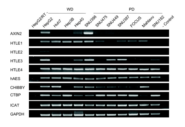
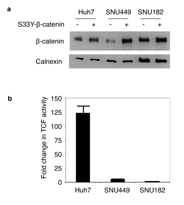
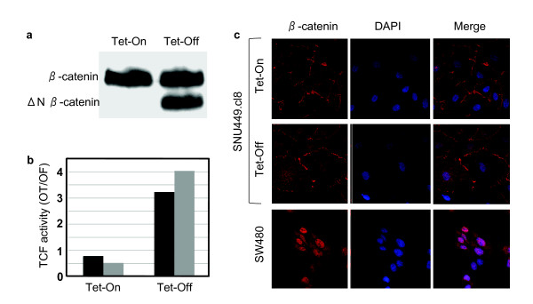
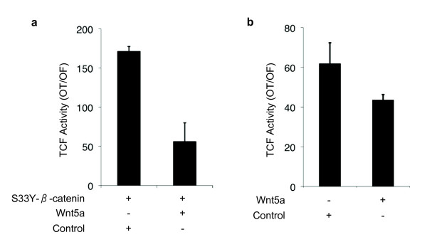
Background: β-catenin mutations that constitutively activate the canonical Wnt signaling have been observed in a subset of hepatocellular carcinomas (HCCs). These mutations are associated with chromosomal stability, low histological grade, low tumor invasion and better patient survival. We hypothesized that canonical Wnt signaling is selectively activated in well-differentiated, but repressed in poorly differentiated HCCs. To this aim, we characterized differentiation status of HCC cell lines and compared their expression status of Wnt pathway genes, and explored their activity of canonical Wnt signaling. Results: We classified human HCC cell lines into "well-differentiated" and "poorly differentiated" subtypes, based on the expression of hepatocyte lineage, epithelial and mesenchymal markers. Poorly differentiated cell lines lost epithelial and hepatocyte lineage markers, and overexpressed mesenchymal markers. Also, they were highly motile and invasive. We compared the expression of 45 Wnt pathway genes between two subtypes. TCF1 and TCF4 factors, and LRP5 and LRP6 co-receptors were ubiquitously expressed. Likewise, six Frizzled receptors, and canonical Wnt3 ligand were expressed in both subtypes. In contrast, canonical ligand Wnt8b and noncanonical ligands Wnt4, Wnt5a, Wnt5b and Wnt7b were expressed selectively in well- and poorly differentiated cell lines, respectively. Canonical Wnt signaling activity, as tested by a TCF reporter assay was detected in 80% of well-differentiated, contrary to 14% of poorly differentiated cell lines. TCF activity generated by ectopic mutant β-catenin was weak in poorly differentiated SNU449 cell line, suggesting a repressive mechanism. We tested Wnt5a as a candidate antagonist. It strongly inhibited canonical Wnt signaling that is activated by mutant β-catenin in HCC cell lines. Conclusion: Differential expression of Wnt ligands in HCC cells is associated with selective activation of canonical Wnt signaling in well-differentiated, and its repression in poorly differentiated cell lines. One potential mechanism of repression involved Wnt5a, acting as an antagonist of canonical Wnt signaling. Our observations support the hypothesis that Wnt pathway is selectively activated or repressed depending on differentiation status of HCC cells. We propose that canonical and noncanonical Wnt pathways have complementary roles in HCC, where the canonical signaling contributes to tumor initiation, and noncanonical signaling to tumor progression.
Overview
The claims and statistics of this paper haven’t been checked yet.
Run the check — free, no account needed. It reads the open-access text and checks the sources it cites.
Integrity signals
Registry
Cites
Statistics
Figures
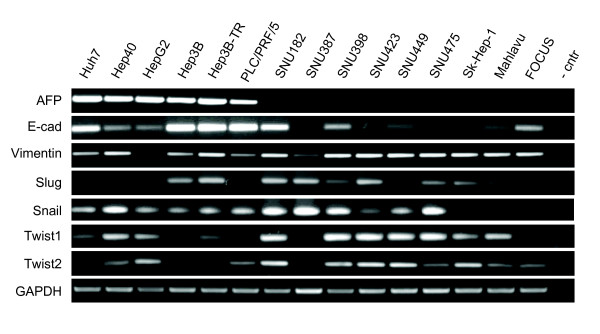
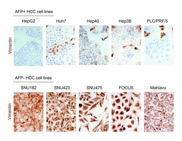
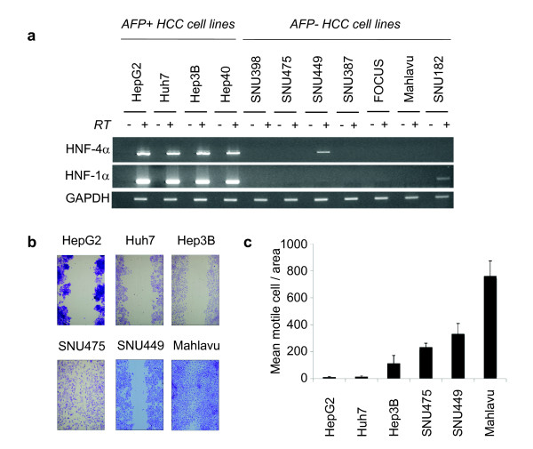
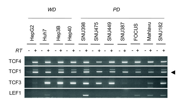
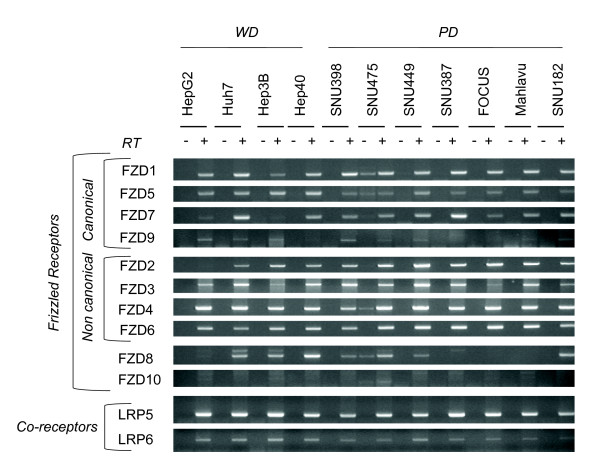
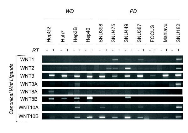
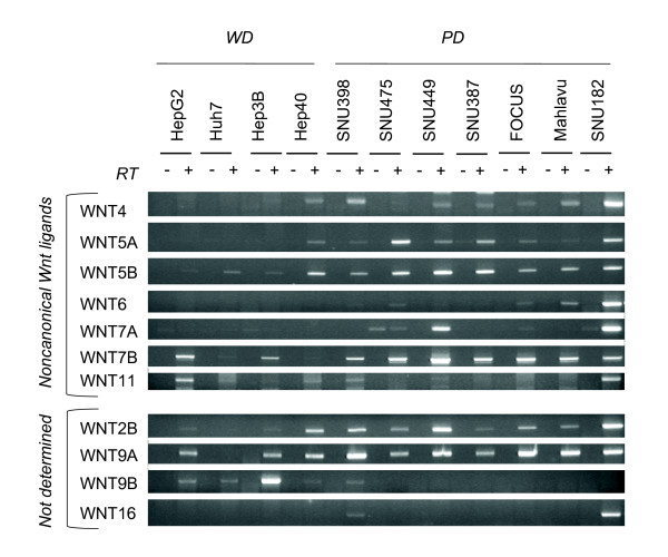
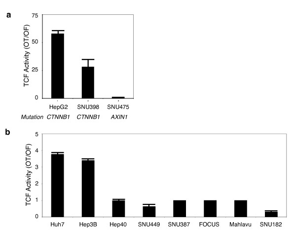
Verification
Claims the paper makes
The claims and statistics of this paper haven’t been checked yet.
Run the check — free, no account needed. It reads the open-access text and checks the sources it cites.
Claims other papers make about this one
Each is checked against this paper’s own text: does it say what the citing paper says it does?
The source backs part of the claim, not all of it — or backs it for a different population, condition or comparison.
In the citing paper
“Huh7 and SNU387 have no known mutations in β-catenin or other Wnt-related gens (27, 28).”
From Wnt-Pathway Activation in Two Molecular Classes of Hepatocellular Carcinoma and Experimental Modulation by Sorafenib (2012) · cites this paper as [27]
Experimental — accuracy not yet measured
Self-check
Percentages, sums and reported differences re-computed from the paper’s own numbers.
The self-check needs the paper’s full text.
Citations
Cites
The basics of epithelial-mesenchymal transition
Kalluri, R., & Weinberg, R. A. (2009) · Journal of Clinical Investigation · doi:10.1172/jci39104 · Cited by 10,196
Activation of β-Catenin-Tcf Signaling in Colon Cancer by Mutations in β-Catenin or APC
Morin, P. J., Sparks, A. B., Kořínek, V., Barker, N., Clevers, H. C., Vogelstein, B., & Kinzler, K. W. (1997) · Science · doi:10.1126/science.275.5307.1787 · Cited by 4,000
Wnt signalling in stem cells and cancer
Reya, T., & Clevers, H. (2005) · Nature · doi:10.1038/nature03319 · Cited by 3,666
Molecular pathogenesis of human hepatocellular carcinoma
Thorgeirsson, S. S., & Grisham, J. W. (2002) · Nature Genetics · doi:10.1038/ng0802-339 · Cited by 1,456
Purified Wnt5a Protein Activates or Inhibits β-Catenin–TCF Signaling Depending on Receptor Context
Mikels, A. J., & Nusse, R. (2006) · PLoS Biology · doi:10.1371/journal.pbio.0040115 · Cited by 1,244
Intestinal polyposis in mice with a dominant stable mutation of the β‐catenin gene
Harada, N., Tamai, Y., Ishikawa, T., Sauer, B., Takaku, K., Oshima, M., & Taketo, M. M. (1999) · The EMBO Journal · doi:10.1093/emboj/18.21.5931 · Cited by 1,212
Yamashita, T., Ji, J., Budhu, A., Forgues, M., Yang, W., Wang, H., Jia, H., Ye, Q., Qin, L., Wauthier, E., et al. (2008) · Gastroenterology · doi:10.1053/j.gastro.2008.12.004 · Cited by 1,142
Munemitsu, S. M., Albert, I., Souza, B., Rubinfeld, B., & Polakis, P. G. (1995) · Proceedings of the National Academy of Sciences · doi:10.1073/pnas.92.7.3046 · Cited by 1,021
Satoh, S., Daigo, Y., Furukawa, Y., Kato, T., Miwa, N., Nishiwaki, T., Kawasoe, T., Ishiguro, H., Fujita, M., Tokino, T., et al. (2000) · Nature Genetics · doi:10.1038/73448 · Cited by 960
Mining the Wnt pathway for cancer therapeutics
Barker, N., & Clevers, H. C. (2006) · Nature Reviews Drug Discovery · doi:10.1038/nrd2154 · Cited by 752
Cited by
Sorted by citations
WNT/β-catenin signaling in the development of liver cancers
He, S., & Tang, S. (2020) · Biomedicine & Pharmacotherapy · doi:10.1016/j.biopha.2020.110851 · Cited by 367
Lachenmayer, A., Alsinet, C., Savic, R., Cabellos, L., Toffanin, S., Hoshida, Y., Villanueva, A., Minguez, B., Newell, P., Tsai, H.-W., et al. (2012) · Clinical Cancer Research · doi:10.1158/1078-0432.ccr-11-2322 · Cited by 306
Pez, F., Lopez, A., Kim, M., Wands, J. R., de Fromentel, C. C., & Merle, P. (2013) · Journal of Hepatology · doi:10.1016/j.jhep.2013.07.001 · Cited by 302
Convergence between Wnt-β-catenin and EGFR signaling in cancer
Hu, T., & Li, C. (2010) · Molecular Cancer · doi:10.1186/1476-4598-9-236 · Cited by 291
Şentürk, Ş., Mumcuoğlu, M., Gürsoy-Yüzügüllü, Ö., Cingoz, B., Akçalı, K. C., & Öztürk, M. (2010) · Hepatology · doi:10.1002/hep.23769 · Cited by 243
Qin, K. H., Yu, M. S., Fan, J., Wang, H., Zhao, P., Zhao, G., Zeng, W., Chen, C., Wang, Y., Wang, A., et al. (2023) · Genes & Diseases · doi:10.1016/j.gendis.2023.01.030 · Cited by 243
Genetic alterations in hepatocellular carcinoma: An update
Niu, Z.-S., Niu, X.-J., & Wang, W. (2016) · World Journal of Gastroenterology · doi:10.3748/wjg.v22.i41.9069 · Cited by 193
Systemic Delivery of a miR34a Mimic as a Potential Therapeutic for Liver Cancer
Daige, C. L., Wiggins, J. F., Priddy, L., Nelligan-Davis, T., Zhao, J. Y., & Brown, D. (2014) · Molecular Cancer Therapeutics · doi:10.1158/1535-7163.mct-14-0209 · Cited by 168
Nwosu, Z. C., Battello, N., Rothley, M., Piorońska, W., Sitek, B., Ebert, M. P. A., Hofmann, U., Sleeman, J. P., Wölfl, S., Meyer, C., et al. (2018) · Journal of Experimental & Clinical Cancer Research · doi:10.1186/s13046-018-0872-6 · Cited by 156
Sáez-Rodríguez, J., Alexopoulos, L. G., Zhang, M., Morris, M. K., Lauffenburger, D. A., & Sorger, P. K. (2011) · Cancer Research · doi:10.1158/0008-5472.can-10-4453 · Cited by 150
Mathematical Modelling of Molecular Pathways Enabling Tumour Cell Invasion and Migration
Cohen, D., Martignetti, L., Robine, S., Barillot, E., Zinovyev, A. Y., & Calzone, L. (2015) · PLoS Computational Biology · doi:10.1371/journal.pcbi.1004571 · Cited by 141
Vázquez, F., García-Ocaña, M., Galván, J. A., García–Martínez, J., García-Pravia, C., Menéndez‐Rodríguez, P., del Rey, C. G., Barneo, L., & Toyos, J. R. D. L. (2015) · Tumor Biology · doi:10.1007/s13277-015-3295-4 · Cited by 131
Karagonlar, Z. F., Koç, D., İşcan, E., Erdal, E., & Atabey, N. (2016) · Cancer Science · doi:10.1111/cas.12891 · Cited by 132
Self-citationWnt Signaling in the Regulation of Immune Cell and Cancer Therapeutics
Haseeb, M., Pirzada, R. H., Ain, Q. U., & Choi, S. (2019) · Cells · doi:10.3390/cells8111380 · Cited by 136
Chen, L., Fulcoli, F. G., Ferrentino, R., Martucciello, S., Illingworth, E. A., & Baldini, A. (2012) · PLoS Genetics · doi:10.1371/journal.pgen.1002571 · Cited by 122
Yu, M. S., Qin, K. H., Fan, J., Zhao, G., Zhao, P., Zeng, W., Chen, C., Wang, A., Wang, Y., Zhong, J., et al. (2023) · Genes & Diseases · doi:10.1016/j.gendis.2023.04.042 · Cited by 125
The involvement of noncanonical Wnt signaling in cancers
Chen, Y., Chen, Z., Tang, Y., & Xiao, Q. (2020) · Biomedicine & Pharmacotherapy · doi:10.1016/j.biopha.2020.110946 · Cited by 118
Wnt signaling regulates hepatobiliary repair following cholestatic liver injury in mice
Okabe, H., Yang, J., Sylakowski, K., Yovchev, M. I., Miyagawa, Y., Nagarajan, S., Chikina, M. D., Thompson, M. D., Oertel, M., Baba, H., et al. (2016) · Hepatology · doi:10.1002/hep.28774 · Cited by 108
Intersection of AHR and Wnt Signaling in Development, Health, and Disease
Schneider, A. J., Branam, A. M., & Peterson, R. E. (2014) · International Journal of Molecular Sciences · doi:10.3390/ijms151017852 · Cited by 109
Xu, L., Lin, W., Wen, L., & Li, G. (2019) · Stem Cell Research & Therapy · doi:10.1186/s13287-019-1288-8 · Cited by 108
PDZ domains: the building blocks regulating tumorigenesis
Subbaiah, V. K., Kranjec, C., Thomas, M., & Banks, L. (2011) · Biochemical Journal · doi:10.1042/bj20110903 · Cited by 102
Aberrant regulation of Wnt signaling in hepatocellular carcinoma
Liu, L., Xie, S., Chen, Y.-T., Xue, J.-L., Zhang, C., & Zhu, F. (2016) · World Journal of Gastroenterology · doi:10.3748/wjg.v22.i33.7486 · Cited by 102
Wnt Signaling in Renal Cell Carcinoma
Xu, Q., Krause, M., Samoylenko, A., & Vainio, S. (2016) · Cancers · doi:10.3390/cancers8060057 · Cited by 99
The role of WNT10B in physiology and disease
Wend, P., Wend, K., Krum, S. A., & Miranda‐Carboni, G. A. (2011) · Acta Physiologica · doi:10.1111/j.1748-1716.2011.02296.x · Cited by 97
Wnt/β-Catenin Signaling in Alzheimer’s Disease
Ferrari, G., Avila, M., Medina, M., Perez-Palma, E., Bustos, B., & Alarcon, M. (2014) · CNS & Neurological Disorders - Drug Targets · doi:10.2174/1871527312666131223113900 · Cited by 95
Zhi, X., Lin, L., Yang, S., Bhuvaneshwar, K., Wang, H., Gusev, Y., Lee, M., Kallakury, B., Shivapurkar, N., Cahn, K., et al. (2014) · Hepatology · doi:10.1002/hep.27558 · Cited by 94
Borrell-Pagès, M., Romero, J. C., Juan-Babot, O., & Badimon, L. (2011) · European Heart Journal · doi:10.1093/eurheartj/ehr062 · Cited by 84
The Wnt/β-catenin signaling pathway in liver biology and disease
Behari, J. (2010) · Expert Review of Gastroenterology & Hepatology · doi:10.1586/egh.10.74 · Cited by 90
Wang, W., Pan, Q., Fuhler, G. M., Smits, R., & Peppelenbosch, M. P. (2016) · Journal of Gastroenterology · doi:10.1007/s00535-016-1299-5 · Cited by 89
Zhang, Y., Li, Q., Zhou, D., & Chen, H. (2012) · British Journal Of Nutrition · doi:10.1017/s0007114512000876 · Cited by 85
Emerging Themes in PDZ Domain Signaling
Liu, X., & Fuentes, E. J. (2018) · International review of cytology · doi:10.1016/bs.ircmb.2018.05.013 · Cited by 86
Regulation of Wnt Signaling Pathways at the Plasma Membrane and Their Misregulation in Cancer
Azbazdar, Y., Karabicici, M., Erdal, E., & Özhan, G. (2021) · Frontiers in Cell and Developmental Biology · doi:10.3389/fcell.2021.631623 · Cited by 84
Self-citationTewari, D., Bawari, S., Sharma, S., DeLiberto, L. K., & Bishayee, A. (2021) · Pharmacology & Therapeutics · doi:10.1016/j.pharmthera.2021.107876 · Cited by 81
Witzigmann, D., Quagliata, L., Schenk, S., Quintavalle, C., Terracciano, L., & Huwyler, J. (2015) · Hepatology Research · doi:10.1111/hepr.12599 · Cited by 81
Cooperative interaction of MUC1 with the HGF/c-Met pathway during hepatocarcinogenesis
Bozkaya, G., Korhan, P., Çokaklı, M., Erdal, E., Sağol, Ö., Karademır, S., Korch, C. T., & Atabey, N. (2012) · Molecular Cancer · doi:10.1186/1476-4598-11-64 · Cited by 80
Self-citationDNA methylation and histone modifications of Wnt genes by genistein during colon cancer development
Zhang, Y., Li, Q., & Chen, H. (2013) · Carcinogenesis · doi:10.1093/carcin/bgt129 · Cited by 79
Qi, L., Sun, B., Liu, Z., Li, H., Gao, J., & Leng, X. (2012) · Cancer Science · doi:10.1111/j.1349-7006.2012.02222.x · Cited by 79
Ma, Y., Lv, Z., Yu, F., Chang, Z., Cong, X., Zhong, X., Lu, G., Zhu, J., & Fu, D. (2018) · Journal of Experimental & Clinical Cancer Research · doi:10.1186/s13046-018-0927-8 · Cited by 74
Cevik, D. (2015) · World Journal of Gastroenterology · doi:10.3748/wjg.v21.i1.311 · Cited by 76
Paracrine WNT5A Signaling Inhibits Expansion of Tumor-Initiating Cells
Borcherding, N. C., Kusner, D. M., Kolb, R. H., Xie, Q. M., Li, W., Yuan, F., Velez, G., Askeland, R. W., Weigel, R. J., & Zhang, W. (2015) · Cancer Research · doi:10.1158/0008-5472.can-14-2761 · Cited by 72
Identification and Characterization of Wnt Signaling Pathway in Keloid Pathogenesis
Igota, S., Tosa, M., Murakami, M., Egawa, S., Shimizu, H., Hyakusoku, H., & Ghazizadeh, M. (2013) · International Journal of Medical Sciences · doi:10.7150/ijms.5349 · Cited by 69
Zimmerli, D., Hausmann, G., Cantù, C., & Basler, K. (2017) · British Journal of Pharmacology · doi:10.1111/bph.13864 · Cited by 65
WNT5B in Physiology and Disease
Suthon, S., Perkins, R. S., Bryja, V., Miranda‐Carboni, G. A., & Krum, S. A. (2021) · Frontiers in Cell and Developmental Biology · doi:10.3389/fcell.2021.667581 · Cited by 72
Fernandes, L. M., Khan, N. M., Medrano-Trochez, C., Duan, M., Díaz‐Hernández, M. E., Presciutti, S. M., Gibson, G. C., & Drissi, H. (2020) · Scientific Reports · doi:10.1038/s41598-020-72261-7 · Cited by 64
Pourreyron, C., Reilly, L., Proby, C. M., Panteleyev, A. A., Fleming, C., McLean, K., South, A. P., & Foerster, J. (2012) · PLoS ONE · doi:10.1371/journal.pone.0031827 · Cited by 63
Pathogenesis of Type 2 Epithelial to Mesenchymal Transition (EMT) in Renal and Hepatic Fibrosis
Tennakoon, A. H., Izawa, T., Kuwamura, M., & Yamate, J. (2015) · Journal of Clinical Medicine · doi:10.3390/jcm5010004 · Cited by 68
Yıldız, G., Arslan-Ergül, A., Baǧışlar, S., Konu, Ö., Yuzugullu, H., Gürsoy-Yüzügüllü, Ö., Öztürk, N., Özen, Ç., Özdağ, H., Erdal, E., et al. (2013) · PLoS ONE · doi:10.1371/journal.pone.0064016 · Cited by 61
Self-citationYap Promotes Noncanonical Wnt Signals From Cardiomyocytes for Heart Regeneration
Liu, S., Tang, L., Zhao, X., Nguyen, B., Heallen, T. R., Li, M., Wang, J., Wang, J., & Martin, J. F. (2021) · Circulation Research · doi:10.1161/circresaha.121.318966 · Cited by 63
Wnt Signaling Cascades and the Roles of Syndecan Proteoglycans
Pataki, C. A., Couchman, J., & Brábek, J. (2015) · Journal of Histochemistry & Cytochemistry · doi:10.1369/0022155415586961 · Cited by 58
Guadecitabine (SGI‐110) priming sensitizes hepatocellular carcinoma cells to oxaliplatin
Kuang, Y., El-Khoueiry, A. B., Taverna, P., Ljungman, M. E., & Neamati, N. (2015) · Molecular Oncology · doi:10.1016/j.molonc.2015.06.002 · Cited by 57
Differential expression of canonical and non‐canonical Wnt ligands in ameloblastoma
Siar, C. H., Nagatsuka, H., Han, P. P., Buery, R. R., Tsujigiwa, H., Nakano, K., Ng, K. H., & Kawakami, T. (2011) · Journal of Oral Pathology and Medicine · doi:10.1111/j.1600-0714.2011.01104.x · Cited by 56
Liu, A., Chen, S., Cai, S., Dong, L., Liu, L., Yang, Y., Guo, F., Lu, X., He, H., Chen, Q., et al. (2014) · PLoS ONE · doi:10.1371/journal.pone.0090229 · Cited by 56
Latasa, M. U., Salis, F., Urtasun, R., García‐Irigoyen, O., Elizalde, M., Uriarte, I., Santamaría, M., Feo, F., Pascale, R. M., Prìeto, J., et al. (2012) · PLoS ONE · doi:10.1371/journal.pone.0052711 · Cited by 54
Karabicici, M., Azbazdar, Y., Özhan, G., Şentürk, Ş., Karagonlar, Z. F., & Erdal, E. (2021) · Frontiers in Cell and Developmental Biology · doi:10.3389/fcell.2021.639779 · Cited by 52
Self-citationZhang, Y., Du, J., Zheng, J., Liu, J., Xu, R., Shen, T. H., Zhu, Y., Chang, J., Wang, H., Zhang, Z., et al. (2015) · Oncotarget · doi:10.18632/oncotarget.3133 · Cited by 53
Mao, J., Wang, D., Wang, Z., Tian, W., Li, X., Duan, J., Wang, Y., Yang, H., You, L., Cheng, Y., et al. (2016) · Cancer Letters · doi:10.1016/j.canlet.2016.06.020 · Cited by 53
Topel, H., Bağırsakçı, E., Çömez, D., Bağcı, G., Cakan‐Akdogan, G., & Atabey, N. (2020) · Cell Communication and Signaling · doi:10.1186/s12964-020-00602-0 · Cited by 52
Self-citationGiakoustidis, A. E., Giakoustidis, D. E., Mudan, S., Sklavos, A., & Williams, R. L. (2015) · Canadian Journal of Gastroenterology and Hepatology · doi:10.1155/2015/172356 · Cited by 54
Cooperative Wnt-Nodal Signals Regulate the Patterning of Anterior Neuroectoderm
Yaguchi, J., Takeda, N., Inaba, K., & Yaguchi, S. (2016) · PLoS Genetics · doi:10.1371/journal.pgen.1006001 · Cited by 50
Fang, Y., Xiao, X., Wang, J., Dasari, S., Pépin, D., Nephew, K. P., Zamarin, D., & Mitra, A. K. (2024) · npj Precision Oncology · doi:10.1038/s41698-023-00495-5 · Cited by 52
FZD1 Regulates Cumulus Expansion Genes and Is Required for Normal Female Fertility in Mice1
Lapointe, É., Boyer, A., Rico, C., Paquet, M., Franco, H. L., Gossen, J. A., DeMayo, F. J., Richards, J. S., & Boerboom, D. (2012) · Biology of Reproduction · doi:10.1095/biolreprod.112.102608 · Cited by 48
Kim, S. S., Cho, H. J., Lee, H. Y., Park, J. H., Noh, C., Shin, S. J., Lee, K. M., Yoo, B. M., Lee, K. J., Cho, S. W., et al. (2016) · Clinical Biochemistry · doi:10.1016/j.clinbiochem.2016.01.025 · Cited by 47
The mechanisms of genome-wide target gene regulation by TCF7L2 in liver cells
Norton, L., Chen, X., Fourcaudot, M. J., Acharya, N., DeFronzo, R. A., & Heikkinen, S. (2014) · Nucleic Acids Research · doi:10.1093/nar/gku1225 · Cited by 48
Wang, W., Xu, L., Liu, P., Jairam, K., Yin, Y., Chen, K., Sprengers, D., Peppelenbosch, M. P., Pan, Q., & Smits, R. (2016) · Neoplasia · doi:10.1016/j.neo.2016.10.004 · Cited by 47
Liu, J., Wang, Z., Tang, J., Tang, R., Shan, X., Zhang, W., Chen, Q., Zhou, F., Chen, K., Huang, A., et al. (2011) · Archives of Virology · doi:10.1007/s00705-011-0943-x · Cited by 46
Bogaerts, E., Heindryckx, F., Vandewynckel, Y., van Grunsven, L. A., & Vlierberghe, H. V. (2014) · International Journal of Oncology · doi:10.3892/ijo.2014.2286 · Cited by 43
Saito, S., Tampe, B., Muller, G. A., & Zeisberg, M. (2015) · Fibrogenesis & Tissue Repair · doi:10.1186/s13069-015-0024-y · Cited by 42
Whole-genome sequencing of matched primary and metastatic hepatocellular carcinomas
Ouyang, L., Lee, J., Park, C.-K., Mao, M., Shi, Y., Gong, Z., Zheng, H., Li, Y., Zhao, Y., Wang, G., et al. (2014) · BMC Medical Genomics · doi:10.1186/1755-8794-7-2 · Cited by 42
Dedifferentiation of hepatocellular carcinoma: molecular mechanisms and therapeutic implications.
Chao, J., Zhao, S., & Sun, H. (2020) · PubMed Central · Cited by 41
Liver cancer cells are sensitive to Lanatoside C induced cell death independent of their PTEN status
Durmaz, İ., Güven, E. B., Erşahin, T., Öztürk, M., Çalış, İ., & Çetin-Atalay, R. (2015) · Phytomedicine · doi:10.1016/j.phymed.2015.11.012 · Cited by 41
Xing, M., Li, P., Wang, X., Li, J., Shi, J., Qin, J., Zhang, X., Ma, Y., Francia, G., & Zhang, J.-Y. (2019) · Cancers · doi:10.3390/cancers12010007 · Cited by 39
Rawson, J. B., Mrkonjic, M., Daftary, D., Dicks, E. L., Buchanan, D. D., Younghusband, H. B., Parfrey, P. S., Young, J. P., Pollett, A. F., Green, R. C., et al. (2011) · British Journal of Cancer · doi:10.1038/bjc.2011.165 · Cited by 39
The role of usnic acid-induced apoptosis and autophagy in hepatocellular carcinoma
Yurdacan, B., Egelí, Ü., Eskiler, G. G., Eryılmaz, I. E., Çeçener, G., & Tunca, B. (2018) · Human & Experimental Toxicology · doi:10.1177/0960327118792052 · Cited by 38
Camcı, İ. Y., Balçık-Erçin, P., Çetįn, M., Odabas, G., Tokay, N., Sayan, E., & Yağcı, T. (2019) · Molecular Carcinogenesis · doi:10.1002/mc.22994 · Cited by 38
Zhao, Y., Zhu, C., Chang, Q., Peng, P., Yang, J., Liu, C., Liu, Y., Chen, X., Liu, Y., Cheng, R., et al. (2020) · PLoS ONE · doi:10.1371/journal.pone.0242179 · Cited by 38
Shi, W., Xu, C., Gong, Y., Wang, J., Ren, Q., Yan, Z., Mei, L., Tang, C., Ji, X., Hu, X., et al. (2021) · Cell Regeneration · doi:10.1186/s13619-020-00071-3 · Cited by 38
WNT5A Inhibits Hepatocyte Proliferation and Concludes β-Catenin Signaling in Liver Regeneration
Yang, J., Cusimano, A., Monga, J. K., Preziosi, M., Pullara, F., Calero, G. A., Lang, R. A., Yamaguchi, T. P., Nejak‐Bowen, K. N., & Monga, S. P. (2015) · American Journal Of Pathology · doi:10.1016/j.ajpath.2015.04.021 · Cited by 37
Liu, H., Zhang, C., Ma, Y., He, H., Wang, J.-P., & Shao, R. (2016) · Life Sciences · doi:10.1016/j.lfs.2016.02.098 · Cited by 40
Titration of RAS alters senescent state and influences tumour initiation
Chan, A. S. L., Zhu, H., Narita, M., Cassidy, L. D., Young, A. R. J., Bermejo‐Rodríguez, C., Janowska, A. T., Chen, H., Gough, S., Oshimori, N., et al. (2024) · Nature · doi:10.1038/s41586-024-07797-z · Cited by 38
Chuang, T. Y., Wu, H. L., Min, J., Diamond, M. P., Azziz, R., & Chen, Y. (2017) · Cancer Cell International · doi:10.1186/s12935-017-0429-3 · Cited by 36
Kim, H.-S., Kim, J. S., Park, N. R., Nam, H., Sung, P. S., Bae, S. H., Choi, J. Y., Yoon, S. K., Hur, W., & Jang, J. W. (2021) · Frontiers in Oncology · doi:10.3389/fonc.2021.637247 · Cited by 34
miR-362-3p targets nemo-like kinase and functions as a tumor suppressor in renal cancer cells
Zou, X., Zhong, J., Li, J., SU, Z., Chen, Y., Deng, W., Li, Y., Lu, S., Lin, Y.-C., Luo, L., et al. (2015) · Molecular Medicine Reports · doi:10.3892/mmr.2015.4632 · Cited by 36
Wnt Signaling in the Development of Bone Metastasis
Ben-Ghedalia-Peled, N., & Vago, R. (2022) · Cells · doi:10.3390/cells11233934 · Cited by 33
Dysregulation of signaling pathways and putative biomarkers in liver cancer stem cells (Review)
Song, K., Wu, J., & Jiang, C. (2012) · Oncology Reports · doi:10.3892/or.2012.2082 · Cited by 33
Kahraman, E., & Göker, E. (2022) · Journal of Trace Elements in Medicine and Biology · doi:10.1016/j.jtemb.2022.127043 · Cited by 33
Balçık-Erçin, P., & Sever, B. (2022) · Chemico-Biological Interactions · doi:10.1016/j.cbi.2022.110152 · Cited by 33
Development of a novel zebrafish xenograft model in ache mutants using liver cancer cell lines
Avcı, M. E., Keşküş, A. G., Targen, S., Işılak, M. E., Öztürk, M., Çetin-Atalay, R., Adams, M. M., & Konu, Ö. (2018) · Scientific Reports · doi:10.1038/s41598-018-19817-w · Cited by 31
Sun, Y., Wang, W., & Zhao, C. (2020) · Oncology Research Featuring Preclinical and Clinical Cancer Therapeutics · doi:10.3727/096504020x16014648664459 · Cited by 31
A Wnt5a-Cdc42 axis controls aging and rejuvenation of hair-follicle stem cells
Tiwari, R. L., Mishra, P., Martin, N., George, N. O., Sakk, V., Soller, K., Nalapareddy, K., Nattamai, K., Scharffetter‐Kochanek, K., Florian, M. C., et al. (2021) · Aging · doi:10.18632/aging.202694 · Cited by 31
Xu, P., Zhang, X., Ni, W., Fan, H., Xu, J., Chen, Y., Zhu, J. Y., Gu, X., Yang, L., Ni, R., et al. (2015) · Digestive Diseases and Sciences · doi:10.1007/s10620-015-3774-x · Cited by 31
Βeta-catenin N-terminal domain: An enigmatic region prone to cancer causing mutations
Dar, M. S., Singh, P., Mir, R. A., & Dar, M. J. (2017) · Mutation Research/Reviews in Mutation Research · doi:10.1016/j.mrrev.2017.06.001 · Cited by 30
Identification of TAF1, SAT1, and ARHGEF9 as DNA methylation biomarkers for hepatocellular carcinoma
Cai, C., Xie, X., Zhou, J., Fang, X., Wang, F., & Wang, M. (2019) · Journal of Cellular Physiology · doi:10.1002/jcp.28999 · Cited by 30
Targeting PDZ domains as potential treatment for viral infections, neurodegeneration and cancer
Nardella, C., Visconti, L., Malagrinò, F., Pagano, L., Bufano, M., Nalli, M., Coluccia, A., Regina, G. L., Silvestri, R., Gianni, S., et al. (2021) · Biology Direct · doi:10.1186/s13062-021-00303-9 · Cited by 31
WNT4 mediates the autocrine effects of growth hormone in mammary carcinoma cells
Vouyovitch, C. M., Perry, J. K., Liu, D. X., Bezin, L., Vilain, E. J. N., Diaz, J., Lobie, P. E., & Mertani, H. C. (2016) · Endocrine Related Cancer · doi:10.1530/erc-15-0528 · Cited by 31
Indian Hedgehog links obesity to development of hepatocellular carcinoma
Chong, Y. C., Lim, T. E., Fu, Y., Shin, E. M., Tergaonkar, V., & Han, W. (2019) · Oncogene · doi:10.1038/s41388-018-0585-5 · Cited by 29
Li, Y., Huan, C., Sun, H., Zhang, W., Guo, Z., Li, C., Yao, J., Zhang, Z., Yu, S., Dong, Q., et al. (2025) · Advanced Science · doi:10.1002/advs.202514661 · Cited by 28
Stucchi, M., Cairati, S., Çetin-Atalay, R., Christodoulou, M. S., Grazioso, G., Pescitelli, G., Silvani, A., Kahraman, D., & Lesma, G. (2015) · Organic & Biomolecular Chemistry · doi:10.1039/c5ob00218d · Cited by 28
Stuckenholz, C., Lu, L., Thakur, P. C., Choi, T. Y., Shin, D., & Bahary, N. (2013) · PLoS ONE · doi:10.1371/journal.pone.0062470 · Cited by 27
LRP 5 negatively regulates differentiation of monocytes through abrogation of Wnt signalling
Borrell-Pagés, M., Romero, J. C., & Badimón, L. (2013) · Journal of Cellular and Molecular Medicine · doi:10.1111/jcmm.12190 · Cited by 26
Strubberg, A. M., Veronese-Paniagua, D., Zhao, T., Dublin, L., Pritchard, T., Bayguinov, P. O., Fitzpatrick, J. A. J., & Madison, B. (2018) · Stem Cell Reports · doi:10.1016/j.stemcr.2018.06.009 · Cited by 24
Metadata
- Authors
- Yuzugullu, H., Benhaj, K., Ozturk, N., Senturk, S., Celik, E., Toylu, A., Tasdemir, N., Yilmaz, M., Erdal, E., Akcali, K. C., Atabey, N., & Ozturk, M.
- Affiliations
- Bilkent University (TR)
- Dokuz Eylül University (TR)
- Venue
- Molecular Cancer
- Published
- 2009-10-22
- Volume
- 8
- Issue
- 1
- Pages
- 90-90
- ISSN
- 1476-4598 (ISSN-L)
- PMC
- PMC2770486
- OpenAlex
- W2152347253
- Cited by
- 215
- References
- 76
- Citations by year
- 2026: 5 · 2025: 12 · 2024: 9 · 2023: 16 · 2022: 14 · 2021: 21 · 2020: 19 · 2019: 9 · 2018: 13 · 2017: 9 · 2016: 17 · 2015: 21 · 2014: 13 · 2013: 9 · 2012: 13
- FWCI
- 3.76
- Type
- journal article
- Language
- en
- Open access
- gold
- doi.org
- Licence
- cc-by
- Locations
- Molecular Cancer (publishedVersion, cc-by)
- PubMed Central (publishedVersion, cc-by)
- PubMed (publishedVersion)
- Bilkent University Institutional Repository (Bilkent University) (submittedVersion)
- Carolina Digital Repository (University of North Carolina at Chapel Hill) (submittedVersion)
- HAL (submittedVersion, other-oa)
- Carolina Digital Repository (University of North Carolina at Chapel Hill) (submittedVersion, cc-by)
- DOAJ (DOAJ: Directory of Open Access Journals) (submittedVersion)
- UNC Libraries
- Funders
- Kræftens Bekæmpelse
- Türkiye Bilimsel ve Teknolojik Araştırma Kurumu
- Türkiye Bilimler Akademisi
- Topics
- Life Sciences › Biochemistry, Genetics and Molecular Biology › Molecular Biology › Wnt/β-catenin signaling in development and cancer
- Health Sciences › Medicine › Oncology › Cancer Cells and Metastasis
- Life Sciences › Biochemistry, Genetics and Molecular Biology › Molecular Biology › Signaling Pathways in Disease
- Keywords
- hepatocellular carcinoma, Wnt/β-catenin signaling, non-canonical Wnt signaling, Wnt5a, CTNNB1 mutation, tumor differentiation, TCF transcription factors, Frizzled receptors, tumor invasion, tumor progression
- MeSH terms
- Wnt-5a Protein, Cell Differentiation, Cell Nucleus, Carcinoma, Hepatocellular, Humans, Immunohistochemistry, Ligands, Liver Neoplasms, Mutation, Neoplasm Proteins, Proto-Oncogene Proteins, Receptors, LDL, Biomarkers, Tumor, Signal Transduction, Gene Expression Regulation, Neoplastic, Genes, Reporter, Autocrine Communication, Cell Line, Tumor, Wnt Proteins, Frizzled Receptors, TCF Transcription Factors
About these checks
Registry, retraction, Cites and statistics results are computed, not judged, with one exception: a printed reference that no index lists is read once by a model before it counts as a citation error. Claims are extracted by a model and kept only when their quote appears verbatim in the text; each cited source is then checked, with a model’s help, for whether it says what the claim relies on. Those verdicts can be wrong — read the claim’s quote and the cited source before relying on one.
Statistics and arithmetic are recomputed, not judged.
Confidence bands are shown only for fields calibrated on our annotated test set. None is calibrated yet, so no band is shown.
What counts as a claim ›How we measure confidence ›How we check this ›
Are you an author, or do you see a problem with this page? Request a review
