

The DOI matches its Crossref record; 40 of the 41 references it cites were found in a registry or an index.
Early correlation of microglial activation with enhanced tumor necrosis factor-alpha and monocyte chemoattractant protein-1 expression specifically within the entorhinal cortex of triple transgenic Alzheimer's disease mice
Open accessAbstract
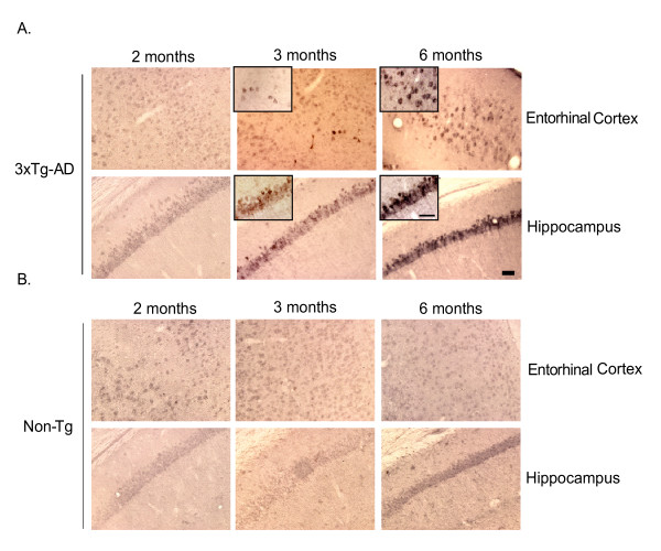
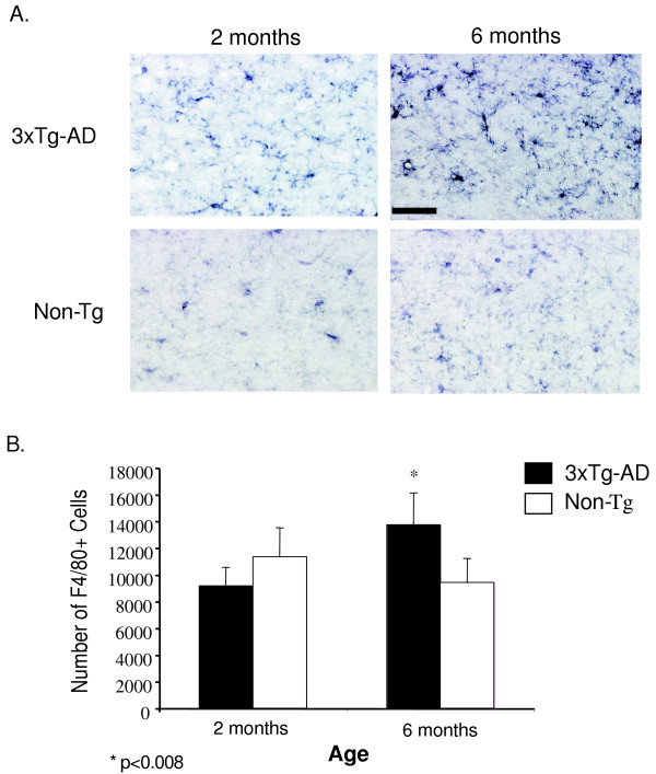
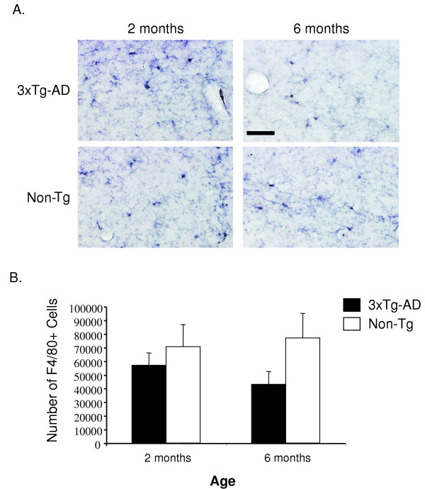
Background: Alzheimer's disease is a complex neurodegenerative disorder characterized pathologically by a temporal and spatial progression of beta-amyloid (Aβ) deposition, neurofibrillary tangle formation, and synaptic degeneration. Inflammatory processes have been implicated in initiating and/or propagating AD-associated pathology within the brain, as inflammatory cytokine expression and other markers of inflammation are pronounced in individuals with AD pathology. The current study examines whether inflammatory processes are evident early in the disease process in the 3xTg-AD mouse model and if regional differences in inflammatory profiles exist. Methods: Coronal brain sections were used to identify Aβ in 2, 3, and 6-month 3xTg-AD and non-transgenic control mice. Quantitative real-time RT-PCR was performed on microdissected entorhinal cortex and hippocampus tissue of 2, 3, and 6-month 3xTg-AD and non-transgenic mice. Microglial/macrophage cell numbers were quantified using unbiased stereology in 3xTg-AD and non-transgenic entorhinal cortex and hippocampus containing sections. Results: We observed human Aβ deposition at 3 months in 3xTg-AD mice which is enhanced by 6 months of age. Interestingly, we observed a 14.8-fold up-regulation of TNF-α and 10.8-fold up-regulation of MCP-1 in the entorhinal cortex of 3xTg-AD mice but no change was detected over time in the hippocampus or in either region of non-transgenic mice. Additionally, this increase correlated with a specific increase in F4/80-positive microglia and macrophages in 3xTg-AD entorhinal cortex. Conclusion: Our data provide evidence for early induction of inflammatory processes in a model that develops amyloid and neurofibrillary tangle pathology. Additionally, our results link inflammatory processes within the entorhinal cortex, which represents one of the earliest AD-affected brain regions.
Overview
The claims and statistics of this paper haven’t been checked yet.
Run the check — free, no account needed. It reads the open-access text and checks the sources it cites.
Integrity signals
Registry
Cites
Statistics
Figures
Verification
Claims the paper makes
The claims and statistics of this paper haven’t been checked yet.
Run the check — free, no account needed. It reads the open-access text and checks the sources it cites.
Claims other papers make about this one
Each is checked against this paper’s own text: does it say what the citing paper says it does?
The cited source says what the claim attributes to it.
In the citing paper
“Similar to previous findings in AD brains and mouse models, 39 -42 the levels of CCL2 and TNF␣ mRNAs were elevated in the APPPS1;Cx3cr1 ϩ/ϩ mouse brain.”
From CX3CR1 Deficiency Alters Microglial Activation and Reduces Beta-Amyloid Deposition in Two Alzheimer's Disease Mouse Models (2010) · cites this paper as [42]
Self-check
Percentages, sums and reported differences re-computed from the paper’s own numbers.
The self-check needs the paper’s full text.
Citations
Cites
Neuropathological stageing of Alzheimer-related changes.
Braak, H., & Braak, E. (1991) · Acta Neuropathologica · doi:10.1007/bf00308809 · Cited by 16,123
Amyloid oligomers exacerbate tau pathology in a mouse model of tauopathy
Selenica, M.-L. B., Brownlow, M., Jimenez, J. P., Lee, D. C., Pena, G., Dickey, C. A., Gordon, M. N., & Morgan, D. (2013) · Neurodegenerative Diseases · doi:10.1159/000337230 · Cited by 10,359
Glutamate neurotoxicity and diseases of the nervous system
Choi, D. W. (1988) · Neuron · doi:10.1016/0896-6273(88)90162-6 · Cited by 8,188
Alzheimer's Disease Is a Synaptic Failure
Selkoe, D. J. (2002) · Science · doi:10.1126/science.1074069 · Cited by 4,420
Triple-Transgenic Model of Alzheimer’s Disease with Plaques and Tangles
Oddo, S., Caccamo, A., Shepherd, J. D., Murphy, M. P., Golde, T. E., Kayed, R., Metherate, R., Mattson, M. P., Akbari, Y., & LaFerla, F. M. (2003) · Neuron · doi:10.1016/s0896-6273(03)00434-3 · Cited by 4,126
Self-citationStaging of alzheimer's disease-related neurofibrillary changes
Braak, H., & Braak, E. (1995) · Neurobiology of Aging · doi:10.1016/0197-4580(95)00021-6 · Cited by 2,357
Profound Loss of Layer II Entorhinal Cortex Neurons Occurs in Very Mild Alzheimer’s Disease
Gómez-Isla, T., Price, J. L., McKeel Jr., D. W., Morris, J. C., Growdon, J. H., & Hyman, B. T. (1996) · The Journal of Neuroscience · doi:10.1523/jneurosci.16-14-04491.1996 · Cited by 1,796 · cited 1×
“Moreover, individuals diagnosed with mild cognitive impairment, a forme fruste of AD, display decreased entorhinal and hippocampal volume, primarily associated with diminished neuron number as compared to non-cognitively impaired controls [7-10].”
Microglia as a source and target of cytokines
Hanisch, U. (2002) · Glia · doi:10.1002/glia.10161 · Cited by 1,645
Nonsteroidal Antiinflammatory Drugs and the Risk of Alzheimer's Disease
in 't Veld, B. A., Ruitenberg, A., Hofman, A., Launer, L. J., van Duijn, C. M., Stijnen, T., Breteler, M. M. B., & Stricker, B. H. C. (2001) · New England Journal of Medicine · doi:10.1056/nejmoa010178 · Cited by 1,196
Risk of Alzheimer's disease and duration of NSAID use
Stewart, W. F., Kawas, C., Corrada, M., & Metter, E. J. (1997) · Neurology · doi:10.1212/wnl.48.3.626 · Cited by 1,132
Plaque-independent disruption of neural circuits in Alzheimer’s disease mouse models
Hsia, A. Y., Masliah, E., McConlogue, L., Yu, G.-Q., Tatsuno, G., Hu, K., Kholodenko, D., Malenka, R. C., Nicoll, R. A., & Mucke, L. (1999) · Proceedings of the National Academy of Sciences · doi:10.1073/pnas.96.6.3228 · Cited by 1,123
Amyloid deposition precedes tangle formation in a triple transgenic model of Alzheimer's disease
Oddo, S. (2003) · Neurobiology of Aging · doi:10.1016/j.neurobiolaging.2003.08.012 · Cited by 980
Self-citationStaging of Alzheimer-Related Cortical Destruction
Braak, H., Braak, E., & Bohl, J. (2008) · European Neurology · doi:10.1159/000116984 · Cited by 836 · cited 1×
“The entorhinal cortex and hippocampus were the regions chosen because of the abundance of evidence implicating these regions in the earliest stages of disease [6,7,10,22].”
Praticò, D., Uryu, K., Leight, S., Trojanoswki, J. Q., & Lee, V. M.-Y. (2001) · The Journal of Neuroscience · doi:10.1523/jneurosci.21-12-04183.2001 · Cited by 777
The role of inflammation in Alzheimer’s disease
Tuppo, E. E., & Arias, H. R. (2005) · The International Journal of Biochemistry & Cell Biology · doi:10.1016/j.biocel.2004.07.009 · Cited by 734
Kitazawa, M., Oddo, S., Yamasaki, T. R., Green, K. N., & LaFerla, F. M. (2005) · The Journal of Neuroscience · doi:10.1523/jneurosci.2868-05.2005 · Cited by 697
Self-citationHippocampus and entorhinal cortex in mild cognitive impairment and early AD
Pennanen, C., Kivipelto, M., Tuomainen, S., Hartikainen, P. H., Hänninen, T., Laakso, M. P., Hallikainen, M., Vanhanen, M., Nissinen, A. M., Helkala, E., et al. (2003) · Neurobiology of Aging · doi:10.1016/s0197-4580(03)00084-8 · Cited by 668
Davies, C. A., Mann, D. M. A., Sumpter, P. Q., & Yates, P. O. (1987) · Journal of the Neurological Sciences · doi:10.1016/0022-510x(87)90057-8 · Cited by 628
Advances in the early detection of Alzheimer's disease
Nestor, P. J., Scheltens, P., & Hodges, J. R. (2004) · Nature Medicine · doi:10.1038/nrn1433 · Cited by 480
Koenigsknecht-Talboo, J., & Landreth, G. E. (2005) · The Journal of Neuroscience · doi:10.1523/jneurosci.1808-05.2005 · Cited by 477
Inflammatory processes in Alzheimer's disease
McGeer, E. G., & McGeer, P. L. (2003) · Progress in Neuro-Psychopharmacology and Biological Psychiatry · doi:10.1016/s0278-5846(03)00124-6 · Cited by 475
Liao, Y.-F., Wang, B.-J., Cheng, H.-T., Kuo, L.-H., & Wolfe, M. S. (2004) · Journal of Biological Chemistry · doi:10.1074/jbc.m402034200 · Cited by 389
Microglia and Alzheimer's disease pathogenesis
Streit, W. J. (2004) · Journal of Neuroscience Research · doi:10.1002/jnr.20093 · Cited by 335
MRI-derived entorhinal volume is a good predictor of conversion from MCI to AD
deToledo‐Morrell, L., Stoub, T. R., Bulgakova, M., Wilson, R. S., Bennett, D. A., Leurgans, S. E., Wuu, J., & Turner, D. A. (2004) · Neurobiology of Aging · doi:10.1016/j.neurobiolaging.2003.12.007 · Cited by 330
The role of TNF and its receptors in Alzheimer's disease
Perry, R. (2001) · Neurobiology of Aging · doi:10.1016/s0197-4580(01)00291-3 · Cited by 301
Haroutunian, V. (1998) · Archives of Neurology · doi:10.1001/archneur.55.9.1185 · Cited by 289
Blasko, I., Veerhuis, R., Stampfer-Kountchev, M., Saurwein-Teissl, M., Eikelenboom, P., & Grubeck-Loebenstein, B. (2000) · Neurobiology of Disease · doi:10.1006/nbdi.2000.0321 · Cited by 258
Immune reactive cells in senile plaques and cognitive decline in Alzheimer’s disease
Vehmas, A. K., Kawas, C. H., Stewart, W. F., & Troncoso, J. C. (2003) · Neurobiology of Aging · doi:10.1016/s0197-4580(02)00090-8 · Cited by 230
Localization and Cell Association of C1q in Alzheimer's Disease Brain
Afagh, A., Cummings, B. J., Cribbs, D. H., Cotman, C. W., & Tenner, A. J. (1996) · Experimental Neurology · doi:10.1006/exnr.1996.0043 · Cited by 224
Staging of Alzheimer-Related Cortical Destruction
Braak, H., & Braak, E. (1997) · International Psychogeriatrics · doi:10.1017/s1041610297004973 · Cited by 217
Zhao, B., & Schwartz, J. P. (1998) · Journal of Neuroscience Research · doi:10.1002/(sici)1097-4547(19980401)52:1 · Cited by 207
Neurofibrillary tangles in nondemented elderly subjects and mild Alzheimer disease
Haroutunian, V., Purohit, D. P., Perl, D. P., Marin, D., Khan, K., Lantz, M., Davis, K. L., & Mohs, R. C. (1999) · Archives of Neurology · doi:10.1001/archneur.56.6.713 · Cited by 201
Yamamoto, M., Horiba, M., Buescher, J. L., Huang, D., Gendelman, H. E., Ransohoff, R. M., & Ikezu, T. (2005) · The American Journal of Pathology · doi:10.1016/s0002-9440(10)62364-4 · Cited by 137
Villar, K. D., & Miller, C. A. (2004) · Proceedings of the National Academy of Sciences · doi:10.1073/pnas.0307349101 · Cited by 135
β-Amyloid Protein Structure Determines the Nature of Cytokine Release From Rat Microglia
Lindberg, C., Selenica, M. B., Westlind‐Danielsson, A., & Schultzberg, M. (2005) · Journal of Molecular Neuroscience · doi:10.1385/jmn:27:1:001 · Cited by 76
Molecular and cellular mediators of Alzheimer's disease inflammation
Strohmeyer, R., & Rogers, J. (2001) · Journal of Alzheimer s Disease · doi:10.3233/jad-2001-3118 · Cited by 52
Li, M., Pisalyaput, K., Galvan, M. D., & Tenner, A. J. (2004) · Journal of Neurochemistry · doi:10.1111/j.1471-4159.2004.02765.x · Cited by 38
Microglial responses to amyloid β peptide opsonization and indomethacin treatment
Strohmeyer, R., Kovelowski, C. J., Mastroeni, D., Leonard, B. W., Grover, A., & Rogers, J. (2005) · Journal of Neuroinflammation · doi:10.1186/1742-2094-2-18 · Cited by 26
EFFECTIVENESS OF GROUP ANXIETY MANAGEMENT WITH ELDERLY CLIENTS OF A COMMUNITY PSYCHOGERIATRIC TEAM
RADLEY, M., REDSTON, C., BATES, F., PONTEFRACT, M., & Lindesay, J. E. B. (1997) · International Journal of Geriatric Psychiatry · doi:10.1002/(sici)1099-1166(199701)12:1 · Cited by 26
Role for peroxisome proliferator-activated receptor-gamma in Alzheimer's disease.
van Geel (2001) · Open Repository and Bibliography (University of Luxembourg) · doi:10.1002/1531-8249(20010201)49:2 · Cited by 7
Cited by
Sorted by citations
Microglial Dysfunction and Defective β-Amyloid Clearance Pathways in Aging Alzheimer's Disease Mice
Hickman, S. E., Allison, E. K., & El Khoury, J. (2008) · The Journal of Neuroscience · doi:10.1523/jneurosci.0616-08.2008 · Cited by 1,329
“In addition, TNF is upregulated in the cortex of triple transgenic AD mice (Janelsins et al., 2005).”
El Khoury, J., Toft, M., Hickman, S. E., Means, T. K., Terada, K., Geula, C., & Luster, A. D. (2007) · Nature Medicine · doi:10.1038/nm1555 · Cited by 915
Neutrophils promote Alzheimer's disease–like pathology and cognitive decline via LFA-1 integrin
Zenaro, E., Pietronigro, E., Bianca, V. D., Piacentino, G., Marongiu, L., Budui, S., Turano, E., Rossi, B., Angiari, S., Dusi, S., et al. (2015) · Nature Medicine · doi:10.1038/nm.3913 · Cited by 865
McCoy, M. K., & Tansey, M. G. (2008) · Journal of Neuroinflammation · doi:10.1186/1742-2094-5-45 · Cited by 870
Microglia, neuroinflammation, and beta-amyloid protein in Alzheimer’s disease
Cai, Z., Hussain, M. D., & Yan, L.-J. (2014) · International Journal of Neuroscience · doi:10.3109/00207454.2013.833510 · Cited by 583
Metadata
- Authors
- Janelsins, M. C., Mastrangelo, M. A., Oddo, S., LaFerla, F. M., Federoff, H. J., & Bowers, W. J.
- Affiliations
- University of Rochester Medicine (US)
- University of California, Irvine (US)
- Corresponding author
- Bowers, William J
- Venue
- Journal of Neuroinflammation
- Published
- 2005-10-18
- Volume
- 2
- Issue
- 1
- Pages
- 23-23
- ISSN
- 1742-2094 (ISSN-L)
- PMC
- PMC1276812
- OpenAlex
- W2163115168
- Cited by
- 260
- References
- 41
- Citations by year
- 2026: 2 · 2025: 9 · 2024: 22 · 2023: 13 · 2022: 13 · 2021: 8 · 2020: 18 · 2019: 13 · 2018: 14 · 2017: 14 · 2016: 12 · 2015: 18 · 2014: 11 · 2013: 13 · 2012: 15
- FWCI
- 4.44
- Type
- journal article
- Language
- en
- Open access
- gold
- doi.org
- Licence
- cc-by
- Locations
- Journal of Neuroinflammation (publishedVersion, cc-by)
- PubMed Central (publishedVersion, cc-by)
- PubMed (publishedVersion)
- eScholarship (California Digital Library) (submittedVersion)
- DOAJ (DOAJ: Directory of Open Access Journals) (submittedVersion, cc-by-sa)
- Funders
- National Institutes of Health
- National Institute on Aging (R01AG020204, R01AG023593)
- National Institute of Neurological Disorders and Stroke (F31NS049995)
- Topics
- Life Sciences › Neuroscience › Neurology › Neuroinflammation and Neurodegeneration Mechanisms
- Life Sciences › Neuroscience › Cellular and Molecular Neuroscience › Alzheimer's disease research and treatments
- Life Sciences › Neuroscience › Neurology › Neurological Disease Mechanisms and Treatments
- Keywords
- Alzheimer's disease, microglial activation, neuroinflammation, 3xTg-AD mice, entorhinal cortex, hippocampus, amyloid-beta deposition, TNF-α, MCP-1, microglia/macrophages, F4/80, neurofibrillary tangles
About these checks
Registry, retraction, Cites and statistics results are computed, not judged, with one exception: a printed reference that no index lists is read once by a model before it counts as a citation error. Claims are extracted by a model and kept only when their quote appears verbatim in the text; each cited source is then checked, with a model’s help, for whether it says what the claim relies on. Those verdicts can be wrong — read the claim’s quote and the cited source before relying on one.
Statistics and arithmetic are recomputed, not judged.
Confidence bands are shown only for fields calibrated on our annotated test set. None is calibrated yet, so no band is shown.
What counts as a claim ›How we measure confidence ›How we check this ›
Are you an author, or do you see a problem with this page? Request a review
