

The DOI matches its Crossref record; 82 of the 83 references it cites were found in a registry or an index.
A novel p38α MAPK inhibitor suppresses brain proinflammatory cytokine up-regulation and attenuates synaptic dysfunction and behavioral deficits in an Alzheimer's disease mouse model
Open accessAbstract
BACKGROUND: An accumulating body of evidence is consistent with the hypothesis that excessive or prolonged increases in proinflammatory cytokine production by activated glia is a contributor to the progression of pathophysiology that is causally linked to synaptic dysfunction and hippocampal behavior deficits in neurodegenerative diseases such as Alzheimer's disease (AD). This raises the opportunity for the development of new classes of potentially disease-modifying therapeutics. A logical candidate CNS target is p38 alpha MAPK, a well-established drug discovery molecular target for altering proinflammatory cytokine cascades in peripheral tissue disorders. Activated p38 MAPK is seen in human AD brain tissue and in AD-relevant animal models, and cell culture studies strongly implicate p38 MAPK in the increased production of proinflammatory cytokines by glia activated with human amyloid-beta (A beta) and other disease-relevant stressors. However, the vast majority of small molecule drugs do not have sufficient penetrance of the blood-brain barrier to allow their use as in vivo research tools or as therapeutics for neurodegenerative disorders. The goal of this study was to test the hypothesis that brain p38 alpha MAPK is a potential in vivo target for orally bioavailable, small molecules capable of suppressing excessive cytokine production by activated glia back towards homeostasis, allowing an improvement in neurologic outcomes. METHODS: A novel synthetic small molecule based on a molecular scaffold used previously was designed, synthesized, and subjected to analyses to demonstrate its potential in vivo bioavailability, metabolic stability, safety and brain uptake. Testing for in vivo efficacy used an AD-relevant mouse model. RESULTS: A novel, CNS-penetrant, non-toxic, orally bioavailable, small molecule inhibitor of p38 alpha MAPK (MW01-2-069A-SRM) was developed. Oral administration of the compound at a low dose (2.5 mg/kg) resulted in attenuation of excessive proinflammatory cytokine production in the hippocampus back towards normal in the animal model. Animals with attenuated cytokine production had reductions in synaptic dysfunction and hippocampus-dependent behavioral deficits. CONCLUSION: The p38 alpha MAPK pathway is quantitatively important in the A beta-induced production of proinflammatory cytokines in hippocampus, and brain p38 alpha MAPK is a viable molecular target for future development of potential disease-modifying therapeutics in AD and related neurodegenerative disorders.
Overview
The claims and statistics of this paper haven’t been checked yet.
Run the check — free, no account needed. It reads the open-access text and checks the sources it cites.
Integrity signals
Registry
Cites
Statistics
Figures
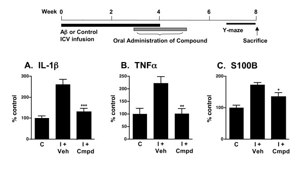
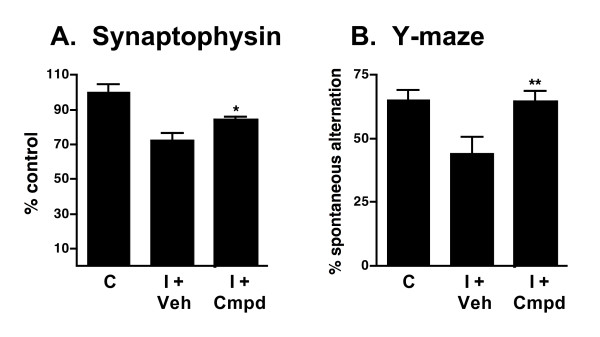
Verification
Claims the paper makes
The claims and statistics of this paper haven’t been checked yet.
Run the check — free, no account needed. It reads the open-access text and checks the sources it cites.
Claims other papers make about this one
Each is checked against this paper’s own text: does it say what the citing paper says it does?
The source backs part of the claim, not all of it — or backs it for a different population, condition or comparison.
In the citing paper
“First, p38α MAPK has been shown to mediate Aβ-induced expression of various proinflammatory cytokines/chemokines from microglial cells that causes neuroinflammation [25, 26, 29, 35].”
From Selective suppression of the α isoform of p38 MAPK rescues late-stage tau pathology (2016) · cites this paper as [35]
Experimental — accuracy not yet measuredThe source backs part of the claim, not all of it — or backs it for a different population, condition or comparison.
In the citing paper
“Targeted inactivation of p38α MAPK with small molecule inhibitors prevents the production of proinflammatory cytokines, reduces neurodegeneration, and improves neuronal and cognitive functions in rodent models of amyloid pathology [25, 26, 29, 35].”
From Selective suppression of the α isoform of p38 MAPK rescues late-stage tau pathology (2016) · cites this paper as [35]
Experimental — accuracy not yet measured
Self-check
Percentages, sums and reported differences re-computed from the paper’s own numbers.
The self-check needs the paper’s full text.
Citations
Cites
Amyloid oligomers exacerbate tau pathology in a mouse model of tauopathy
Selenica, M.-L. B., Brownlow, M., Jimenez, J. P., Lee, D. C., Pena, G., Dickey, C. A., Gordon, M. N., & Morgan, D. (2013) · Neurodegenerative Diseases · doi:10.1159/000337230 · Cited by 10,359
Kyriakis, J. M., & Avruch, J. (2001) · Physiological Reviews · doi:10.1152/physrev.2001.81.2.807 · Cited by 3,400
Hopkins, A. L., & Groom, C. R. (2002) · Nature Reviews Drug Discovery · doi:10.1038/nrd892 · Cited by 3,305
Antiinflammatory Action of Glucocorticoids — New Mechanisms for Old Drugs
Rhen, T., & Cidlowski, J. A. (2005) · New England Journal of Medicine · doi:10.1056/nejmra050541 · Cited by 3,149
Mechanisms involved in the side effects of glucocorticoids
SCHACKE, H. (2002) · Pharmacology & Therapeutics · doi:10.1016/s0163-7258(02)00297-8 · Cited by 1,847
Brain interleukin 1 and S-100 immunoreactivity are elevated in Down syndrome and Alzheimer disease.
Griffin, W. S., Stanley, L. C., Ling, C., White, L., MacLeod, V, Perrot, L. J., White, C. L., & Araoz, C. (1989) · Proceedings of the National Academy of Sciences · doi:10.1073/pnas.86.19.7611 · Cited by 1,777
Kaminska, B. (2005) · Biochimica et Biophysica Acta (BBA) - Proteins and Proteomics · doi:10.1016/j.bbapap.2005.08.017 · Cited by 1,285
p38 MAP kinases: key signalling molecules as therapeutic targets for inflammatory diseases
Kumar, S., Boehm, J. C., & Lee, J. C. (2003) · Nature Reviews Drug Discovery · doi:10.1038/nrd1177 · Cited by 1,199
Pardridge, W. M. (2007) · Drug Discovery Today · doi:10.1016/j.drudis.2006.10.013 · Cited by 1,191
Bhat, N. R., Zhang, P., Lee, J. C., & Hogan, E. L. (1998) · Journal of Neuroscience · doi:10.1523/jneurosci.18-05-01633.1998 · Cited by 714
Cited by
Sorted by citations
Pathological roles of MAPK signaling pathways in human diseases
Kim, E. K., & Choi, E.-J. (2010) · Biochimica et Biophysica Acta (BBA) - Molecular Basis of Disease · doi:10.1016/j.bbadis.2009.12.009 · Cited by 2,472
Rajmohan, R., & Reddy, P. H. (2016) · Journal of Alzheimer s Disease · doi:10.3233/jad-160612 · Cited by 730
Wild-type and mutant SOD1 share an aberrant conformation and a common pathogenic pathway in ALS
Bosco, D. A., Morfini, G. A., Karabacak, M., Song, Y., Gros‐Louis, F., Pasinelli, P., Goolsby, H., Fontaine, B. A., Lemay, N., McKenna-Yasek, D. M., et al. (2010) · Nature Neuroscience · doi:10.1038/nn.2660 · Cited by 685
Synaptic degeneration in Alzheimer disease
Tzioras, M., McGeachan, R. I., Durrant, C. S., & Spires-Jones, T. L. (2023) · Nature Reviews Neurology · doi:10.1038/s41582-022-00749-z · Cited by 504
Targeting p38 MAPK pathway for the treatment of Alzheimer's disease
Munoz, L., & Ammit, A. J. (2009) · Neuropharmacology · doi:10.1016/j.neuropharm.2009.11.010 · Cited by 381
Self-citation
Metadata
- Authors
- Munoz, L., Ranaivo, H. R., Roy, S. M., Hu, W., Craft, J. M., McNamara, L. K., Chico, L. W., Van Eldik, L. J., & Watterson, D. M.
- Affiliations
- Northwestern University (US)
- Corresponding author
- Watterson, D Martin
- Venue
- Journal of Neuroinflammation
- Published
- 2007-09-04
- Volume
- 4
- Issue
- 1
- Pages
- 21-21
- ISSN
- 1742-2094 (ISSN-L)
- PMC
- PMC2014744
- OpenAlex
- W2029663297
- Cited by
- 224
- References
- 83
- Citations by year
- 2026: 4 · 2025: 4 · 2024: 11 · 2023: 10 · 2022: 9 · 2021: 12 · 2020: 11 · 2019: 13 · 2018: 9 · 2017: 20 · 2016: 15 · 2015: 13 · 2014: 10 · 2013: 14 · 2012: 21
- FWCI
- 6.26
- Type
- journal article
- Language
- en
- Open access
- gold
- doi.org
- Licence
- cc-by
- Locations
- Journal of Neuroinflammation (publishedVersion, cc-by)
- PubMed Central (publishedVersion, cc-by)
- PubMed (publishedVersion)
- DOAJ (DOAJ: Directory of Open Access Journals) (submittedVersion, cc-by-sa)
- Funders
- Northwestern University
- National Institutes of Health
- National Institute on Aging (R01AG031311)
- Topics
- Life Sciences › Neuroscience › Cellular and Molecular Neuroscience › Alzheimer's disease research and treatments
- Life Sciences › Neuroscience › Neurology › Neuroinflammation and Neurodegeneration Mechanisms
- Life Sciences › Neuroscience › Cellular and Molecular Neuroscience › Neuroscience and Neuropharmacology Research
- Keywords
- Alzheimer's disease, inflammatory cytokines, glial activation, synaptic dysfunction, amyloid-beta, small molecule inhibitors, blood-brain barrier penetration, orally bioavailable drugs, disease-modifying therapy, neuroinflammation
- MeSH terms
- Alzheimer Disease, Animals, Behavior, Animal, Brain, Disease Models, Animal, Humans, Mice, Inbred C57BL, Pyridazines, Pyrimidines, Synapses, Drugs, Investigational, Up-Regulation, Cytokines, Inflammation Mediators, MAP Kinase Signaling System, Protein Kinase Inhibitors, Mitogen-Activated Protein Kinase 14, Mice
About these checks
Registry, retraction, Cites and statistics results are computed, not judged, with one exception: a printed reference that no index lists is read once by a model before it counts as a citation error. Claims are extracted by a model and kept only when their quote appears verbatim in the text; each cited source is then checked, with a model’s help, for whether it says what the claim relies on. Those verdicts can be wrong — read the claim’s quote and the cited source before relying on one.
Statistics and arithmetic are recomputed, not judged.
Confidence bands are shown only for fields calibrated on our annotated test set. None is calibrated yet, so no band is shown.
What counts as a claim ›How we measure confidence ›How we check this ›
Are you an author, or do you see a problem with this page? Request a review
