

The DOI matches its Crossref record; 91 of the 91 references it cites were found in a registry or an index.
Concurrent hippocampal induction of MHC II pathway components and glial activation with advanced aging is not correlated with cognitive impairment
Open accessAbstract
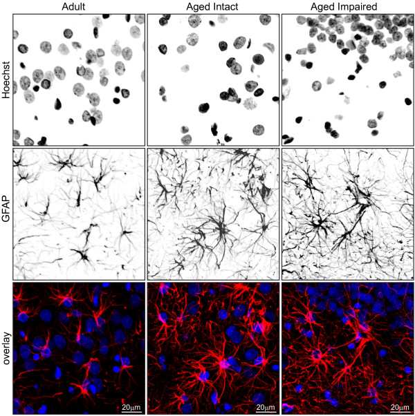
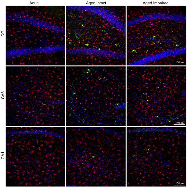
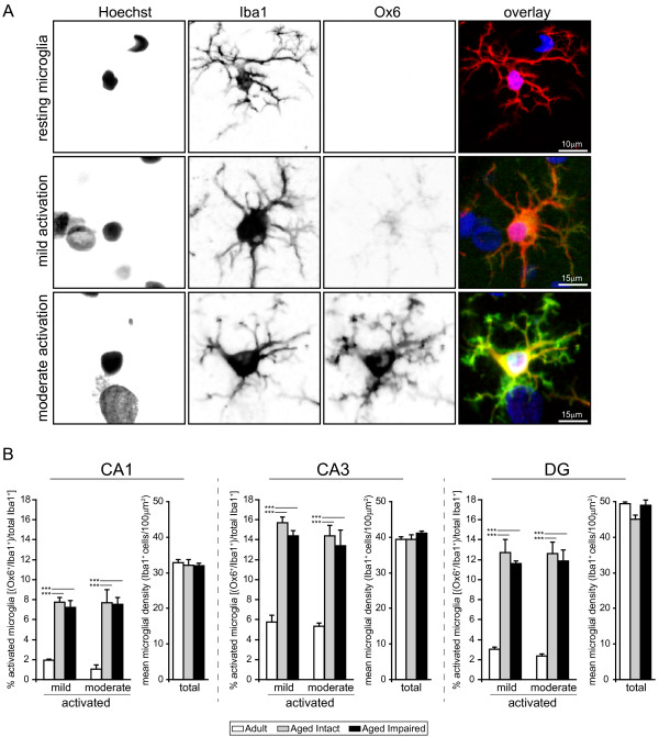
Background: Age-related cognitive dysfunction, including impairment of hippocampus-dependent spatial learning and memory, affects approximately half of the aged population. Induction of a variety of neuroinflammatory measures has been reported with brain aging but the relationship between neuroinflammation and cognitive decline with non-neurodegenerative, normative aging remains largely unexplored. This study sought to comprehensively investigate expression of the MHC II immune response pathway and glial activation in the hippocampus in the context of both aging and age-related cognitive decline. Methods: Three independent cohorts of adult (12-13 months) and aged (26-28 months) F344xBN rats were behaviorally characterized by Morris water maze testing. Expression of MHC II pathway-associated genes identified by transcriptomic analysis as upregulated with advanced aging was quantified by qPCR in synaptosomal fractions derived from whole hippocampus and in hippocampal subregion dissections (CA1, CA3, and DG). Activation of astrocytes and microglia was assessed by GFAP and Iba1 protein expression, and by immunohistochemical visualization of GFAP and both CD74 (Ox6) and Iba1. Results: We report a marked age-related induction of neuroinflammatory signaling transcripts (i.e., MHC II components, toll-like receptors, complement, and downstream signaling factors) throughout the hippocampus in all aged rats regardless of cognitive status. Astrocyte and microglial activation was evident in CA1, CA3 and DG of intact and impaired aged rat groups, in the absence of differences in total numbers of GFAP+astrocytes or Iba1+microglia. Both mild and moderate microglial activation was significantly increased in all three hippocampal subregions in aged cognitively intact and cognitively impaired rats compared to adults. Neither induction of MHCII pathway gene expression nor glial activation correlated to cognitive performance. Conclusions: These data demonstrate a novel, coordinated age-related induction of the MHC II immune response pathway and glial activation in the hippocampus, indicating an allostatic shift toward a para-inflammatory phenotype with advancing age. Our findings demonstrate that age-related induction of these aspects of hippocampal neuroinflammation, while a potential contributing factor, is not sufficient by itself to elicit impairment of spatial learning and memory in models of normative aging. Future efforts are needed to understand how neuroinflammation may act synergistically with cognitive-decline specific alterations to cause cognitive impairment.
Overview
The claims and statistics of this paper haven’t been checked yet.
Run the check — free, no account needed. It reads the open-access text and checks the sources it cites.
Integrity signals
Registry
Cites
Statistics
Figures
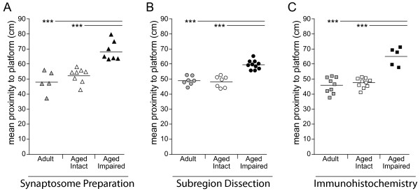
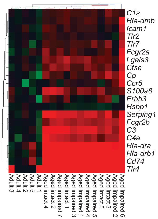
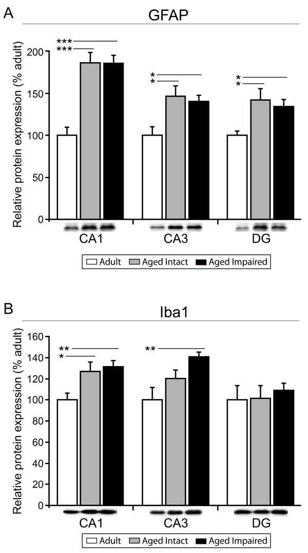
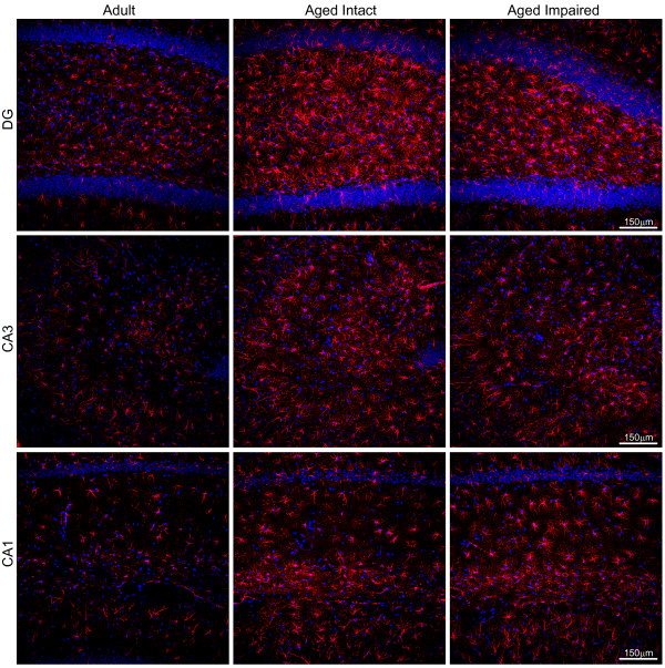
Verification
Claims the paper makes
The claims and statistics of this paper haven’t been checked yet.
Run the check — free, no account needed. It reads the open-access text and checks the sources it cites.
Claims other papers make about this one
Each is checked against this paper’s own text: does it say what the citing paper says it does?
In the citing paper
“Microglia under normal aging (ie, aging in absence of neurodegeneration) showed upregulation of genes associated with antigen presentation, such as MHCII, CD86, and CIITA (Frank et al, 2006;Godbout et al, 2005;VanGuilder et al, 2011), and suppression of anti-inflammatory genes, such as IL-10 and CD200 (Frank et al, 2006).”
From Microglia Priming with Aging and Stress (2017) · cites this paper as VanGuilder et al, 2011
Self-check
Percentages, sums and reported differences re-computed from the paper’s own numbers.
The self-check needs the paper’s full text.
Citations
Cites
Origin and physiological roles of inflammation
Medzhitov, R. (2008) · Nature · doi:10.1038/nature07201 · Cited by 6,618
Resting microglial cells are highly dynamic surveillants of brain parenchyma in vivo
Nimmerjahn, A., Kirchhoff, F., & Helmchen, F. (2005) · Science · doi:10.1126/science.1110647 · Cited by 5,827
Microglia: Active sensor and versatile effector cells in the normal and pathologic brain
Hanisch, U.-K., & Kettenmann, H. (2007) · Nature Neuroscience · doi:10.1038/nn1997 · Cited by 3,676
DAMPs, PAMPs and alarmins: all we need to know about danger
Bianchi, M. E. (2007) · Journal of Leukocyte Biology · doi:10.1189/jlb.0306164 · Cited by 2,794
Insights into the ageing mind: a view from cognitive neuroscience
Hedden, T., & Gabrieli, J. D. E. (2004) · Nature Reviews Neuroscience · doi:10.1038/nrn1323 · Cited by 2,223
The ageing systemic milieu negatively regulates neurogenesis and cognitive function
Villeda, S. A., Luo, J., Mosher, K. I., Zou, B., Britschgi, M., Bieri, G., Stan, T. M., Fainberg, N., Ding, Z., Eggel, A., et al. (2011) · Nature · doi:10.1038/nature10357 · Cited by 1,763
Immune modulation of learning, memory, neural plasticity and neurogenesis
Yirmiya, R., & Goshen, I. (2011) · Brain, Behavior, and Immunity · doi:10.1016/j.bbi.2010.10.015 · Cited by 1,483
Debris clearance by microglia: an essential link between degeneration and regeneration
Neumann, H., Kotter, M. R., & Franklin, R. J. M. (2008) · Brain · doi:10.1093/brain/awn109 · Cited by 1,111
Gene-expression profile of the ageing brain in mice
Lee, C.-K., Weindruch, R., & Prolla, T. A. (2000) · Nature Genetics · doi:10.1038/77046 · Cited by 1,055
Godbout, J. P., Chen, J., Abraham, J., Richwine, A. F., Berg, B. M., Kelley, K. W., & Johnson, R. W. (2005) · The FASEB Journal · doi:10.1096/fj.05-3776fje · Cited by 813
Cited by
Sorted by citations
Items 101–135 of 135
Wang, X. L., Shao, X., Yu, L., Sun, J., Yin, X., Chen, Z., Xu, Y., Wang, N., Zhang, D., Qiu, W., et al. (2025) · Molecular Brain · doi:10.1186/s13041-025-01180-3 · Cited by 10
Role of Autophagy in HIV-1 and Drug Abuse-Mediated Neuroinflammaging
Sil, S., Thangaraj, A., Oladapo, A., Hu, G., Kutchy, N. A., Liao, K., Buch, S. J., & Periyasamy, P. (2022) · Viruses · doi:10.3390/v15010044 · Cited by 10
Shishkina, G. T., Kalinina, T. S., Lanshakov, D. A., Bulygina, V. V., Komysheva, N. P., Bannova, A. V., Drozd, U. S., & Дыгало, Н. Н. (2023) · Biomedicines · doi:10.3390/biomedicines11102595 · Cited by 11
Kneynsberg, A., & Kanaan, N. M. (2017) · eNeuro · doi:10.1523/eneuro.0043-17.2017 · Cited by 11
Huang, Y., Liu, Z., Li, M., Wang, D., Ye, J., Hu, Q., Zhang, Q., Lin, Y., Chen, R., Liang, X., et al. (2024) · Immunity & Ageing · doi:10.1186/s12979-024-00452-1 · Cited by 8
The Age-related Gliosis and Accompanying Deficit in Spatial Learning are Unaffected by Dimebon
Cowley, T. R., González-Reyes, R. E., Richardson, J. C., Virley, D. J., Upton, N., & Lynch, M. A. (2012) · Neurochemical Research · doi:10.1007/s11064-012-0884-0 · Cited by 6
Krejčová, L. V., Bento‐Torres, J., Diniz, D. G., Pereira, A., Hornsby, M. B.-D.-O., Morais, A., Mendes-da-Silva, R. F., Abadie‐Guedes, R., dos Santos, Â. A., de Lima, D. S. C., et al. (2024) · Brain Sciences · doi:10.3390/brainsci14050497 · Cited by 5
Ebersole, J., Rose, G. M., Eid, T., Behar, K. L., & Patrylo, P. R. (2021) · Neurobiology of Aging · doi:10.1016/j.neurobiolaging.2021.02.015 · Cited by 5
Neurobiological Mechanisms of Cognitive Decline Correlated with Brain Aging
Zhang, X., An, H., Chen, Y., & Shu, N. (2023) · Advances in experimental medicine and biology · doi:10.1007/978-981-99-1627-6_10 · Cited by 4
Lai, K. O., Wong, J. H., Tham, N., Fairley, L. H., Naik, R. R., Wang, Y., Langley, S. R., & Barron, A. M. (2025) · Aging Cell · doi:10.1111/acel.70039 · Cited by 4
Contemporary insights into neuroimmune interactions across development and aging
Yeo, X. Y., Choi, Y., Hong, Y., Kwon, H. N., & Jung, S. (2025) · Frontiers in Neurology · doi:10.3389/fneur.2025.1611124 · Cited by 5
Microglial Hemoxygenase-1 Deletion Reduces Inflammation in the Retina of Old Mice with Tauopathy
Fernández‐Albarral, J. A., Salobrar‐García, E., Matamoros, J. A., Fernández‐Mendívil, C., del Sastre, E., Chen, L., de Hoz, R., López‐Cuenca, I., Sánchez-Puebla, L., Ramirez, J. M., et al. (2022) · Antioxidants · doi:10.3390/antiox11112151 · Cited by 4
Plantera, L., Immig, K., Fritsche, A., Reinicke, M., Zemer, A., Monsonego, A., Ceglarek, U., & Bechmann, I. (2025) · Molecular Nutrition & Food Research · doi:10.1002/mnfr.70168 · Cited by 3
Active immunization: A novel therapeutic strategy for treating Alzheimer's disease
Mazahir, F. (2025) · European Journal of Pharmacology · doi:10.1016/j.ejphar.2025.178288 · Cited by 3
Microglial morphology reflects cognitive status in the aging rat brain
Myers, S. J., Roseborough, A. D., Bayona, C. X., Carrese, C., Allman, B. L., & Whitehead, S. N. (2026) · Brain Behavior and Immunity · doi:10.1016/j.bbi.2026.106533 · Cited by 1
Molecular Aspects of Hippocampal Aging
Temido‐Ferreira, M., & Lopes, L. V. (2020) · Springer eBooks · doi:10.1007/978-3-030-40955-5_3 · Cited by 1
Malik, S., Xavier, S., Soch, A., Younesi, S., Yip, J. L. K., Slayo, M., Barrientos, R. M., Sominsky, L., & Spencer, S. J. (2024) · Neurobiology of Aging · doi:10.1016/j.neurobiolaging.2024.04.010 · Cited by 1
Fenn, A. M., Norden, D. M., & Godbout, J. P. (2015) · Wiley eBooks · doi:10.1002/9781118732748.ch6 · Cited by 1
Martin, S. A. (2013) · Illinois Digital Environment for Access to Learning and Scholarship (University of Illinois at Urbana-Champaign)
Aging and Microglial Activation in Neurodegenerative Diseases
Oyarzabal, E. A., Shih, Y. I., & Hong, J. (2016) · Oxidative stress in applied basic research and clinical practice · doi:10.1007/978-3-319-33486-8_7
Caffeine Consumption and Prevention of Cognitive Decline
Góngora-Alfaro, J. L., Álvarez-Cervera, F. J., & Bata-García, J. L. (2014) · Elsevier eBooks · doi:10.1016/b978-0-12-407824-6.00081-1
Long-Lasting Memory Impairments after Subchronic Immune Challenge
Tchessalova, D. (2019) · Deep Blue (University of Michigan)
Koch, B. M. E. (2019) · eDiss (Georg-August-Universität Göttingen) · doi:10.53846/goediss-7716
Jadhav, V. (2021) · IUScholarWorks (Indiana University) · doi:10.7912/c2/2086
Fletcher, S. (2019) · Faculty Opinions – Post-Publication Peer Review of the Biomedical Literature · doi:10.3410/f.732900592.793560211
Rommy von Bernhardi, M., Flores, B., & Nakanishi, H. (2014) · Springer eBooks · doi:10.1007/978-1-4939-1429-6_13
Rombaut, B. (2024) · Research Publications (Maastricht University) · doi:10.26481/dis.20241211br
Age-related brain functional changes and microglia
Nakamura, Y., Hisaoka‐Nakashima, K., & Morioka, N. (2025) · Folia Pharmacologica Japonica · doi:10.1254/fpj.25033
Huang, Y., Liu, Z., Li, M., Wang, D., Ye, J., Hu, Q., Zhang, Q., Lin, Y., Chen, R., Liang, X., et al. (2024) · Figshare · doi:10.6084/m9.figshare.26333395
Huang, Y., Liu, Z., Li, M., Wang, D., Ye, J., Hu, Q., Zhang, Q., Lin, Y., Chen, R., Liang, X., et al. (2024) · Figshare · doi:10.6084/m9.figshare.26333189
Huang, Y., Liu, Z., Li, M., Wang, D., Ye, J., Hu, Q., Zhang, Q., Lin, Y., Chen, R., Liang, X., et al. (2024) · Figshare · doi:10.6084/m9.figshare.26750735
Huang, Y., Liu, Z., Li, M., Wang, D., Ye, J., Hu, Q., Zhang, Q., Lin, Y., Chen, R., Liang, X., et al. (2024) · Figshare · doi:10.6084/m9.figshare.26750738
Chataigner, M. (2021) · theses.fr (ABES) · doi:10.70675/a4317690zc4c8z4ba6za738zd457c6bb9b74
Guerin, S. P., Sanchez, K., Verma, A., Salem, N. A., Mayfield, R. D., & Fonken, L. K. (2026) · Journal of Neuroimmunology · doi:10.1016/j.jneuroim.2026.578947
Metadata
- Authors
- VanGuilder, H. D., Bixler, G. V., Brucklacher, R. M., Farley, J. A., Yan, H., Warrington, J. P., Sonntag, W. E., & Freeman, W. M.
- Affiliations
- Pennsylvania State University (US)
- University of Oklahoma Health Sciences Center (US)
- Corresponding author
- Freeman, Willard M
- Venue
- Journal of Neuroinflammation
- Published
- 2011-10-11
- Volume
- 8
- Issue
- 1
- Pages
- 138-138
- ISSN
- 1742-2094 (ISSN-L)
- PMC
- PMC3216278
- OpenAlex
- W2081626361
- Cited by
- 132
- References
- 91
- Citations by year
- 2026: 3 · 2025: 7 · 2024: 7 · 2023: 4 · 2022: 11 · 2021: 12 · 2020: 10 · 2019: 11 · 2018: 10 · 2017: 15 · 2016: 11 · 2015: 9 · 2014: 7 · 2013: 10 · 2012: 5
- FWCI
- 3.89
- Type
- journal article
- Language
- en
- Open access
- gold
- doi.org
- Licence
- cc-by
- Locations
- Journal of Neuroinflammation (publishedVersion, cc-by)
- Europe PMC (PubMed Central) (publishedVersion, cc-by)
- PubMed Central (publishedVersion, cc-by)
- PubMed (publishedVersion)
- DOAJ (DOAJ: Directory of Open Access Journals) (submittedVersion)
- Funders
- Donald W. Reynolds Foundation
- National Institutes of Health (5r01ag026607-06)
- National Institute on Aging (#R01AG026607, 5R01AG026607, P01AG011370)
- Topics
- Life Sciences › Neuroscience › Neurology › Neuroinflammation and Neurodegeneration Mechanisms
- Life Sciences › Neuroscience › Neurology › Neurological Disease Mechanisms and Treatments
- Life Sciences › Neuroscience › Developmental Neuroscience › Neurogenesis and neuroplasticity mechanisms
- Keywords
- hippocampal inflammation, glial activation, microglial activation, astrocytes, cognitive aging, age-related cognitive decline, spatial learning and memory, Toll-like receptors, complement system, hippocampal subfields
- MeSH terms
- Aging, Animals, Histocompatibility Antigens Class II, Behavior, Animal, Cognition Disorders, Genes, MHC Class II, Hippocampus, Humans, Major Histocompatibility Complex, Male, Neuroglia, Rats, Inbred F344, Biomarkers, Maze Learning, Gene Expression Profiling, Microarray Analysis, Rats, Transcriptome
About these checks
Registry, retraction, Cites and statistics results are computed, not judged, with one exception: a printed reference that no index lists is read once by a model before it counts as a citation error. Claims are extracted by a model and kept only when their quote appears verbatim in the text; each cited source is then checked, with a model’s help, for whether it says what the claim relies on. Those verdicts can be wrong — read the claim’s quote and the cited source before relying on one.
Statistics and arithmetic are recomputed, not judged.
Confidence bands are shown only for fields calibrated on our annotated test set. None is calibrated yet, so no band is shown.
What counts as a claim ›How we measure confidence ›How we check this ›
Are you an author, or do you see a problem with this page? Request a review
