

The DOI matches its Crossref record; 91 of the 91 references it cites were found in a registry or an index.
Concurrent hippocampal induction of MHC II pathway components and glial activation with advanced aging is not correlated with cognitive impairment
Open accessAbstract
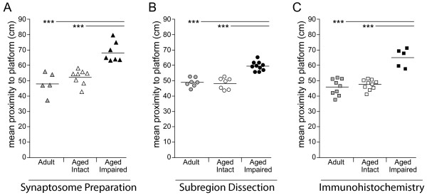
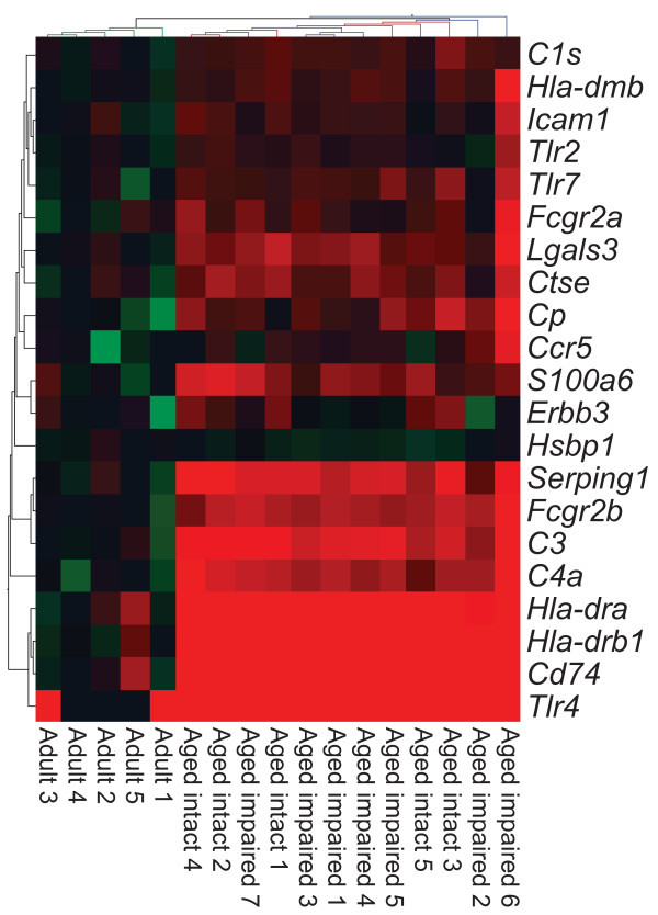
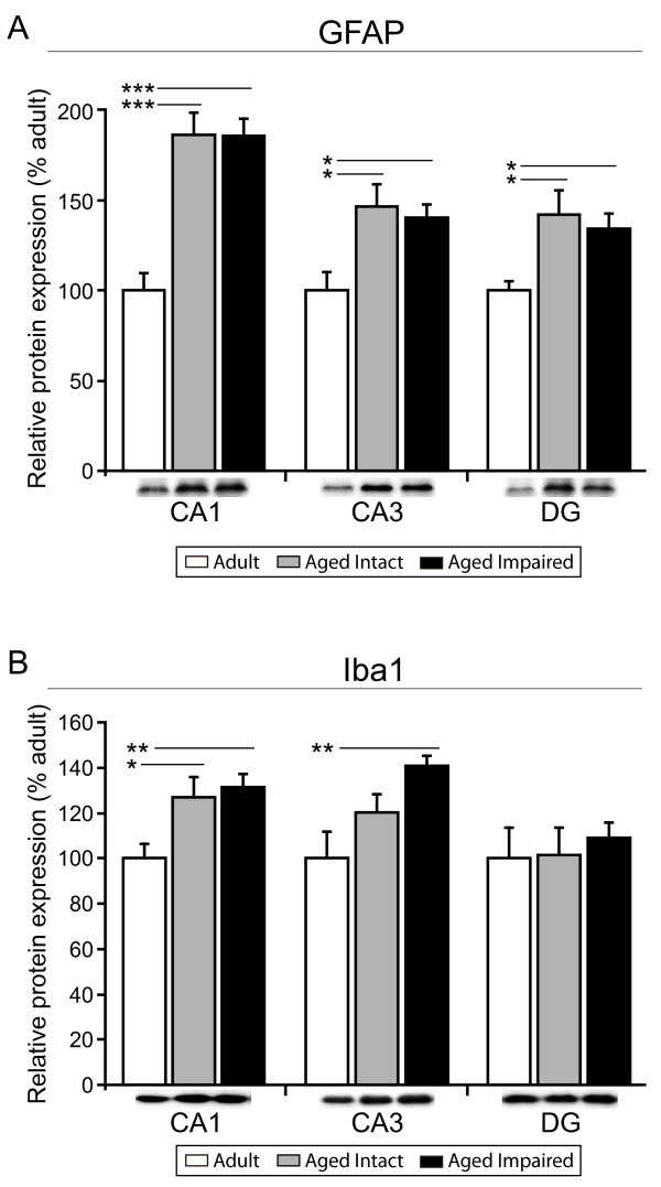
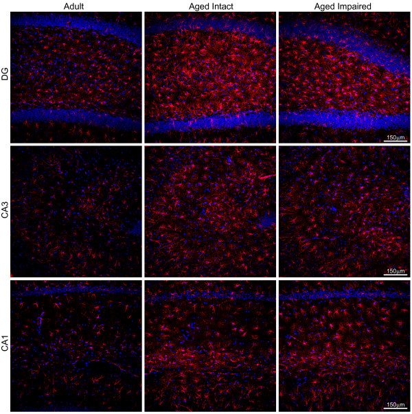
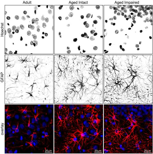
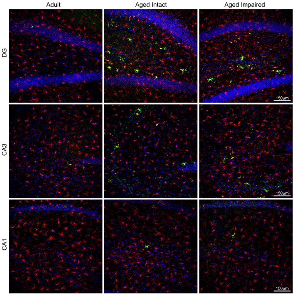
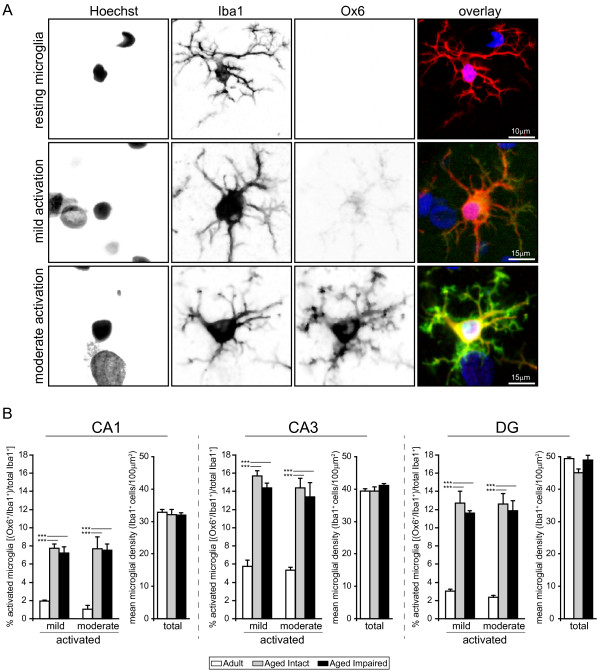
Background: Age-related cognitive dysfunction, including impairment of hippocampus-dependent spatial learning and memory, affects approximately half of the aged population. Induction of a variety of neuroinflammatory measures has been reported with brain aging but the relationship between neuroinflammation and cognitive decline with non-neurodegenerative, normative aging remains largely unexplored. This study sought to comprehensively investigate expression of the MHC II immune response pathway and glial activation in the hippocampus in the context of both aging and age-related cognitive decline. Methods: Three independent cohorts of adult (12-13 months) and aged (26-28 months) F344xBN rats were behaviorally characterized by Morris water maze testing. Expression of MHC II pathway-associated genes identified by transcriptomic analysis as upregulated with advanced aging was quantified by qPCR in synaptosomal fractions derived from whole hippocampus and in hippocampal subregion dissections (CA1, CA3, and DG). Activation of astrocytes and microglia was assessed by GFAP and Iba1 protein expression, and by immunohistochemical visualization of GFAP and both CD74 (Ox6) and Iba1. Results: We report a marked age-related induction of neuroinflammatory signaling transcripts (i.e., MHC II components, toll-like receptors, complement, and downstream signaling factors) throughout the hippocampus in all aged rats regardless of cognitive status. Astrocyte and microglial activation was evident in CA1, CA3 and DG of intact and impaired aged rat groups, in the absence of differences in total numbers of GFAP+astrocytes or Iba1+microglia. Both mild and moderate microglial activation was significantly increased in all three hippocampal subregions in aged cognitively intact and cognitively impaired rats compared to adults. Neither induction of MHCII pathway gene expression nor glial activation correlated to cognitive performance. Conclusions: These data demonstrate a novel, coordinated age-related induction of the MHC II immune response pathway and glial activation in the hippocampus, indicating an allostatic shift toward a para-inflammatory phenotype with advancing age. Our findings demonstrate that age-related induction of these aspects of hippocampal neuroinflammation, while a potential contributing factor, is not sufficient by itself to elicit impairment of spatial learning and memory in models of normative aging. Future efforts are needed to understand how neuroinflammation may act synergistically with cognitive-decline specific alterations to cause cognitive impairment.
Overview
The claims and statistics of this paper haven’t been checked yet.
Run the check — free, no account needed. It reads the open-access text and checks the sources it cites.
Integrity signals
Registry
Cites
Statistics
Figures
Verification
Claims the paper makes
The claims and statistics of this paper haven’t been checked yet.
Run the check — free, no account needed. It reads the open-access text and checks the sources it cites.
Claims other papers make about this one
Each is checked against this paper’s own text: does it say what the citing paper says it does?
In the citing paper
“Microglia under normal aging (ie, aging in absence of neurodegeneration) showed upregulation of genes associated with antigen presentation, such as MHCII, CD86, and CIITA (Frank et al, 2006;Godbout et al, 2005;VanGuilder et al, 2011), and suppression of anti-inflammatory genes, such as IL-10 and CD200 (Frank et al, 2006).”
From Microglia Priming with Aging and Stress (2017) · cites this paper as VanGuilder et al, 2011
Self-check
Percentages, sums and reported differences re-computed from the paper’s own numbers.
The self-check needs the paper’s full text.
Citations
Cites
Origin and physiological roles of inflammation
Medzhitov, R. (2008) · Nature · doi:10.1038/nature07201 · Cited by 6,618
Resting microglial cells are highly dynamic surveillants of brain parenchyma in vivo
Nimmerjahn, A., Kirchhoff, F., & Helmchen, F. (2005) · Science · doi:10.1126/science.1110647 · Cited by 5,827
Microglia: Active sensor and versatile effector cells in the normal and pathologic brain
Hanisch, U.-K., & Kettenmann, H. (2007) · Nature Neuroscience · doi:10.1038/nn1997 · Cited by 3,676
DAMPs, PAMPs and alarmins: all we need to know about danger
Bianchi, M. E. (2007) · Journal of Leukocyte Biology · doi:10.1189/jlb.0306164 · Cited by 2,794
Insights into the ageing mind: a view from cognitive neuroscience
Hedden, T., & Gabrieli, J. D. E. (2004) · Nature Reviews Neuroscience · doi:10.1038/nrn1323 · Cited by 2,223
The ageing systemic milieu negatively regulates neurogenesis and cognitive function
Villeda, S. A., Luo, J., Mosher, K. I., Zou, B., Britschgi, M., Bieri, G., Stan, T. M., Fainberg, N., Ding, Z., Eggel, A., et al. (2011) · Nature · doi:10.1038/nature10357 · Cited by 1,763
Immune modulation of learning, memory, neural plasticity and neurogenesis
Yirmiya, R., & Goshen, I. (2011) · Brain, Behavior, and Immunity · doi:10.1016/j.bbi.2010.10.015 · Cited by 1,483
Debris clearance by microglia: an essential link between degeneration and regeneration
Neumann, H., Kotter, M. R., & Franklin, R. J. M. (2008) · Brain · doi:10.1093/brain/awn109 · Cited by 1,111
Gene-expression profile of the ageing brain in mice
Lee, C.-K., Weindruch, R., & Prolla, T. A. (2000) · Nature Genetics · doi:10.1038/77046 · Cited by 1,055
Godbout, J. P., Chen, J., Abraham, J., Richwine, A. F., Berg, B. M., Kelley, K. W., & Johnson, R. W. (2005) · The FASEB Journal · doi:10.1096/fj.05-3776fje · Cited by 813
Methods of Behavior Analysis in Neuroscience
(2008) · CRC Press eBooks · doi:10.1201/9780367802622 · Cited by 777
Meta-analysis of age-related gene expression profiles identifies common signatures of aging
de Magalhães, J. P., Curado, J., & Church, G. M. (2009) · Bioinformatics · doi:10.1093/bioinformatics/btp073 · Cited by 737
Immune activation in brain aging and neurodegeneration: too much or too little?
Lucin, K. M., & Wyss-Coray, T. (2009) · Neuron · doi:10.1016/j.neuron.2009.08.039 · Cited by 679
Intellectual Development in Adulthood: The Seattle Longitudinal Study
Schaie, K. W., & Baltes, P. B. (1996) · Medical Entomology and Zoology · Cited by 674
Cytokines and cognition–the case for a head-to-toe inflammatory paradigm.
Wilson, C. J., Finch, C. E., & Cohen, H. J. (2002) · Journal of the American Geriatrics Society · doi:10.1046/j.1532-5415.2002.50619.x · Cited by 637
Gene expression changes in the course of normal brain aging are sexually dimorphic
Berchtold, N. C., Cribbs, D. H., Coleman, P. D., Rogers, J., Head, E., Kim, R., Beach, T., Miller, C., Troncoso, J., Trojanowski, J. Q., et al. (2008) · Proceedings of the National Academy of Sciences · doi:10.1073/pnas.0806883105 · Cited by 626
Blalock, E. M., Chen, K.-C., Sharrow, K., Herman, J. P., Porter, N. M., Foster, T. C., & Landfield, P. W. (2003) · The Journal of Neuroscience · doi:10.1523/jneurosci.23-09-03807.2003 · Cited by 570
Oxidative stress and protein aggregation during biological aging
Squier, T. C. (2001) · Experimental Gerontology · doi:10.1016/s0531-5565(01)00139-5 · Cited by 519
Microglial control of neuronal death and synaptic properties
Bessis, A., Béchade, C., Bernard, D., & Roumier, A. (2006) · Glia · doi:10.1002/glia.20459 · Cited by 486
THE USE OF ANIMAL MODELS TO STUDY THE EFFECTS OF AGING ON COGNITION
Gallagher, M., & Rapp, P. R. (1997) · Annual Review of Psychology · doi:10.1146/annurev.psych.48.1.339 · Cited by 445
Brain Aging: Models, Methods, and Mechanisms
Root-Bernstein, R. S. (2007) · JAMA · doi:10.1001/jama.298.23.2798 · Cited by 396
Multistability of cognitive maps in the hippocampus of old rats
Barnes, C. A., Suster, M. S., Shen, J., & McNaughton, B. L. (1997) · Nature · doi:10.1038/40859 · Cited by 370
Gorelick, P. B. (2010) · Annals of the New York Academy of Sciences · doi:10.1111/j.1749-6632.2010.05726.x · Cited by 361
Methods of Behavior Analysis in Neuroscience
(2000) · CRC Press eBooks · doi:10.1201/9781420041811 · Cited by 345
Conde, J. R., & Streit, W. J. (2006) · Journal of Neuropathology and Experimental Neurology · doi:10.1097/01.jnen.0000202887.22082.63 · Cited by 344
mRNA up-regulation of MHC II and pivotal pro-inflammatory genes in normal brain aging
Frank, M. G., Barrientos, R. M., Biedenkapp, J. C., Rudy, J. W., Watkins, L. R., & Maier, S. F. (2006) · Neurobiology of Aging · doi:10.1016/j.neurobiolaging.2005.03.013 · Cited by 327
Sonntag, W. E., Lynch, C. D., Cooney, P. T., & Hutchins, P. M. (1997) · Endocrinology · doi:10.1210/endo.138.8.5330 · Cited by 307
Self-citationAloisi, F., Ria, F., Penna, G., & Adorini, L. (1998) · The Journal of Immunology · doi:10.4049/jimmunol.160.10.4671 · Cited by 276
Glia: The many ways to modulate synaptic plasticity
Achour, S. B., & Pascual, O. (2010) · Neurochemistry International · doi:10.1016/j.neuint.2010.02.013 · Cited by 240
Sustained hippocampal IL-1β overexpression impairs contextual and spatial memory in transgenic mice
Hein, A. M., Stasko, M. R., Matousek, S. B., Scott-McKean, J. J., Maier, S. F., Olschowka, J. A., Costa, A. C. S., & O’Banion, M. K. (2009) · Brain Behavior and Immunity · doi:10.1016/j.bbi.2009.10.002 · Cited by 235
Microglial Expression of MHC Class II Increases in Normal Aging of Nonhuman Primates
Sheffield, L. G., & Berman, N. E. J. (1998) · Neurobiology of Aging · doi:10.1016/s0197-4580(97)00168-1 · Cited by 234
What is the most sensitive measure of water maze probe test performance?
Maei, H. R. (2009) · Frontiers in Integrative Neuroscience · doi:10.3389/neuro.07.004.2009 · Cited by 228
Huang, Y., Henry, C. J., Dantzer, R., Johnson, R. W., & Godbout, J. P. (2007) · Neurobiology of Aging · doi:10.1016/j.neurobiolaging.2007.04.012 · Cited by 226
(2007) · CRC Press eBooks · doi:10.1201/9781420005523 · Cited by 222
Landfield, P. W., Rose, G., Sandles, L., Wohlstadter, T. C., & LYNCH, G. S. (1977) · Journal of Gerontology · doi:10.1093/geronj/32.1.3 · Cited by 221
Neniškytė, U., Neher, J. J., & Brown, G. C. (2011) · Journal of Biological Chemistry · doi:10.1074/jbc.m111.267583 · Cited by 215
Rowe, W. B., Blalock, E. M., Chen, K.-C., Kadish, I., Wang, D., Barrett, J. E., Thibault, O., Porter, N. M., Rose, G. M., & Landfield, P. W. (2007) · Journal of Neuroscience · doi:10.1523/jneurosci.4163-06.2007 · Cited by 215
Neuroinflammation and Cognitive Aging
Ownby, R. L. (2010) · Current Psychiatry Reports · doi:10.1007/s11920-009-0082-1 · Cited by 212
Increased microglial activation and protein nitration in white matter of the aging monkey☆
Sloane, J. A., Hollander, W., Moss, M. B., Rosene, D. L., & Abraham, C. R. (1999) · Neurobiology of Aging · doi:10.1016/s0197-4580(99)00066-4 · Cited by 208
Cellular environment facilitates protein accumulation in aged rat hippocampus
Gavilán, M. P., Vela, J. M., Castaño, A., Ramos, B., del Río, J. C., Vitórica, J., & Ruano, D. (2005) · Neurobiology of Aging · doi:10.1016/j.neurobiolaging.2005.05.010 · Cited by 201
Hippocampal Subfield Volumes: Age, Vascular Risk, and Correlation with Associative Memory
Shing, Y. L., Rodrigue, K. M., Kennedy, K. M., Fandakova, Y., Bodammer, N., Werkle‐Bergner, M., Lindenberger, U., & Raz, N. (2011) · Frontiers in Aging Neuroscience · doi:10.3389/fnagi.2011.00002 · Cited by 200
Brooks, W. M., Stidley, C. A., Petropoulos, H., Jung, R. E., Weers, D. C., Friedman, S. D., Barlow, M. A., Sibbitt, W. L., & Yeo, R. A. (2000) · Journal of Neurotrauma · doi:10.1089/089771500415382 · Cited by 193
Dash, P. K., Orsi, S. A., Zhang, M., Grill, R. J., Pati, S., Zhao, J., & Moore, A. N. (2010) · PLoS ONE · doi:10.1371/journal.pone.0011383 · Cited by 193
Effects of ageing on microglia in the normal rat brain: immunohistochemical observations
Ogura, K., Ogawa, M., & Yoshida, M. (1994) · Neuroreport · doi:10.1097/00001756-199406020-00016 · Cited by 181
Signatures of hippocampal oxidative stress in aged spatial learning-impaired rodents
Nicolle, M. M., Gonzalez, J., Sugaya, K., Baskerville, K. A., Bryan, D., Lund, K. C., Gallagher, M., & McKinney, M. (2001) · Neuroscience · doi:10.1016/s0306-4522(01)00374-8 · Cited by 178
A role for microglia in synaptic plasticity?
Tremblay, M.-È., & Majewska, A. K. (2011) · Communicative & Integrative Biology · doi:10.4161/cib.4.2.14506 · Cited by 176 · cited 1×
“roles in neuronal function by metabolizing glutamate, providing nutritional support, and removing potentially toxic cell debris [15,34,35] In the aged hippocampus, cellular debris detected by activated microglia and astrocytes may include degenerating synaptic terminals [5,11,36], myelin fragments [37], and misfolded…”
BackgroundPhagocytic Clearance of Apoptotic Neurons by Microglia/Brain Macrophages In Vitro
Witting, A., Müller, P. K., Herrmann, A., Kettenmann, H., & Nolte, C. (2000) · Journal of Neurochemistry · doi:10.1046/j.1471-4159.2000.0751060.x · Cited by 175
Innate immune receptor expression in normal brain aging
Letiembre, M., Hao, W., Liu, Y., Walter, S., Mihaljevic, I., Rivest, S., Hartmann, T., & Fassbender, K. (2007) · Neuroscience · doi:10.1016/j.neuroscience.2007.01.004 · Cited by 175
MHC class II molecules on the move for successful antigen presentation
Rocha, N., & Neefjes, J. (2007) · The EMBO Journal · doi:10.1038/sj.emboj.7601945 · Cited by 167
Fuller, S., Steele, M. L., & Münch, G. (2010) · Mutation research · doi:10.1016/j.mrfmmm.2009.08.016 · Cited by 164
Stereological analysis of astrocyte and microglia in aging mouse hippocampus
Long, J. M., Kalehua, A. N., Muth, N. J., Calhoun, M. E., Jucker, M., Hengemihle, J. M., Ingram, D. K., & Mouton, P. R. (1998) · Neurobiology of Aging · doi:10.1016/s0197-4580(98)00088-8 · Cited by 163
Calabrese, V. (2004) · Mechanisms of Ageing and Development · doi:10.1016/j.mad.2004.01.003 · Cited by 162
Kadish, I., Thibault, O., Blalock, E. M., Chen, K.-C., Gant, J. C., Porter, N. M., & Landfield, P. W. (2009) · Journal of Neuroscience · doi:10.1523/jneurosci.4599-08.2009 · Cited by 155
VanGuilder, H. D., Yan, H., Farley, J. A., Sonntag, W. E., & Freeman, W. M. (2010) · Journal of Neurochemistry · doi:10.1111/j.1471-4159.2010.06719.x · Cited by 154
Self-citationThe concentration of glial fibrillary acidic protein increases with age in the mouse and rat brain
O'Callaghan, J. P., & Miller, D. B. (1991) · Neurobiology of Aging · doi:10.1016/0197-4580(91)90057-q · Cited by 152
Lee, S. C., Moore, G. R. W., Golenwsky, G., & Raine, C. S. (1990) · Journal of Neuropathology & Experimental Neurology · doi:10.1097/00005072-199003000-00005 · Cited by 149
Microglial activation in white matter lesions and nonlesional white matter of ageing brains
Simpson, J. E., Ince, P. G., Higham, C. E., Gelsthorpe, C., Fernando, M. S., Matthews, F. E., Forster, G., O'Brien, J. T., Barber, R., Kalaria, R. N., et al. (2007) · Neuropathology and Applied Neurobiology · doi:10.1111/j.1365-2990.2007.00890.x · Cited by 146
O'Callaghan, R. M., Griffin, É. W., & Kelly, Á. M. (2009) · Hippocampus · doi:10.1002/hipo.20591 · Cited by 146
Glial reaction in the hippocampal formation is highly correlated with aging in human brain
David, J., Ghozali, F., Fallet‐Bianco, C., Wattez, A., Delaine, S. C., Boniface, B., Menza, C. D., & Delacourte, A. (1997) · Neuroscience Letters · doi:10.1016/s0304-3940(97)00708-8 · Cited by 145
VanGuilder, H. D., Farley, J. A., Han, Y., Kirk, C. A. V., Mitschelen, M. C., Sonntag, W. E., & Freeman, W. M. (2011) · Neurobiology of Disease · doi:10.1016/j.nbd.2011.03.012 · Cited by 144
Self-citationPoon, H. F., Shepherd, H. M., Reed, T. T., Calabrese, V., Stella, A. G., Pennisi, G., Cai, J., Pierce, W. M. J., Klein, J. B., & Butterfield, D. A. (2005) · Neurobiology of Aging · doi:10.1016/j.neurobiolaging.2005.05.014 · Cited by 129
Cognitive and neuroinflammatory consequences of mild repeated stress are exacerbated in aged mice
Buchanan, J. B., Sparkman, N. L., Chen, J., & Johnson, R. W. (2008) · Psychoneuroendocrinology · doi:10.1016/j.psyneuen.2008.02.013 · Cited by 128
Spatial Navigation (Water Maze) Tasks
Terry, A. V. (2008) · Frontiers in neuroscience · doi:10.1201/noe1420052343.ch13 · Cited by 116
Immune response gene expression increases in the aging murine hippocampus
Terao, A., Apte-Deshpande, A. D., Dousman, L., Morairty, S. R., Eynon, B. P., Kilduff, T. S., & Freund, Y. R. (2002) · Journal of Neuroimmunology · doi:10.1016/s0165-5728(02)00317-x · Cited by 111
Brucklacher, R. M., Patel, K. M., VanGuilder, H. D., Bixler, G. V., Barber, A. J., Antonetti, D. A., Lin, C., LaNoue, K. F., Gardner, T. W., Bronson, S. K., et al. (2008) · BMC Medical Genomics · doi:10.1186/1755-8794-1-26 · Cited by 110
Self-citationMicroglial—Neuronal interactions
Streit, W. J. (1993) · Journal of Chemical Neuroanatomy · doi:10.1016/0891-0618(93)90047-8 · Cited by 110
Bernal, G. M., & Peterson, D. A. (2011) · Aging Cell · doi:10.1111/j.1474-9726.2011.00694.x · Cited by 109
Image analysis of GFA-positive astrocytes from adolescence to senescence
Bj�rklund, H., Eriksdotter-Nilsson, M., Dahl, D. R., Rose, G., Hoffer, B. J., & Olson, L. (1985) · Experimental Brain Research · doi:10.1007/bf00238964 · Cited by 109
Hypertrophy of astroglial processes in the dentate gyrus of the senescent rat
Geinisman, Y., Bondareff, W., & Dodge, J. T. (1978) · American Journal of Anatomy · doi:10.1002/aja.1001530405 · Cited by 96
Differential Effects of Aging and Insulin-like Growth Factor-1 on Synapses in CA1 of Rat Hippocampus
Shi, L., Linville, M. C., Tucker, E. W., Sonntag, W. E., & Brunso‐Bechtold, J. K. (2004) · Cerebral Cortex · doi:10.1093/cercor/bhh158 · Cited by 95
Self-citationSloane, J. A., Hollander, W., Rosene, D. L., Moss, M. B., Kemper, T. L., & Abraham, C. R. (2000) · Brain Research · doi:10.1016/s0006-8993(00)02059-x · Cited by 85
Gavilán, M. P., Revilla, E., Pintado, C., Castaño, A., Vizuete, M., Moreno‐González, I., Baglietto‐Vargas, D., Sanchez-Varo, R. M., Vitórica, J., Gutiérrez, A., et al. (2007) · Journal of Neurochemistry · doi:10.1111/j.1471-4159.2007.04787.x · Cited by 79
The Hippocampal Neuroproteome with Aging and Cognitive Decline: Past Progress and Future Directions
VanGuilder, H. D., & Freeman, W. M. (2011) · Frontiers in Aging Neuroscience · doi:10.3389/fnagi.2011.00008 · Cited by 78
Self-citationAstrocyte Function is Modified by Alzheimer's Disease-like Pathology in Aged Mice
Peters, O., Schipke, C. G., Philipps, A., Haas, B., Pannasch, U., Wang, L. P., Benedetti, B., Kingston, A. E., & Kettenmann, H. (2009) · Journal of Alzheimer's Disease · doi:10.3233/jad-2009-1140 · Cited by 76
Kumar, A., Thinschmidt, J. S., Foster, T. C., & King, M. A. (2007) · Journal of Neurophysiology · doi:10.1152/jn.00249.2007 · Cited by 68
Blalock, E. M., Grondin, R., Chen, K.-C., Thibault, O., Thibault, V., Pandya, J. D., Dowling, A. L. S., Zhang, Z., Sullivan, P. G., Porter, N. M., et al. (2010) · Journal of Neuroscience · doi:10.1523/jneurosci.3956-09.2010 · Cited by 61
Caloric restriction does not reverse aging-related changes in hippocampal BDNF
Newton, I. G., Forbes, M. E., Legault, C., Johnson, J. E., Brunso‐Bechtold, J. K., & Riddle, D. R. (2004) · Neurobiology of Aging · doi:10.1016/j.neurobiolaging.2004.06.005 · Cited by 55
Hamo, L., Stohlman, S. A., Otto‐Duessel, M., & Bergmann, C. C. (2007) · Glia · doi:10.1002/glia.20538 · Cited by 54
VanGuilder, H. D., Bixler, G. V., Kutzler, L., Brucklacher, R. M., Bronson, S. K., Kimball, S. R., & Freeman, W. M. (2011) · PLoS ONE · doi:10.1371/journal.pone.0016271 · Cited by 51
Self-citationPoe, B., Linville, C., Riddle, D. R., Sonntag, W. E., & Brunso-Bechtold, J. K. (2001) · Neuroscience · doi:10.1016/s0306-4522(01)00341-4 · Cited by 49
Self-citationEffects of age on the glucose metabolic changes in mild cognitive impairment
Kantarci, K., Senjem, M. L., Lowe, V. J., Wiste, H. J., Weigand, S. D., Kemp, B. J., Frank, A. R., Shiung, M. M., Boeve, B. F., Knopman, D. S., et al. (2010) · American Journal of Neuroradiology · doi:10.3174/ajnr.a2070 · Cited by 47
Neurophysiology of Old Neurons and Synapses
Kumar, A., & Foster, T. C. (2007) · CRC Press eBooks · doi:10.1201/9781420005523-10 · Cited by 46
Cognitive Impairment in the Septic Brain
Comim, C. M., Constantino, L. C., Barichello, T., STRECK, E. L., Quevedo, J., & Dal‐Pizzol, F. (2009) · Current Neurovascular Research · doi:10.2174/156720209788970045 · Cited by 45
Cognitive performance and age-related changes in the hippocampal proteome
Freeman, W. M., VanGuilder, H. D., Bennett, C., & Sonntag, W. E. (2008) · Neuroscience · doi:10.1016/j.neuroscience.2008.12.004 · Cited by 42
Self-citationMitschelen, M. C., Garteiser, P., Carnes, B. A., Farley, J. A., Doblas, S., DeMoe, J. H., Warrington, J. P., Yan, H., Nicolle, M. M., Towner, R. A., et al. (2009) · Neuroscience · doi:10.1016/j.neuroscience.2009.08.070 · Cited by 36
Self-citationAge-related alterations in retinal neurovascular and inflammatory transcripts.
Kirk, C. A. V., VanGuilder, H. D., Young, M. M., Farley, J. A., Sonntag, W. E., & Freeman, W. M. (2011) · Europe PMC (PubMed Central) · Cited by 30
Self-citationBixler, G. V., VanGuilder, H. D., Brucklacher, R. M., Kimball, S. R., Bronson, S. K., & Freeman, W. M. (2011) · BMC Medical Genomics · doi:10.1186/1755-8794-4-40 · Cited by 28
Self-citationCognitive and Functional Impairment After Severe Sepsis
Khandaker, G. M. (2011) · JAMA · doi:10.1001/jama.2011.142 · Cited by 24
Spatial Navigation (Water Maze) Tasks -- Methods of Behavior Analysis in Neuroscience
Jj, B. (2009) · Cited by 15
Neurophysiology of Old Neurons and Synapses -- Brain Aging: Models, Methods, and Mechanisms
Dr, R. (2007) · Cited by 3
Mattson, M. (1999) · doi:10.1002/(sici)1097-4547(19991001)58:1
Cited by
Sorted by citations
Review: microglia of the aged brain: primed to be activated and resistant to regulation
Norden, D. M., & Godbout, J. P. (2013) · Neuropathology and Applied Neurobiology · doi:10.1111/j.1365-2990.2012.01306.x · Cited by 792
Tay, T. L., Savage, J. C., Hui, C. W., Bisht, K., & Tremblay, M. (2017) · The Journal of Physiology · doi:10.1113/jp272134 · Cited by 562
Norden, D. M., Muccigrosso, M. M., & Godbout, J. P. (2015) · Neuropharmacology · doi:10.1016/j.neuropharm.2014.10.028 · Cited by 413
Microglia Priming with Aging and Stress
Niraula, A., Sheridan, J. F., & Godbout, J. P. (2017) · Neuropsychopharmacology · doi:10.1038/npp.2016.185 · Cited by 398
Microglia Phenotypes in Aging and Neurodegenerative Diseases
Wendimu, M. Y., & Hooks, S. B. (2022) · Cells · doi:10.3390/cells11132091 · Cited by 336
Metadata
- Authors
- VanGuilder, H. D., Bixler, G. V., Brucklacher, R. M., Farley, J. A., Yan, H., Warrington, J. P., Sonntag, W. E., & Freeman, W. M.
- Affiliations
- Pennsylvania State University (US)
- University of Oklahoma Health Sciences Center (US)
- Corresponding author
- Freeman, Willard M
- Venue
- Journal of Neuroinflammation
- Published
- 2011-10-11
- Volume
- 8
- Issue
- 1
- Pages
- 138-138
- ISSN
- 1742-2094 (ISSN-L)
- PMC
- PMC3216278
- OpenAlex
- W2081626361
- Cited by
- 132
- References
- 91
- Citations by year
- 2026: 3 · 2025: 7 · 2024: 7 · 2023: 4 · 2022: 11 · 2021: 12 · 2020: 10 · 2019: 11 · 2018: 10 · 2017: 15 · 2016: 11 · 2015: 9 · 2014: 7 · 2013: 10 · 2012: 5
- FWCI
- 3.89
- Type
- journal article
- Language
- en
- Open access
- gold
- doi.org
- Licence
- cc-by
- Locations
- Journal of Neuroinflammation (publishedVersion, cc-by)
- Europe PMC (PubMed Central) (publishedVersion, cc-by)
- PubMed Central (publishedVersion, cc-by)
- PubMed (publishedVersion)
- DOAJ (DOAJ: Directory of Open Access Journals) (submittedVersion)
- Funders
- Donald W. Reynolds Foundation
- National Institutes of Health (5r01ag026607-06)
- National Institute on Aging (#R01AG026607, 5R01AG026607, P01AG011370)
- Topics
- Life Sciences › Neuroscience › Neurology › Neuroinflammation and Neurodegeneration Mechanisms
- Life Sciences › Neuroscience › Neurology › Neurological Disease Mechanisms and Treatments
- Life Sciences › Neuroscience › Developmental Neuroscience › Neurogenesis and neuroplasticity mechanisms
- Keywords
- hippocampal inflammation, glial activation, microglial activation, astrocytes, cognitive aging, age-related cognitive decline, spatial learning and memory, Toll-like receptors, complement system, hippocampal subfields
- MeSH terms
- Aging, Animals, Histocompatibility Antigens Class II, Behavior, Animal, Cognition Disorders, Genes, MHC Class II, Hippocampus, Humans, Major Histocompatibility Complex, Male, Neuroglia, Rats, Inbred F344, Biomarkers, Maze Learning, Gene Expression Profiling, Microarray Analysis, Rats, Transcriptome
About these checks
Registry, retraction, Cites and statistics results are computed, not judged, with one exception: a printed reference that no index lists is read once by a model before it counts as a citation error. Claims are extracted by a model and kept only when their quote appears verbatim in the text; each cited source is then checked, with a model’s help, for whether it says what the claim relies on. Those verdicts can be wrong — read the claim’s quote and the cited source before relying on one.
Statistics and arithmetic are recomputed, not judged.
Confidence bands are shown only for fields calibrated on our annotated test set. None is calibrated yet, so no band is shown.
What counts as a claim ›How we measure confidence ›How we check this ›
Are you an author, or do you see a problem with this page? Request a review
