Cited by 135Neuroinflammation and Neurodegeneration MechanismsNeurological Disease Mechanisms and TreatmentsNeurogenesis and neuroplasticity mechanisms

The DOI matches its Crossref record; 91 of the 91 references it cites were found in a registry or an index.

Concurrent hippocampal induction of MHC II pathway components and glial activation with advanced aging is not correlated with cognitive impairment

Open access

Abstract

Background: Age-related cognitive dysfunction, including impairment of hippocampus-dependent spatial learning and memory, affects approximately half of the aged population. Induction of a variety of neuroinflammatory measures has been reported with brain aging but the relationship between neuroinflammation and cognitive decline with non-neurodegenerative, normative aging remains largely unexplored. This study sought to comprehensively investigate expression of the MHC II immune response pathway and glial activation in the hippocampus in the context of both aging and age-related cognitive decline. Methods: Three independent cohorts of adult (12-13 months) and aged (26-28 months) F344xBN rats were behaviorally characterized by Morris water maze testing. Expression of MHC II pathway-associated genes identified by transcriptomic analysis as upregulated with advanced aging was quantified by qPCR in synaptosomal fractions derived from whole hippocampus and in hippocampal subregion dissections (CA1, CA3, and DG). Activation of astrocytes and microglia was assessed by GFAP and Iba1 protein expression, and by immunohistochemical visualization of GFAP and both CD74 (Ox6) and Iba1. Results: We report a marked age-related induction of neuroinflammatory signaling transcripts (i.e., MHC II components, toll-like receptors, complement, and downstream signaling factors) throughout the hippocampus in all aged rats regardless of cognitive status. Astrocyte and microglial activation was evident in CA1, CA3 and DG of intact and impaired aged rat groups, in the absence of differences in total numbers of GFAP+astrocytes or Iba1+microglia. Both mild and moderate microglial activation was significantly increased in all three hippocampal subregions in aged cognitively intact and cognitively impaired rats compared to adults. Neither induction of MHCII pathway gene expression nor glial activation correlated to cognitive performance. Conclusions: These data demonstrate a novel, coordinated age-related induction of the MHC II immune response pathway and glial activation in the hippocampus, indicating an allostatic shift toward a para-inflammatory phenotype with advancing age. Our findings demonstrate that age-related induction of these aspects of hippocampal neuroinflammation, while a potential contributing factor, is not sufficient by itself to elicit impairment of spatial learning and memory in models of normative aging. Future efforts are needed to understand how neuroinflammation may act synergistically with cognitive-decline specific alterations to cause cognitive impairment.

Overview

The claims and statistics of this paper haven’t been checked yet.

Run the check — free, no account needed. It reads the open-access text and checks the sources it cites.

A few free checks a day · papers already verified open instantly · results are public

Integrity signals

Registry

Crossref · 10.1186/1742-2094-8-138

Cites

91 of 91 references verified0 citation errors · 0 not checkable

Statistics

Figures

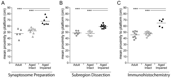
Figure 1 Stratification of aged rats by cognitive performance. Three independent cohorts of adult (12-13 months) and aged (26-28 months) male Fischer × Brown Norway hybrid (F1) rats were assessed for hippocampus-dependent spatial learning and memory using the Morris water maze. Using mean proximity to the escape platform location as the dependent variable, aged rats performing within the range of adults were classified as cognitively intact, while those with poorer performance were classified as cognitively impaired. Based on this stratification, the performance of both adult and aged intact rats was significantly superior to aged impaired rats in (A) set 1: utilized for preparation of whole-hippocampus synaptosomal fractions used in transcriptomic profiling and mRNA quantitation, (B) set 2: utilized for dissection of CA1, CA3 and DG used in mRNA and protein quantitation, and (C) set 3: perfusion-fixed for immunohistochemical assessments. Points represent individual animals and horizontal bars indicate group means; *** p < 0.001, ANOVA SNK post hoc test. See Table 1 for cohort information. Behavioral data for cohorts 1 and 3 are adapted from reference 27.
Figure 1 Stratification of aged rats by cognitive performance. Three independent cohorts of adult (12-13 months) and aged (26-28 months) male Fischer × Brown Norway hybrid (F1) rats were assessed for hippocampus-dependent spatial learning and memory using the Morris water maze. Using mean proximity to the escape platform location as the dependent variable, aged rats performing within the range of adults were classified as cognitively intact, while those with poorer performance were classified as cognitively impaired. Based on this stratification, the performance of both adult and aged intact rats was significantly superior to aged impaired rats in (A) set 1: utilized for preparation of whole-hippocampus synaptosomal fractions used in transcriptomic profiling and mRNA quantitation, (B) set 2: utilized for dissection of CA1, CA3 and DG used in mRNA and protein quantitation, and (C) set 3: perfusion-fixed for immunohistochemical assessments. Points represent individual animals and horizontal bars indicate group means; *** p < 0.001, ANOVA SNK post hoc test. See Table 1 for cohort information. Behavioral data for cohorts 1 and 3 are adapted from reference 27.CC BY · PubMed Central
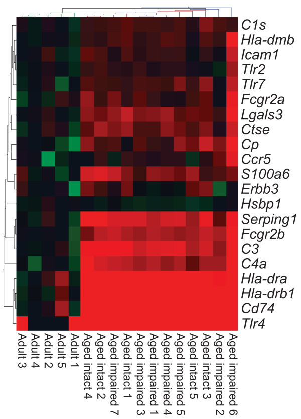
Figure 2 Age-related induction of MHC II and inflammation-response transcripts. Bioinformatic analysis of transcriptomic expression data identified significant upregulation the MHC II antigen presentation pathway and associated inflammatory signaling factors in hippocampal synaptosomes derived from both cognitively intact and cognitively impaired aged rats compared to adults (ANOVA with SNK post hoc testing, p < 0.05). Notably, no differences were observed between aged intact and aged impaired rats, which clustered separately from adults but did not cluster by cognitive status. Log-scaled gene expression is presented relative to the adult group mean per transcript (green: decreased; black: unchanged; red: increased).
Figure 2 Age-related induction of MHC II and inflammation-response transcripts. Bioinformatic analysis of transcriptomic expression data identified significant upregulation the MHC II antigen presentation pathway and associated inflammatory signaling factors in hippocampal synaptosomes derived from both cognitively intact and cognitively impaired aged rats compared to adults (ANOVA with SNK post hoc testing, p < 0.05). Notably, no differences were observed between aged intact and aged impaired rats, which clustered separately from adults but did not cluster by cognitive status. Log-scaled gene expression is presented relative to the adult group mean per transcript (green: decreased; black: unchanged; red: increased).CC BY · PubMed Central
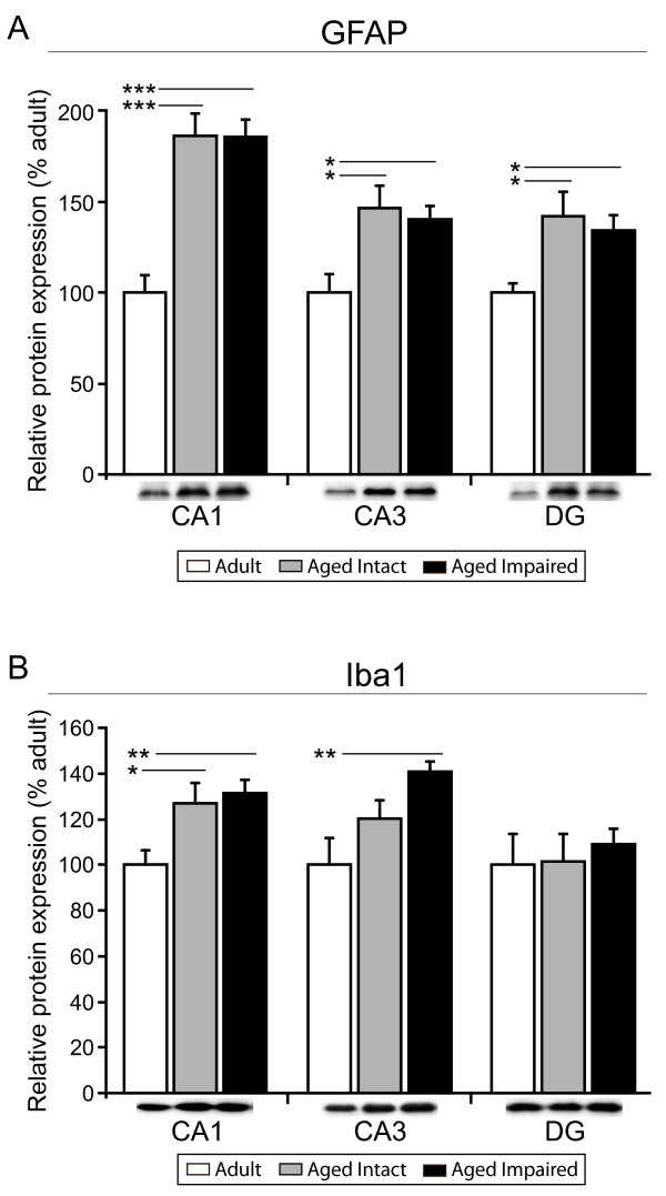
Figure 3 Age-related upregulation of hippocampal astrocyte and microglial activation protein markers. (A) Immunoblotting for GFAP, an intermediate filament protein expressed by astrocytes and upregulated with inflammation, is significantly increased in CA1, CA3 and DG of both aged cognitively intact and aged cognitively impaired rats compared to adults. (B) Iba1, a microglia-specific calcium binding and actin-bundling protein, is significantly induced in CA1 and CA3, but not DG of aged animals, regardless of cognitive status, compared to adults. Insets depict representative immunoblot images. *p < 0.05, **p < 0.01, ***p < 0.001, one-way ANOVA with Student Newman Keuls post hoc testing, n = 7-10/group.
Figure 3 Age-related upregulation of hippocampal astrocyte and microglial activation protein markers. (A) Immunoblotting for GFAP, an intermediate filament protein expressed by astrocytes and upregulated with inflammation, is significantly increased in CA1, CA3 and DG of both aged cognitively intact and aged cognitively impaired rats compared to adults. (B) Iba1, a microglia-specific calcium binding and actin-bundling protein, is significantly induced in CA1 and CA3, but not DG of aged animals, regardless of cognitive status, compared to adults. Insets depict representative immunoblot images. *p < 0.05, **p < 0.01, ***p < 0.001, one-way ANOVA with Student Newman Keuls post hoc testing, n = 7-10/group.CC BY · PubMed Central
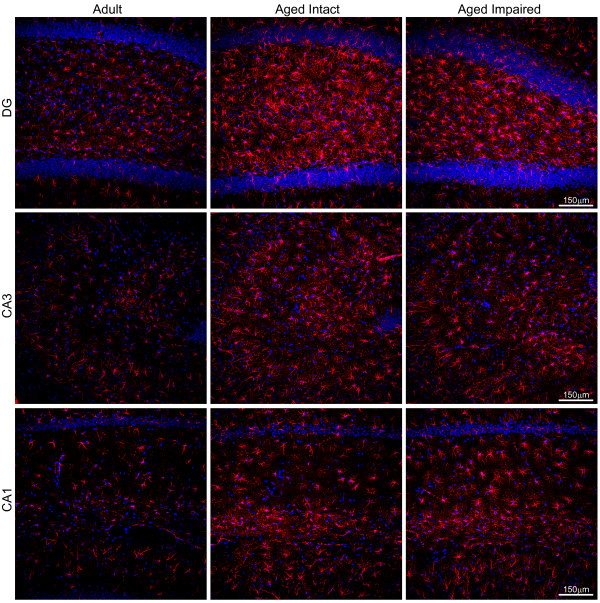
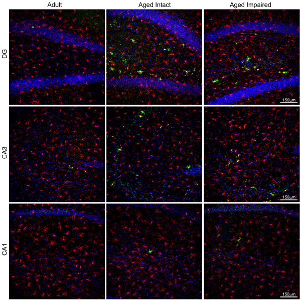
Figure 4 Age-induced activation of hippocampal astrocytes. Immunohistochemical visualization of GFAP revealed low-to-moderate expression in astrocytes distributed throughout DG, CA1 and CA3 of adults. GFAP immunoreactivity was markedly increased, and astrocyte morphology indicated activation, throughout the hippocampal formation of aged rats. No qualitative differences between aged intact and aged impaired rats were evident, and no subregion-specific alterations in astrocyte activation were observed. Increased GFAP immunoreactivity was associated with astrocyte hypertrophy, but not astrocyte proliferation, as total populations of GFAP+ glia were not changed between groups in CA1, CA3 or DG. Blue: Hoechst; Red: GFAP.
Figure 4 Age-induced activation of hippocampal astrocytes. Immunohistochemical visualization of GFAP revealed low-to-moderate expression in astrocytes distributed throughout DG, CA1 and CA3 of adults. GFAP immunoreactivity was markedly increased, and astrocyte morphology indicated activation, throughout the hippocampal formation of aged rats. No qualitative differences between aged intact and aged impaired rats were evident, and no subregion-specific alterations in astrocyte activation were observed. Increased GFAP immunoreactivity was associated with astrocyte hypertrophy, but not astrocyte proliferation, as total populations of GFAP+ glia were not changed between groups in CA1, CA3 or DG. Blue: Hoechst; Red: GFAP.CC BY · PubMed Central
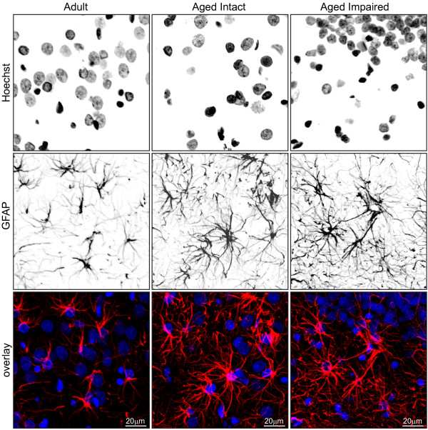
Figure 5 Morphological characteristics of activated astrocytes in aged hippocampus. Higher magnification imaging revealed a stellate morphology and low level of GFAP immunoreactivity in hippocampal astrocytes of adult rats. In hippocampus of both cognitively intact and cognitively impaired aged rats, astrocytes exhibited intense GFAP immunoreactivity and adopted a highly ramified morphology with hypertrophic processes, while maintaining spatial compartmentalization.
Figure 5 Morphological characteristics of activated astrocytes in aged hippocampus. Higher magnification imaging revealed a stellate morphology and low level of GFAP immunoreactivity in hippocampal astrocytes of adult rats. In hippocampus of both cognitively intact and cognitively impaired aged rats, astrocytes exhibited intense GFAP immunoreactivity and adopted a highly ramified morphology with hypertrophic processes, while maintaining spatial compartmentalization.CC BY · PubMed Central
Figure 6 Activation of hippocampal microglia with aging. Immunohistochemical co-localization of the microglia-specific marker Iba1 and the activation-specific marker CD74 (MHC II invariant chain) identified an increase in activated microglia in the aged hippocampus regardless of cognitive status. In adults, Iba1+ microglia were distributed throughout hippocampal subregions, with a greater population evident in CA3 than in CA1 or DG. A small fraction of microglia exhibited mild activation indicated by low levels of CD74 immunoreactivity. In both aged cognitively intact and aged cognitively impaired rats, a marked increase in the number of Iba1+/CD74+ microglia was observed in all three subregions, in the form of both mildly-activated and moderately-activated microglia. Activated microglia were associated with both synaptic and cell body-containing hippocampal strata. Blue: Hoechst; Red: Iba1; Green: CD74; Orange: Iba1/weak CD74 co-expression (mild activation); Yellow: Iba1/high CD74 co-expression (moderate activation).
Figure 6 Activation of hippocampal microglia with aging. Immunohistochemical co-localization of the microglia-specific marker Iba1 and the activation-specific marker CD74 (MHC II invariant chain) identified an increase in activated microglia in the aged hippocampus regardless of cognitive status. In adults, Iba1+ microglia were distributed throughout hippocampal subregions, with a greater population evident in CA3 than in CA1 or DG. A small fraction of microglia exhibited mild activation indicated by low levels of CD74 immunoreactivity. In both aged cognitively intact and aged cognitively impaired rats, a marked increase in the number of Iba1+/CD74+ microglia was observed in all three subregions, in the form of both mildly-activated and moderately-activated microglia. Activated microglia were associated with both synaptic and cell body-containing hippocampal strata. Blue: Hoechst; Red: Iba1; Green: CD74; Orange: Iba1/weak CD74 co-expression (mild activation); Yellow: Iba1/high CD74 co-expression (moderate activation).CC BY · PubMed Central
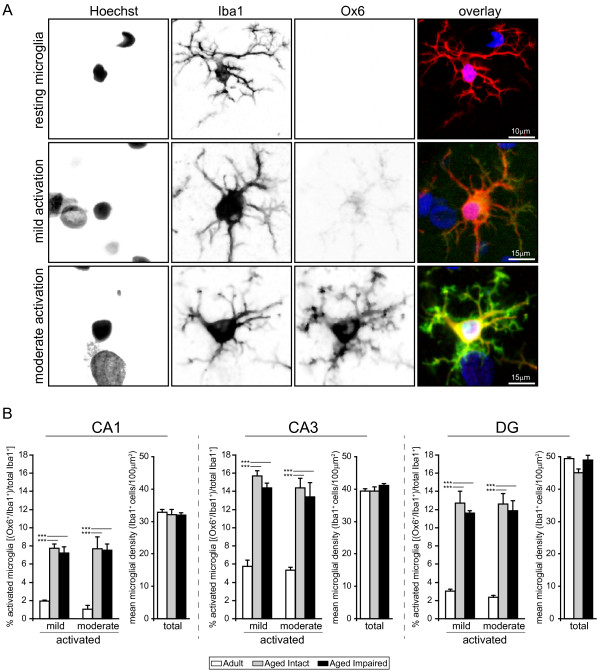
Figure 7 Age-related increase in activated microglial populations. (A) High-magnification visualization of resting/surveilling, or non-activated, and activated microglia reveals differences in both immunoreactivity and morphology with aging. Non-activated microglia expressed Iba1 but not CD74, and had long, thin, branched processes. Microglia undergoing mild activation displayed increased Iba1 immunoreactivity, weak CD74 immunoreactivity, and enlarged somata, while moderately activated microglia demonstrated increased Iba1 immunoreactivity, intense CD74 immunoreactivity, enlarged somata, thickened proximal processes, and retracted distal processes. Notably, activated microglia maintained a ramified rather than ameboid morphology, indicating that microglial activation had not progressed to a reactive, phagocytic phenotype. (B) Quantitation of activated microglia (calculated as the ratio of Iba1+/CD74+ to total Iba1+ cells) revealed significant increases in all three hippocampal subregions studied. In adult rats, a small fraction of total microglia were mildly or moderately activated in CA3, with far fewer activated microglia evident in CA1 and DG. The percentage of microglia undergoing mild or moderate activation was significantly increased, by several fold, throughout the hippocampus of both aged cognitively intact and aged cognitively impaired rats compared to adults. No differences between the degree of microglial activation (i.e., mild versus moderate) were observed between aged intact and aged impaired rats in CA1, CA3 or DG. Increased numbers of activated microglia in aged rats were not due to proliferation/infiltration, as total populations of microglia (Iba1+ cells) were not different between groups. ***p < 0.001, one-way ANOVA with SNK post hoc testing.
Figure 7 Age-related increase in activated microglial populations. (A) High-magnification visualization of resting/surveilling, or non-activated, and activated microglia reveals differences in both immunoreactivity and morphology with aging. Non-activated microglia expressed Iba1 but not CD74, and had long, thin, branched processes. Microglia undergoing mild activation displayed increased Iba1 immunoreactivity, weak CD74 immunoreactivity, and enlarged somata, while moderately activated microglia demonstrated increased Iba1 immunoreactivity, intense CD74 immunoreactivity, enlarged somata, thickened proximal processes, and retracted distal processes. Notably, activated microglia maintained a ramified rather than ameboid morphology, indicating that microglial activation had not progressed to a reactive, phagocytic phenotype. (B) Quantitation of activated microglia (calculated as the ratio of Iba1+/CD74+ to total Iba1+ cells) revealed significant increases in all three hippocampal subregions studied. In adult rats, a small fraction of total microglia were mildly or moderately activated in CA3, with far fewer activated microglia evident in CA1 and DG. The percentage of microglia undergoing mild or moderate activation was significantly increased, by several fold, throughout the hippocampus of both aged cognitively intact and aged cognitively impaired rats compared to adults. No differences between the degree of microglial activation (i.e., mild versus moderate) were observed between aged intact and aged impaired rats in CA1, CA3 or DG. Increased numbers of activated microglia in aged rats were not due to proliferation/infiltration, as total populations of microglia (Iba1+ cells) were not different between groups. ***p < 0.001, one-way ANOVA with SNK post hoc testing.CC BY · PubMed Central
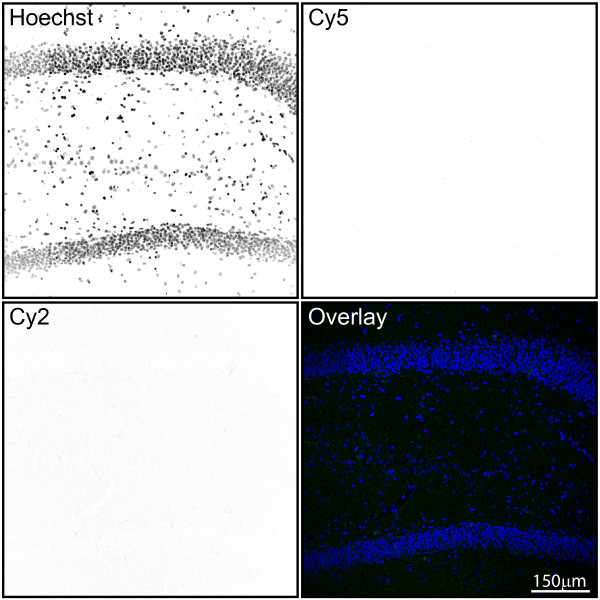
Figure 8 Assessment of negative control signals in immunohistochemical experiments. To ensure that visualization of astrocytes and microglia was not confounded by nonspecific secondary antibody binding, a negative control for the Cy2 (donkey-anti-mouse DyLight 488) and Cy5 (donkey-anti-rabbit DyLight 649) channels was included with primary antibodies omitted. Individual channels are presented as inverted gray scale. Weak, diffuse background signal was observed in the Cy2 channel, and no signal was apparent in the Cy5 channel. No nonspecific cell staining in the absence of primary antibodies occurred. Blue: Hoechst; Green: Cy2; Red: Cy5.
Figure 8 Assessment of negative control signals in immunohistochemical experiments. To ensure that visualization of astrocytes and microglia was not confounded by nonspecific secondary antibody binding, a negative control for the Cy2 (donkey-anti-mouse DyLight 488) and Cy5 (donkey-anti-rabbit DyLight 649) channels was included with primary antibodies omitted. Individual channels are presented as inverted gray scale. Weak, diffuse background signal was observed in the Cy2 channel, and no signal was apparent in the Cy5 channel. No nonspecific cell staining in the absence of primary antibodies occurred. Blue: Hoechst; Green: Cy2; Red: Cy5.CC BY · PubMed Central

Verification

Claims the paper makes

The claims and statistics of this paper haven’t been checked yet.

Run the check — free, no account needed. It reads the open-access text and checks the sources it cites.

A few free checks a day · papers already verified open instantly · results are public

Claims other papers make about this one

Each is checked against this paper’s own text: does it say what the citing paper says it does?

Self-check

Percentages, sums and reported differences re-computed from the paper’s own numbers.

The self-check needs the paper’s full text.

Citations

Cites

Show all 91 references

Cited by

Sorted by citations

See all 135

Metadata

Authors
VanGuilder, H. D., Bixler, G. V., Brucklacher, R. M., Farley, J. A., Yan, H., Warrington, J. P., Sonntag, W. E., & Freeman, W. M.
Affiliations
  • Pennsylvania State University (US)
  • University of Oklahoma Health Sciences Center (US)
Corresponding author
Freeman, Willard M
Venue
Journal of Neuroinflammation
Published
2011-10-11
Volume
8
Issue
1
Pages
138-138
ISSN
1742-2094 (ISSN-L)
OpenAlex
W2081626361
Cited by
132
References
91
Citations by year
2026: 3 · 2025: 7 · 2024: 7 · 2023: 4 · 2022: 11 · 2021: 12 · 2020: 10 · 2019: 11 · 2018: 10 · 2017: 15 · 2016: 11 · 2015: 9 · 2014: 7 · 2013: 10 · 2012: 5
FWCI
3.89
Type
journal article
Language
en
Open access
Licence
cc-by
Locations
  • Journal of Neuroinflammation (publishedVersion, cc-by)
  • Europe PMC (PubMed Central) (publishedVersion, cc-by)
  • PubMed Central (publishedVersion, cc-by)
  • PubMed (publishedVersion)
  • DOAJ (DOAJ: Directory of Open Access Journals) (submittedVersion)
Funders
  • Donald W. Reynolds Foundation
  • National Institutes of Health (5r01ag026607-06)
  • National Institute on Aging (#R01AG026607, 5R01AG026607, P01AG011370)
Topics
  • Life Sciences › Neuroscience › Neurology › Neuroinflammation and Neurodegeneration Mechanisms
  • Life Sciences › Neuroscience › Neurology › Neurological Disease Mechanisms and Treatments
  • Life Sciences › Neuroscience › Developmental Neuroscience › Neurogenesis and neuroplasticity mechanisms
Keywords
hippocampal inflammation, glial activation, microglial activation, astrocytes, cognitive aging, age-related cognitive decline, spatial learning and memory, Toll-like receptors, complement system, hippocampal subfields
MeSH terms
Aging, Animals, Histocompatibility Antigens Class II, Behavior, Animal, Cognition Disorders, Genes, MHC Class II, Hippocampus, Humans, Major Histocompatibility Complex, Male, Neuroglia, Rats, Inbred F344, Biomarkers, Maze Learning, Gene Expression Profiling, Microarray Analysis, Rats, Transcriptome

About these checks

Registry, retraction, Cites and statistics results are computed, not judged, with one exception: a printed reference that no index lists is read once by a model before it counts as a citation error. Claims are extracted by a model and kept only when their quote appears verbatim in the text; each cited source is then checked, with a model’s help, for whether it says what the claim relies on. Those verdicts can be wrong — read the claim’s quote and the cited source before relying on one.

Statistics and arithmetic are recomputed, not judged.

Confidence bands are shown only for fields calibrated on our annotated test set. None is calibrated yet, so no band is shown.

What counts as a claim ›How we measure confidence ›How we check this ›

Are you an author, or do you see a problem with this page? Request a review