

The DOI matches its Crossref record; 91 of the 91 references it cites were found in a registry or an index.
Concurrent hippocampal induction of MHC II pathway components and glial activation with advanced aging is not correlated with cognitive impairment
Open accessAbstract
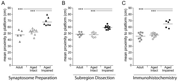
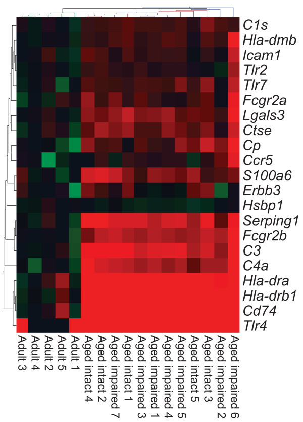
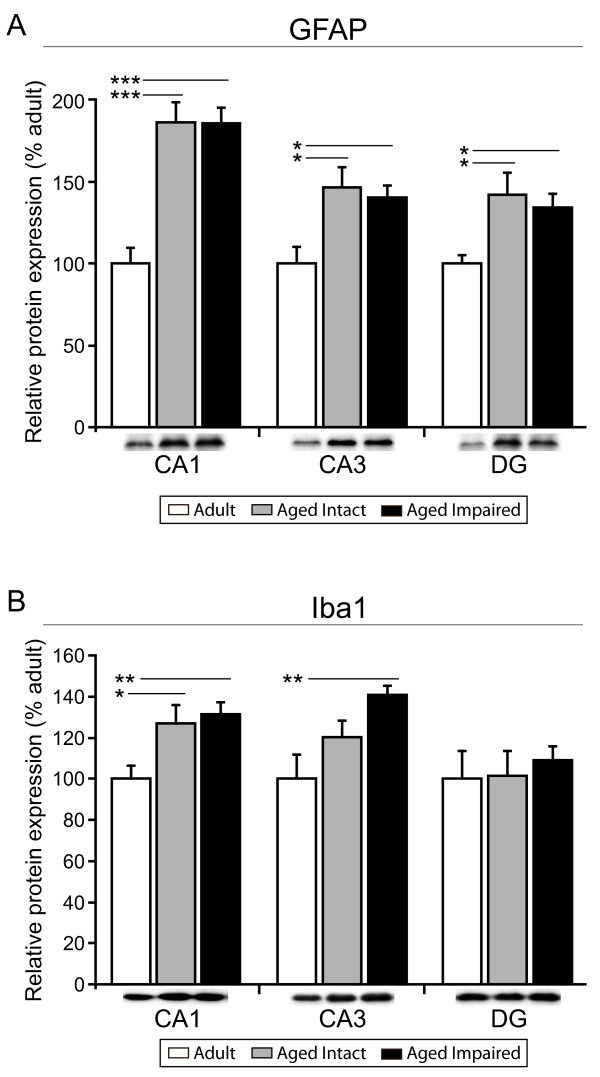
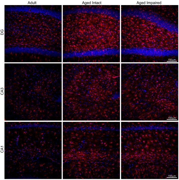
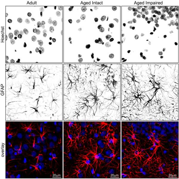
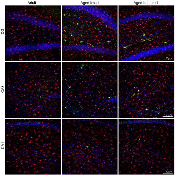
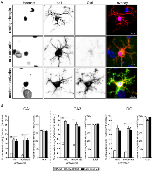
Background: Age-related cognitive dysfunction, including impairment of hippocampus-dependent spatial learning and memory, affects approximately half of the aged population. Induction of a variety of neuroinflammatory measures has been reported with brain aging but the relationship between neuroinflammation and cognitive decline with non-neurodegenerative, normative aging remains largely unexplored. This study sought to comprehensively investigate expression of the MHC II immune response pathway and glial activation in the hippocampus in the context of both aging and age-related cognitive decline. Methods: Three independent cohorts of adult (12-13 months) and aged (26-28 months) F344xBN rats were behaviorally characterized by Morris water maze testing. Expression of MHC II pathway-associated genes identified by transcriptomic analysis as upregulated with advanced aging was quantified by qPCR in synaptosomal fractions derived from whole hippocampus and in hippocampal subregion dissections (CA1, CA3, and DG). Activation of astrocytes and microglia was assessed by GFAP and Iba1 protein expression, and by immunohistochemical visualization of GFAP and both CD74 (Ox6) and Iba1. Results: We report a marked age-related induction of neuroinflammatory signaling transcripts (i.e., MHC II components, toll-like receptors, complement, and downstream signaling factors) throughout the hippocampus in all aged rats regardless of cognitive status. Astrocyte and microglial activation was evident in CA1, CA3 and DG of intact and impaired aged rat groups, in the absence of differences in total numbers of GFAP+astrocytes or Iba1+microglia. Both mild and moderate microglial activation was significantly increased in all three hippocampal subregions in aged cognitively intact and cognitively impaired rats compared to adults. Neither induction of MHCII pathway gene expression nor glial activation correlated to cognitive performance. Conclusions: These data demonstrate a novel, coordinated age-related induction of the MHC II immune response pathway and glial activation in the hippocampus, indicating an allostatic shift toward a para-inflammatory phenotype with advancing age. Our findings demonstrate that age-related induction of these aspects of hippocampal neuroinflammation, while a potential contributing factor, is not sufficient by itself to elicit impairment of spatial learning and memory in models of normative aging. Future efforts are needed to understand how neuroinflammation may act synergistically with cognitive-decline specific alterations to cause cognitive impairment.
Overview
The claims and statistics of this paper haven’t been checked yet.
Run the check — free, no account needed. It reads the open-access text and checks the sources it cites.
Integrity signals
Registry
Cites
Statistics
Figures
Verification
Claims the paper makes
The claims and statistics of this paper haven’t been checked yet.
Run the check — free, no account needed. It reads the open-access text and checks the sources it cites.
Claims other papers make about this one
Each is checked against this paper’s own text: does it say what the citing paper says it does?
In the citing paper
“Microglia under normal aging (ie, aging in absence of neurodegeneration) showed upregulation of genes associated with antigen presentation, such as MHCII, CD86, and CIITA (Frank et al, 2006;Godbout et al, 2005;VanGuilder et al, 2011), and suppression of anti-inflammatory genes, such as IL-10 and CD200 (Frank et al, 2006).”
From Microglia Priming with Aging and Stress (2017) · cites this paper as VanGuilder et al, 2011
Self-check
Percentages, sums and reported differences re-computed from the paper’s own numbers.
The self-check needs the paper’s full text.
Citations
Cites
Origin and physiological roles of inflammation
Medzhitov, R. (2008) · Nature · doi:10.1038/nature07201 · Cited by 6,618
Resting microglial cells are highly dynamic surveillants of brain parenchyma in vivo
Nimmerjahn, A., Kirchhoff, F., & Helmchen, F. (2005) · Science · doi:10.1126/science.1110647 · Cited by 5,827
Microglia: Active sensor and versatile effector cells in the normal and pathologic brain
Hanisch, U.-K., & Kettenmann, H. (2007) · Nature Neuroscience · doi:10.1038/nn1997 · Cited by 3,676
DAMPs, PAMPs and alarmins: all we need to know about danger
Bianchi, M. E. (2007) · Journal of Leukocyte Biology · doi:10.1189/jlb.0306164 · Cited by 2,794
Insights into the ageing mind: a view from cognitive neuroscience
Hedden, T., & Gabrieli, J. D. E. (2004) · Nature Reviews Neuroscience · doi:10.1038/nrn1323 · Cited by 2,223
The ageing systemic milieu negatively regulates neurogenesis and cognitive function
Villeda, S. A., Luo, J., Mosher, K. I., Zou, B., Britschgi, M., Bieri, G., Stan, T. M., Fainberg, N., Ding, Z., Eggel, A., et al. (2011) · Nature · doi:10.1038/nature10357 · Cited by 1,763
Immune modulation of learning, memory, neural plasticity and neurogenesis
Yirmiya, R., & Goshen, I. (2011) · Brain, Behavior, and Immunity · doi:10.1016/j.bbi.2010.10.015 · Cited by 1,483
Debris clearance by microglia: an essential link between degeneration and regeneration
Neumann, H., Kotter, M. R., & Franklin, R. J. M. (2008) · Brain · doi:10.1093/brain/awn109 · Cited by 1,111
Gene-expression profile of the ageing brain in mice
Lee, C.-K., Weindruch, R., & Prolla, T. A. (2000) · Nature Genetics · doi:10.1038/77046 · Cited by 1,055
Godbout, J. P., Chen, J., Abraham, J., Richwine, A. F., Berg, B. M., Kelley, K. W., & Johnson, R. W. (2005) · The FASEB Journal · doi:10.1096/fj.05-3776fje · Cited by 813
Cited by
Sorted by citations
Review: microglia of the aged brain: primed to be activated and resistant to regulation
Norden, D. M., & Godbout, J. P. (2013) · Neuropathology and Applied Neurobiology · doi:10.1111/j.1365-2990.2012.01306.x · Cited by 792
Tay, T. L., Savage, J. C., Hui, C. W., Bisht, K., & Tremblay, M. (2017) · The Journal of Physiology · doi:10.1113/jp272134 · Cited by 562
Norden, D. M., Muccigrosso, M. M., & Godbout, J. P. (2015) · Neuropharmacology · doi:10.1016/j.neuropharm.2014.10.028 · Cited by 413
Microglia Priming with Aging and Stress
Niraula, A., Sheridan, J. F., & Godbout, J. P. (2017) · Neuropsychopharmacology · doi:10.1038/npp.2016.185 · Cited by 398
Microglia Phenotypes in Aging and Neurodegenerative Diseases
Wendimu, M. Y., & Hooks, S. B. (2022) · Cells · doi:10.3390/cells11132091 · Cited by 336
Aging Microglia—Phenotypes, Functions and Implications for Age-Related Neurodegenerative Diseases
Spittau, B. (2017) · Frontiers in Aging Neuroscience · doi:10.3389/fnagi.2017.00194 · Cited by 271
Priming the Inflammatory Pump of the CNS after Traumatic Brain Injury
Witcher, K. G., Eiferman, D. S., & Godbout, J. P. (2015) · Trends in Neurosciences · doi:10.1016/j.tins.2015.08.002 · Cited by 234
Pao, P.-C., Patnaik, D., Watson, L. A., Gao, F., Pan, L., Wang, J., Adaikkan, C., Penney, J., Cam, H. P., Huang, W.-C., et al. (2020) · Nature Communications · doi:10.1038/s41467-020-16361-y · Cited by 216
Sexually divergent induction of microglial-associated neuroinflammation with hippocampal aging
Mangold, C. A., Wronowski, B., Du, M., Masser, D. R., Hadad, N., Bixler, G. V., Brucklacher, R. M., Ford, M. M., Sonntag, W. E., & Freeman, W. M. (2017) · Journal of Neuroinflammation · doi:10.1186/s12974-017-0920-8 · Cited by 203
Self-citationReynolds, L. M., Taylor, J. R., Ding, J., Lohman, K., Johnson, C. W., Siscovick, D. S., Burke, G. L., Post, W. S., Shea, S., Jacobs, D. R., et al. (2014) · Nature Communications · doi:10.1038/ncomms6366 · Cited by 204
Microglia as modulators of cognition and neuropsychiatric disorders
Blank, T., & Prinz, M. (2012) · Glia · doi:10.1002/glia.22372 · Cited by 195
Cellular correlates of cortical thinning throughout the lifespan
Vidal-Pineiro, D., Parker, N., Shin, J., French, L., Grydeland, H., Jackowski, A. P., Mowinckel, A. M., Patel, Y., Pausova, Z., Salum, G., et al. (2020) · Scientific Reports · doi:10.1038/s41598-020-78471-3 · Cited by 154
Inflammation in Traumatic Brain Injury
Postolache, T. T., Wadhawan, A., Can, A., Lowry, C. A., Woodbury, M., Makkar, H., Hoisington, A. J., Scott, A. J., Potocki, E., Benros, M. E., et al. (2020) · Journal of Alzheimer s Disease · doi:10.3233/jad-191150 · Cited by 154
Exploring Sex-Related Differences in Microglia May Be a Game-Changer in Precision Medicine
Lynch, M. A. (2022) · Frontiers in Aging Neuroscience · doi:10.3389/fnagi.2022.868448 · Cited by 146
O’Neil, S. M., Witcher, K. G., McKim, D. B., & Godbout, J. P. (2018) · Acta Neuropathologica Communications · doi:10.1186/s40478-018-0636-8 · Cited by 138
Neuroinflammation as Fuel for Axonal Regeneration in the Injured Vertebrate Central Nervous System
Bollaerts, I., houcke, J. V., Andries, L., Groef, L. D., & Moons, L. (2017) · Mediators of Inflammation · doi:10.1155/2017/9478542 · Cited by 138
Insulin-like growth factor-1 in CNS and cerebrovascular aging
Sonntag, W. E., Deák, F., Ashpole, N. M., Tóth, P., Csiszár, A., Freeman, W. M., & Ungvari, Z. I. (2013) · Frontiers in Aging Neuroscience · doi:10.3389/fnagi.2013.00027 · Cited by 139
Self-citationCholinergic Modulation of Glial Function During Aging and Chronic Neuroinflammation
Gamage, R. V., Wagnon, I. M., Rossetti, I., Childs, R., Niedermayer, G., Chesworth, R., & Gyengési, E. (2020) · Frontiers in Cellular Neuroscience · doi:10.3389/fncel.2020.577912 · Cited by 134
The Inflammatory Continuum of Traumatic Brain Injury and Alzheimer’s Disease
Kokiko-Cochran, O. N., & Godbout, J. P. (2018) · Frontiers in Immunology · doi:10.3389/fimmu.2018.00672 · Cited by 128
O’Loughlin, E., Pakan, J. M. P., Yilmazer-Hanke, D., & McDermott, K. W. (2017) · Journal of Neuroinflammation · doi:10.1186/s12974-017-0981-8 · Cited by 127
Microglia in Aging and Alzheimer’s Disease: A Comparative Species Review
Edler, M. K., Mhatre-Winters, I., & Richardson, J. R. (2021) · Cells · doi:10.3390/cells10051138 · Cited by 126
GFAP expression in injured astrocytes in rats
Zhang, S., Wu, M., Peng, C., Zhao, G., & Gu, R. (2017) · Experimental and Therapeutic Medicine · doi:10.3892/etm.2017.4760 · Cited by 125
Inflammatory mechanisms in neurodegeneration
Nichols, M. R., St‐Pierre, M., Wendeln, A., Makoni, N. J., Gouwens, L. K., Garrad, E. C., Sohrabi, M., Neher, J. J., Tremblay, M., & Combs, C. K. (2019) · Journal of Neurochemistry · doi:10.1111/jnc.14674 · Cited by 129
Sexually dimorphic microglia and ischemic stroke
Kerr, N. A., Dietrich, D. W., Bramlett, H. M., & Raval, A. P. (2019) · CNS Neuroscience & Therapeutics · doi:10.1111/cns.13267 · Cited by 110
Redox Signaling in Neurotransmission and Cognition During Aging
Kumar, A., Yegla, B., & Foster, T. C. (2017) · Antioxidants and Redox Signaling · doi:10.1089/ars.2017.7111 · Cited by 106
Developmental microglial priming in postmortem autism spectrum disorder temporal cortex
Lee, A. S., Azmitia, E. C., & Whitaker‐Azmitia, P. M. (2017) · Brain Behavior and Immunity · doi:10.1016/j.bbi.2017.01.019 · Cited by 100
Glia: victims or villains of the aging brain?
Salas, I. H., Burgado, J., & Allen, N. J. (2020) · Neurobiology of Disease · doi:10.1016/j.nbd.2020.105008 · Cited by 99
Reactive gliosis in traumatic brain injury: a comprehensive review
Amlerova, Z., Chmelová, M., Anděrová, M., & Vargová, L. (2024) · Frontiers in Cellular Neuroscience · doi:10.3389/fncel.2024.1335849 · Cited by 89
Hoeijmakers, L., Heinen, Y., van Dam, A.-M., Lucassen, P. J., & Korosi, A. (2016) · Frontiers in Human Neuroscience · doi:10.3389/fnhum.2016.00398 · Cited by 85
The old guard: Age-related changes in microglia and their consequences
Costa, J., Martins, S., Ferreira, P. A., Cardoso, A. M., Guedes, J. R., Peça, J., & Cardoso, A. L. (2021) · Mechanisms of Ageing and Development · doi:10.1016/j.mad.2021.111512 · Cited by 88
Lee, M. J., Jang, M., Choi, J., Chang, B. S., Kim, D. Y., Kim, S., Kwak, Y.-S., Oh, S., Lee, J., Chang, B., et al. (2015) · Molecular Neurobiology · doi:10.1007/s12035-015-9131-4 · Cited by 83
n-3 Polyunsaturated Fatty Acids and Their Derivates Reduce Neuroinflammation during Aging
Joffre, C., Dinel, A., Chataigner, M., Pallet, V., & Layé, S. (2020) · Nutrients · doi:10.3390/nu12030647 · Cited by 80
Norden, D. M., Trojanowski, P. J., Walker, F. R., & Godbout, J. P. (2016) · Neurobiology of Aging · doi:10.1016/j.neurobiolaging.2016.04.014 · Cited by 73
Inflammation: Bridging Age, Menopause and APOEε4 Genotype to Alzheimer’s Disease
Mishra, A., & Brinton, R. D. (2018) · Frontiers in Aging Neuroscience · doi:10.3389/fnagi.2018.00312 · Cited by 71
Lee, M. J., Jang, M., Choi, J., Lee, G., Min, H. J., Chung, W., Kim, J., Jee, Y., Chae, Y., Kim, S., et al. (2015) · Molecular Neurobiology · doi:10.1007/s12035-014-9012-2 · Cited by 68
Starkey, H. D. V., Kirk, C. A. V., Bixler, G. V., Imperio, C. G., Kale, V. P., Serfass, J. M., Farley, J. A., Han, Y., Warrington, J. P., Han, S. I., et al. (2012) · Journal of Molecular Neuroscience · doi:10.1007/s12031-012-9783-8 · Cited by 67
Self-citationMicroglia recapitulate a hematopoietic master regulator network in the aging human frontal cortex
Wehrspaun, C. C., Haerty, W., & Ponting, C. P. (2015) · Neurobiology of Aging · doi:10.1016/j.neurobiolaging.2015.04.008 · Cited by 64
Ianov, L., Riva, A., Kumar, A., & Foster, T. C. (2017) · Frontiers in Aging Neuroscience · doi:10.3389/fnagi.2017.00249 · Cited by 65
Xu, Y., Au, N. P. B., & Ma, C. H. E. (2022) · Frontiers in Aging Neuroscience · doi:10.3389/fnagi.2022.896852 · Cited by 68
Ageing neuroinflammation and neurodegeneration
Ward, R. J. (2015) · Frontiers in Bioscience-Scholar · doi:10.2741/s433 · Cited by 64
Unraveling Parkinson’s Disease Neurodegeneration: Does Aging Hold the Clues?
Coleman, C., & Martin, I. (2022) · Journal of Parkinson s Disease · doi:10.3233/jpd-223363 · Cited by 65
Age-Dependent Changes in the Activation and Regulation of Microglia
Cornejo, F., & Rommy von Bernhardi, M. (2016) · Advances in experimental medicine and biology · doi:10.1007/978-3-319-40764-7_10 · Cited by 61
Primiani, C. T., Ryan, V. H., Rao, J. S., Cam, M. C., Ahn, K., Modi, H. R., & Rapoport, S. I. (2014) · PLoS ONE · doi:10.1371/journal.pone.0110972 · Cited by 56
Investigation of Sex Differences in the Microglial Response to Binge Ethanol and Exercise
Barton, E. A., Baker, C., & Leasure, J. L. (2017) · Brain Sciences · doi:10.3390/brainsci7100139 · Cited by 55
Transcription Profile of Aging and Cognition-Related Genes in the Medial Prefrontal Cortex
Ianov, L., Rani, A., Beas, B. S., Kumar, A., & Foster, T. C. (2016) · Frontiers in Aging Neuroscience · doi:10.3389/fnagi.2016.00113 · Cited by 55
Navakkode, S., Liu, C., & Soong, T. W. (2018) · Ageing Research Reviews · doi:10.1016/j.arr.2018.01.001 · Cited by 55
Ziebell, J. M., Rowe, R. K., Muccigrosso, M. M., Reddaway, J., Adelson, P. D., Godbout, J. P., & Lifshitz, J. (2016) · Brain Behavior and Immunity · doi:10.1016/j.bbi.2016.03.008 · Cited by 55
Kaneko, Y., Pappas, C. A., Malapira, T., Vale, F. Ĺ., Tajiri, N., & Borlongan, C. V. (2017) · Cellular Physiology and Biochemistry · doi:10.1159/000460513 · Cited by 51
Novel Players in the Aging Synapse: Impact on Cognition
Temido‐Ferreira, M., Coelho, J. E., Pousinha, P. A., & Lopes, L. V. (2019) · Journal of Caffeine and Adenosine Research · doi:10.1089/caff.2019.0013 · Cited by 57
Innate immunity and cellular senescence: The good and the bad in the developmental and aged brain
Santoro, A., Spinelli, C. C., Martucciello, S., Nori, S. L., Capunzo, M., Puca, A. A., & Ciaglia, E. (2018) · Journal of Leukocyte Biology · doi:10.1002/jlb.3mr0118-003r · Cited by 50
Neuroinflammaging and the Immune Landscape: The Role of Autophagy and Senescence in Aging Brain
Tamatta, R., Pai, V., Jaiswal, C., Singh, I., & Singh, A. K. (2025) · Biogerontology · doi:10.1007/s10522-025-10199-x · Cited by 53
VanGuilder, H. D., Bixler, G. V., Sonntag, W. E., & Freeman, W. M. (2012) · Journal of Neurochemistry · doi:10.1111/j.1471-4159.2012.07671.x · Cited by 49
Self-citationAge and Sex Influence the Hippocampal Response and Recovery Following Sepsis
Barter, J., Kumar, A., Stortz, J. A., Hollen, M. K., Nacionales, D. C., Efron, P. A., Moldawer, L. L., & Foster, T. C. (2019) · Molecular Neurobiology · doi:10.1007/s12035-019-01681-y · Cited by 49
Masser, D. R., Bixler, G. V., Brucklacher, R. M., Yan, H., Giles, C. B., Wren, J. D., Sonntag, W. E., & Freeman, W. M. (2014) · The Journals of Gerontology Series A · doi:10.1093/gerona/glu091 · Cited by 52
Self-citationAlmeida-Suhett, C. P., Li, Z., Marini, A. M., Braga, M. F. M., & Eiden, L. E. (2013) · Journal of Neurotrauma · doi:10.1089/neu.2013.3029 · Cited by 47
Brothers, H. M., Bardou, I., Hopp, S. C., Kaercher, R. M., Corona, A. W., Fenn, A. M., Godbout, J. P., & Wenk, G. L. (2013) · Journal of Neuroimmune Pharmacology · doi:10.1007/s11481-013-9476-2 · Cited by 44
Nosi, D., Lana, D., Giovannini, M. G., Delfino, G., & Zecchi‐Orlandini, S. (2021) · Cells · doi:10.3390/cells10051195 · Cited by 41
Wolf, G., Lotan, A., Lifschytz, T., Ben-Ari, H., Kreisel, T., Tatarskyy, P., Valitzky, M., Mernick, B., Avidan, E., Koroukhov, N., et al. (2017) · Frontiers in Aging Neuroscience · doi:10.3389/fnagi.2017.00191 · Cited by 39
Seyedaghamiri, F., Farajdokht, F., Vatandoust, S. M., Mahmoudi, J., Khabbaz, A., & Sadigh‐Eteghad, S. (2021) · Molecular Biology Reports · doi:10.1007/s11033-021-06195-2 · Cited by 38
Aged-Related Changes in Microglia and Neurodegenerative Diseases: Exploring the Connection
Borrajo, A. (2024) · Biomedicines · doi:10.3390/biomedicines12081737 · Cited by 38
Expression of HLA-DR, CD80, and CD86 in Healthy Aging and Alzheimer’s Disease
Busse, S., Steiner, J., Alter, J., Dobrowolny, H., Mawrin, C., Bogerts, B., Hartig, R., & Busse, M. (2015) · Journal of Alzheimer s Disease · doi:10.3233/jad-150217 · Cited by 36
Toonen, L. J. A., Casaca-Carreira, J., Pellisé-Tintoré, M., Mei, H., Temel, Y., Jahanshahi, A., & van Roon‐Mom, W. (2018) · Nucleic Acid Therapeutics · doi:10.1089/nat.2017.0705 · Cited by 38
Senescent accelerated prone 8 (SAMP8) mice as a model of age dependent neuroinflammation
Fernández, A., Quintana, E., Velasco, P., Moreno-Jimenez, B., de Andrés, B., Gaspar, M.-L., Liste, I., Vilar, M., Mira, H., & Cano, E. (2021) · Journal of Neuroinflammation · doi:10.1186/s12974-021-02104-3 · Cited by 38
Naggan, L., Robinson, E., Dinur, E., Goldenberg, H., Kozela, E., & Yirmiya, R. (2023) · Brain Behavior and Immunity · doi:10.1016/j.bbi.2023.02.021 · Cited by 36
Microglia changes associated to Alzheimer's disease pathology in aged chimpanzees
Edler, M. K., Sherwood, C. C., Meindl, R. S., Munger, E. L., Hopkins, W. D., Ely, J. J., ERWIN, J. M., Perl, D. P., Mufson, E. J., Hof, P. R., et al. (2018) · The Journal of Comparative Neurology · doi:10.1002/cne.24484 · Cited by 37
Suthard, R. L., Jellinger, A. L., Surets, M., Shpokayte, M., Pyo, A. Y., Buzharsky, M. D., Senne, R. A., Dorst, K. E., Leblanc, H., & Ramirez, S. (2022) · Neurobiology of Aging · doi:10.1016/j.neurobiolaging.2023.01.007 · Cited by 35
Lana, D., Ugolini, F., Wenk, G. L., Giovannini, M. G., Zecchi‐Orlandini, S., & Nosi, D. (2018) · The FASEB Journal · doi:10.1096/fj.201801539r · Cited by 32
PDE inhibition in distinct cell types to reclaim the balance of synaptic plasticity
Rombaut, B., Kessels, S., Schepers, M., Tiane, A., Paes, D., Solomina, Y., Piccart, E., van den Hove, D., Brône, B., Prickaerts, J., et al. (2020) · Theranostics · doi:10.7150/thno.50701 · Cited by 33
Verkhratsky, A., Zorec, R., Rodriguez, J. J., & Parpura, V. (2019) · Advances in experimental medicine and biology · doi:10.1007/978-981-13-9913-8_8 · Cited by 32
Fernández‐Arjona, M. D. M., León‐Rodríguez, A., Grondona, J. M., & López-Ávalos, M. D. (2022) · Glia · doi:10.1002/glia.24217 · Cited by 28
Viana, L. C., de Lima, C. M., Oliveira, M. A., Borges, R. P., Cardoso, T., Almeida, I., Diniz, D. G., Bento‐Torres, J., Pereira, A., Hornsby, M. B.-D.-O., et al. (2013) · Neuroscience · doi:10.1016/j.neuroscience.2013.02.019 · Cited by 28
Application of mesenchymal stem cells for anti-senescence and clinical challenges
Wang, Y., Gao, T., & Wang, B. (2023) · Stem Cell Research & Therapy · doi:10.1186/s13287-023-03497-z · Cited by 27
Sandoval-Hernández, A. G., Restrepo, A., Cardona‐Gómez, G. P., & Arboleda, G. (2016) · Neuroscience Letters · doi:10.1016/j.neulet.2016.04.007 · Cited by 27
Rodríguez-Callejas, J. D., Fuchs, E., & Perez‐Cruz, C. (2020) · Glia · doi:10.1002/glia.23804 · Cited by 28
de Sousa, A. A., dos Reis, R. R., de Lima, C. M., Augusto‐Oliveira, M., Fernandes, T. N., Gomes, G. F., Diniz, D. G., Magalhães, N. G. D. M., Diniz, C. G., Sosthenes, M. C. K., et al. (2015) · European Journal of Neuroscience · doi:10.1111/ejn.12951 · Cited by 28
Increased hippocampal NgR1 signaling machinery in aged rats with deficits of spatial cognition
Starkey, H. D. V., Sonntag, W. E., & Freeman, W. M. (2013) · European Journal of Neuroscience · doi:10.1111/ejn.12165 · Cited by 25
Self-citationGant, J. C., Blalock, E. M., Chen, K.-C., Kadish, I., Thibault, O., Porter, N. M., & Landfield, P. W. (2017) · Journal of Neuroscience · doi:10.1523/jneurosci.2234-17.2017 · Cited by 25
The Effects of Modified Curcumin Preparations on Glial Morphology in Aging and Neuroinflammation
Ullah, F., Gamage, R. V., Sen, M. K., & Gyengési, E. (2022) · Neurochemical Research · doi:10.1007/s11064-021-03499-4 · Cited by 21
Choi, J. H., Kwon, T. W., Jo, H.-S., Ha, Y., & Cho, I. (2022) · Journal of Ginseng Research · doi:10.1016/j.jgr.2022.11.001 · Cited by 21
The immune marker CD68 correlates with cognitive impairment in normally aged rats
Farso, M. C., Ménard, C., Colby-Milley, J., & Quirion, R. (2013) · Neurobiology of Aging · doi:10.1016/j.neurobiolaging.2013.02.008 · Cited by 21
Shoham, S., Linial, M., & Weinstock, M. (2018) · Journal of Neuroimmune Pharmacology · doi:10.1007/s11481-018-9817-2 · Cited by 21
Microglia-Synapse Pathways: Promising Therapeutic Strategy for Alzheimer’s Disease
Xie, J., Wang, H., Lin, T., & Bi, B. (2017) · BioMed Research International · doi:10.1155/2017/2986460 · Cited by 22
Kékesi, O., Liang, H., Münch, G. W., Morley, J. W., Gyengési, E., & Buskila, Y. (2019) · Brain Structure and Function · doi:10.1007/s00429-019-01905-w · Cited by 19
Navarro, E., Norden, D. M., Trojanowski, P. J., Godbout, J. P., & López, M. G. (2021) · Molecules · doi:10.3390/molecules26082107 · Cited by 20
MP Resulting in Autophagic Cell Death of Microglia through Zinc Changes against Spinal Cord Injury
Li, D., Wang, G., Han, D., Bi, J., Li, C., Wang, H., Liu, Z., Gao, W., Gao, K., Yao, T., et al. (2016) · BioMed Research International · doi:10.1155/2016/6090316 · Cited by 19
Hallmarks of healthy cognitive aging: Inter-individual differences in aging trajectories
Abrous, D. N., Blin, N., Boraxbekk, C., Catheline, G., Fitzsimons, C. P., Hilscher, M. M., Lemoine, M., Lopes, L. V., Maass, A., Nilsson, M., et al. (2026) · Ageing Research Reviews · doi:10.1016/j.arr.2026.103102 · Cited by 18
Dual Roles of Microglia in the Basal Ganglia in Parkinson’s Disease
Choudhury, M. E., Kigami, Y., & Tanaka, J. (2021) · International Journal of Molecular Sciences · doi:10.3390/ijms22083907 · Cited by 18
Starkey, H. D. V., Bixler, G. V., Sonntag, W. E., & Freeman, W. M. (2013) · Cellular and Molecular Neurobiology · doi:10.1007/s10571-013-9929-4 · Cited by 18
Self-citationOperationally defining cognitive reserve genes
Yegla, B., & Foster, T. C. (2021) · Neurobiology of Aging · doi:10.1016/j.neurobiolaging.2021.08.015 · Cited by 19
Transcriptional profiling reveals progeroid Ercc1 -/Δ mice as a model system for glomerular aging
Schermer, B., Bartels, V., Frommolt, P., Habermann, B. H., Braun, F., Schultze, J. L., Roodbergen, M., Hoeijmakers, J. H. J., Schumacher, B., Nürnberg, P., et al. (2013) · BMC Genomics · doi:10.1186/1471-2164-14-559 · Cited by 18
Nheu, D., Ellen, O., Ye, S., Öztürk, E., Pagnin, M., Kertadjaja, S., Theotokis, P., Grigoriadis, N., McLean, C., & Petratos, S. (2022) · Cells · doi:10.3390/cells11233768 · Cited by 16
Expression of the purine biosynthetic enzyme phosphoribosyl formylglycinamidine synthase in neurons
Mangold, C. A., Yao, P. J., Du, M., Freeman, W. M., Benkovic, S. J., & SZPARA, M. L. (2018) · Journal of Neurochemistry · doi:10.1111/jnc.14304 · Cited by 16
Self-citationQuantitative, structural and molecular changes in neuroglia of aging mammals: A review
Pannese, E. (2021) · European Journal of Histochemistry · doi:10.4081/ejh.2021.3249 · Cited by 16
Ding, H. D., Yu, J. H., Chang, W., Liu, F., & He, Z. (2019) · Life Sciences · doi:10.1016/j.lfs.2019.117235 · Cited by 15
Gu, M., Shen, L., Bai, L., Gao, J., Marshall, C., Wu, T., Ding, J., Miao, D., & Xiao, M. (2013) · AGE · doi:10.1007/s11357-013-9552-9 · Cited by 14
Wage, J., Ma, L., Peluso, M. J., Lamont, C., Evens, A. M., Hahnfeldt, P., Hlatky, L., & Beheshti, A. (2015) · Journal of Radiation Research · doi:10.1093/jrr/rrv043 · Cited by 13
Fullerton, E. F., Rubaharan, M., Karom, M. C., Hanberry, R. I., & Murphy, A. Z. (2020) · Neurobiology of Aging · doi:10.1016/j.neurobiolaging.2020.10.020 · Cited by 12
Choi, J. H., Jang, M., Kim, E.-J., Kim, H., Ye, S.-K., & Cho, I. (2016) · Rejuvenation Research · doi:10.1089/rej.2015.1779 · Cited by 11
Israelsson, C., Flygt, J., Ästrand, E., Kiwanuka, O., Bengtsson, H., & Marklund, N. (2014) · Restorative Neurology and Neuroscience · doi:10.3233/rnn-140419 · Cited by 12
Cellular and Molecular Mechanisms for Age-Related Cognitive Decline
Barter, J., & Foster, T. C. (2019) · Cambridge University Press eBooks · doi:10.1017/9781108554350.003 · Cited by 11
Metadata
- Authors
- VanGuilder, H. D., Bixler, G. V., Brucklacher, R. M., Farley, J. A., Yan, H., Warrington, J. P., Sonntag, W. E., & Freeman, W. M.
- Affiliations
- Pennsylvania State University (US)
- University of Oklahoma Health Sciences Center (US)
- Corresponding author
- Freeman, Willard M
- Venue
- Journal of Neuroinflammation
- Published
- 2011-10-11
- Volume
- 8
- Issue
- 1
- Pages
- 138-138
- ISSN
- 1742-2094 (ISSN-L)
- PMC
- PMC3216278
- OpenAlex
- W2081626361
- Cited by
- 132
- References
- 91
- Citations by year
- 2026: 3 · 2025: 7 · 2024: 7 · 2023: 4 · 2022: 11 · 2021: 12 · 2020: 10 · 2019: 11 · 2018: 10 · 2017: 15 · 2016: 11 · 2015: 9 · 2014: 7 · 2013: 10 · 2012: 5
- FWCI
- 3.89
- Type
- journal article
- Language
- en
- Open access
- gold
- doi.org
- Licence
- cc-by
- Locations
- Journal of Neuroinflammation (publishedVersion, cc-by)
- Europe PMC (PubMed Central) (publishedVersion, cc-by)
- PubMed Central (publishedVersion, cc-by)
- PubMed (publishedVersion)
- DOAJ (DOAJ: Directory of Open Access Journals) (submittedVersion)
- Funders
- Donald W. Reynolds Foundation
- National Institutes of Health (5r01ag026607-06)
- National Institute on Aging (#R01AG026607, 5R01AG026607, P01AG011370)
- Topics
- Life Sciences › Neuroscience › Neurology › Neuroinflammation and Neurodegeneration Mechanisms
- Life Sciences › Neuroscience › Neurology › Neurological Disease Mechanisms and Treatments
- Life Sciences › Neuroscience › Developmental Neuroscience › Neurogenesis and neuroplasticity mechanisms
- Keywords
- hippocampal inflammation, glial activation, microglial activation, astrocytes, cognitive aging, age-related cognitive decline, spatial learning and memory, Toll-like receptors, complement system, hippocampal subfields
- MeSH terms
- Aging, Animals, Histocompatibility Antigens Class II, Behavior, Animal, Cognition Disorders, Genes, MHC Class II, Hippocampus, Humans, Major Histocompatibility Complex, Male, Neuroglia, Rats, Inbred F344, Biomarkers, Maze Learning, Gene Expression Profiling, Microarray Analysis, Rats, Transcriptome
About these checks
Registry, retraction, Cites and statistics results are computed, not judged, with one exception: a printed reference that no index lists is read once by a model before it counts as a citation error. Claims are extracted by a model and kept only when their quote appears verbatim in the text; each cited source is then checked, with a model’s help, for whether it says what the claim relies on. Those verdicts can be wrong — read the claim’s quote and the cited source before relying on one.
Statistics and arithmetic are recomputed, not judged.
Confidence bands are shown only for fields calibrated on our annotated test set. None is calibrated yet, so no band is shown.
What counts as a claim ›How we measure confidence ›How we check this ›
Are you an author, or do you see a problem with this page? Request a review
