

The DOI matches its Crossref record; 85 of the 85 references it cites were found in a registry or an index.
Microglia in the aging brain: relevance to neurodegeneration
Open accessAbstract
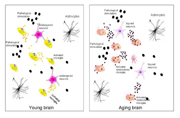
Microglia cells are the brain counterpart of macrophages and function as the first defense in the brain. Although they are neuroprotective in the young brain, microglia cells may be primed to react abnormally to stimuli in the aged brain and to become neurotoxic and destructive during neurodegeneration. Aging-induced immune senescence occurs in the brain as age-associated microglia senescence, which renders microglia to function abnormally and may eventually promote neurodegeneration. Microglia senescence is manifested by both morphological changes and alterations in immunophenotypic expression and inflammatory profile. These changes are likely caused by microinvironmental factors, but intrinsic factors cannot yet be completely excluded. Microglia senescence appears to underlie the switching of microglia from neuroprotective in the young brain to neurotoxic in the aged brain. The hypothesis of microglia senescence during aging offers a novel perspective on their roles in aging-related neurodegeneration. In Parkinson's disease and Alzheimer's disease, over-activation of microglia may play an active role in the pathogenesis because microglia senescence primes them to be neurotoxic during the development of the diseases.
Overview
The claims and statistics of this paper haven’t been checked yet.
Run the check — free, no account needed. It reads the open-access text and checks the sources it cites.
Integrity signals
Registry
Cites
Statistics
Figures
Verification
Claims the paper makes
The claims and statistics of this paper haven’t been checked yet.
Run the check — free, no account needed. It reads the open-access text and checks the sources it cites.
Claims other papers make about this one
Each is checked against this paper’s own text: does it say what the citing paper says it does?
In the citing paper
“The ageing brain is also characterised by structural and functional changes to microglial cells, which are the resident immune cell population of the central nervous system (CNS) and are the CNS equivalent of macrophages. They are activated by brain injury and local and systemic inflammation and become primed (hyper-responsive) to small stimuli with ageing, which can potentially cause damage and”
From Frailty in elderly people (2013) · cites this paper as [26]
Self-check
Percentages, sums and reported differences re-computed from the paper’s own numbers.
The self-check needs the paper’s full text.
Citations
Cites
Resting microglial cells are highly dynamic surveillants of brain parenchyma in vivo
Nimmerjahn, A., Kirchhoff, F., & Helmchen, F. (2005) · Science · doi:10.1126/science.1110647 · Cited by 5,827 · cited 2×
“By using in vivo two-photon imaging techniques, Nimmerjahn et al[37].”
BackgroundMethodInflamm‐aging: An Evolutionary Perspective on Immunosenescence
FRANCESCHI, C., BONAFÈ, M., VALENSIN, S., OLIVIERI, F., DE LUCA, M., OTTAVIANI, E., & DE BENEDICTIS, G. (2000) · Annals of the New York Academy of Sciences · doi:10.1111/j.1749-6632.2000.tb06651.x · Cited by 5,358 · cited 2×
“Extrinsic factors might include neurotransmitter, oxidative stress, neuroendocrine factors[10,68], hormones, and pathological factors such as abnormal protein aggregation [52] that exists in the microenvironment where microglia live.”
BackgroundMicroglia: a sensor for pathological events in the CNS
Kreutzberg, G. W. (1996) · Trends in Neurosciences · doi:10.1016/0166-2236(96)10049-7 · Cited by 4,396 · cited 1×
“When triggered by appropriate stimulation, microglia rapidly transformed from a resting state to an activated state with deramified shape and enhanced antigen presentation, which has been graded to a series of stages[18].”
BackgroundMicroglia: Active sensor and versatile effector cells in the normal and pathologic brain
Hanisch, U.-K., & Kettenmann, H. (2007) · Nature Neuroscience · doi:10.1038/nn1997 · Cited by 3,676 · cited 1×
“Microglia are distributed ubiquitously throughout the brain and function as resident macrophages and antigen-presenting cells in the CNS[35,36].”
BackgroundMicroglia and inflammation-mediated neurodegeneration: Multiple triggers with a common mechanism
Block, M. L., & Hong, J.-S. (2005) · Progress in Neurobiology · doi:10.1016/j.pneurobio.2005.06.004 · Cited by 1,557 · cited 1×
“These studies indicate that microglia are capable of being both neurotrophic and neurotoxic, depending on the specific stimulus, the severity of injury, and the environment [31-34].”
BackgroundMicroglial Dysfunction and Defective β-Amyloid Clearance Pathways in Aging Alzheimer's Disease Mice
Hickman, S. E., Allison, E. K., & El Khoury, J. (2008) · The Journal of Neuroscience · doi:10.1523/jneurosci.0616-08.2008 · Cited by 1,329 · cited 1×
“These genes include Abbinding scavenger receptors A (SRA), CD36, receptor for advanced glycosylation end products (RAGE), Abdegrading enzymes (insulysin, neprilysin) and MMP9 [74].”
BackgroundThe Endocrine Regulation of Aging by Insulin-like Signals
Tatar, M., Bartke, A., & Antebi, A. (2003) · Science · doi:10.1126/science.1081447 · Cited by 1,327 · cited 1×
“Insulin-like signaling pathway has been shown to regulate life span in worms, flies, and mice[ 72 ].”
BackgroundDown-Regulation of the Macrophage Lineage Through Interaction with OX2 (CD200)
Hoek, R. M., Ruuls, S. R., Murphy, C. A., Wright, G. J., Goddard, R., Zurawski, S. M., Blom, B., Homola, M. E., Streit, W. J., Brown, M. H., et al. (2000) · Science · doi:10.1126/science.290.5497.1768 · Cited by 1,022 · cited 1×
“Microglia activation is normally controlled under physiological conditions of the CNS through neuron-glia contact, CD200R-CD200 pathway[19], neuronal electrical activity[20], and/or some soluble neurotransmitters [21].”
BackgroundStreit, W. J., Braak, H., Xue, Q.-S., & Bechmann, I. (2009) · Acta Neuropathologica · doi:10.1007/s00401-009-0556-6 · Cited by 727
Dystrophic microglia in the aging human brain
Streit, W. J., Sammons, N. W., Kuhns, A. J., & Sparks, D. L. (2004) · Glia · doi:10.1002/glia.10319 · Cited by 705 · cited 2×
“”[50] Characteristics of “dystrophic” microglia that were observed in the aged brain include deramification (loss of finely branched cytoplasmic processes), cytoplasmic beading/spheroid formation, shortened and twisted cytoplasmic processes, and instances of partial or complete cytoplasmic fragmentation[50].”
Background
Cited by
Sorted by citations
Clegg, A., Young, J., Iliffe, S., Rikkert, M. O., & Rockwood, K. (2013) · The Lancet · doi:10.1016/s0140-6736(12)62167-9 · Cited by 9,503
Clearance of senescent glial cells prevents tau-dependent pathology and cognitive decline
Bussian, T. J., Aziz, A., Meyer, C. F., Swenson, B. L., van Deursen, J. M., & Baker, D. J. (2018) · Nature · doi:10.1038/s41586-018-0543-y · Cited by 1,289
Tay, T. L., Savage, J. C., Hui, C. W., Bisht, K., & Tremblay, M. (2017) · The Journal of Physiology · doi:10.1113/jp272134 · Cited by 562
“These parameters indicate that as humans and rodents age microglia take on a phenotype consistent with a pro-inflammatory response, even in the absence of overt pathological stimuli (Luo et al. 2010; Choi & Won, 2011; Wong, 2013; Sierra et al. 2014; Ojo et al. 2015; Barrientos et al. 2015a).”
BackgroundAge-related alterations in the dynamic behavior of microglia
Damani, M. R., Zhao, L., Fontainhas, A. M., Amaral, J., Fariss, R. N., & Wong, W. T. (2011) · Aging Cell · doi:10.1111/j.1474-9726.2010.00660.x · Cited by 504
“…aging changes in microglia have been hypothesized to drive the pathogenic progression through a diminution of neuroprotective functions, direct increases in neurotoxicity, and dysregulated responses to signals and perturbations (Flanary et al., 2007; Streit et al., 2008; Luo et al., 2010).”
BackgroundMicroglia during development and aging
Harry, G. J. (2013) · Pharmacology & Therapeutics · doi:10.1016/j.pharmthera.2013.04.013 · Cited by 524
“…hypothesized that changes in the aging microglia drive pathogenic progression of diseases or injury through a diminution of neuroprotective functions, increase in neurotoxicity, and dysregulation of responses to signals and perturbations (Flanary et al., 2007; Lopes et al., 2008; Luo et al., 2010).”
Background
Metadata
- Authors
- Luo, X.-G., Ding, J.-Q., & Chen, S.-D.
- Venue
- Molecular Neurodegeneration
- Published
- 2010-03-24
- Volume
- 5
- Issue
- 1
- Pages
- 12-12
- ISSN
- 1750-1326 (ISSN-L)
- PMC
- PMC2852379
- OpenAlex
- W2156427445
- Cited by
- 236
- References
- 85
- Citations by year
- 2026: 7 · 2025: 12 · 2024: 10 · 2023: 11 · 2022: 17 · 2021: 14 · 2020: 15 · 2019: 11 · 2018: 20 · 2017: 13 · 2016: 18 · 2015: 19 · 2014: 12 · 2013: 22 · 2012: 15
- FWCI
- 10.86
- Type
- journal article
- Language
- en
- Open access
- gold
- doi.org
- Licence
- cc-by
- Locations
- Molecular Neurodegeneration (publishedVersion, cc-by)
- PubMed Central (publishedVersion, cc-by)
- PubMed (publishedVersion)
- DOAJ (DOAJ: Directory of Open Access Journals) (submittedVersion, cc-by-sa)
- Topics
- Life Sciences › Neuroscience › Neurology › Neuroinflammation and Neurodegeneration Mechanisms
- Life Sciences › Biochemistry, Genetics and Molecular Biology › Molecular Biology › Inflammasome and immune disorders
- Life Sciences › Neuroscience › Neurology › Neurological Disease Mechanisms and Treatments
- Keywords
- microglia, brain aging, neurodegeneration, microglial senescence, immunosenescence, neuroinflammation, neuroprotection, Parkinson's disease, Alzheimer's disease, microglial activation, neurotoxicity
About these checks
Registry, retraction, Cites and statistics results are computed, not judged, with one exception: a printed reference that no index lists is read once by a model before it counts as a citation error. Claims are extracted by a model and kept only when their quote appears verbatim in the text; each cited source is then checked, with a model’s help, for whether it says what the claim relies on. Those verdicts can be wrong — read the claim’s quote and the cited source before relying on one.
Statistics and arithmetic are recomputed, not judged.
Confidence bands are shown only for fields calibrated on our annotated test set. None is calibrated yet, so no band is shown.
What counts as a claim ›How we measure confidence ›How we check this ›
Are you an author, or do you see a problem with this page? Request a review
