

The DOI matches its Crossref record; 85 of the 85 references it cites were found in a registry or an index.
Microglia in the aging brain: relevance to neurodegeneration
Open accessAbstract
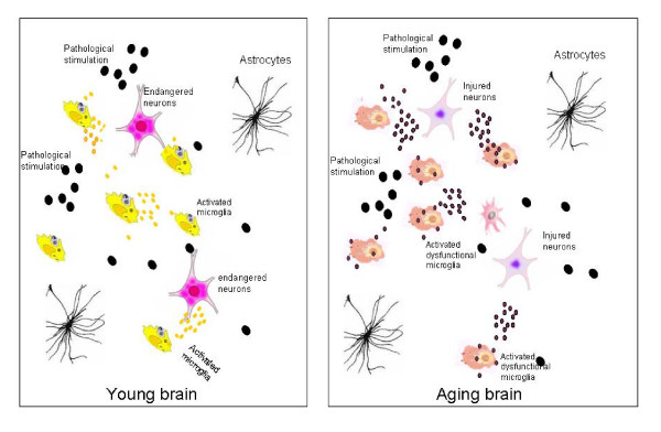
Microglia cells are the brain counterpart of macrophages and function as the first defense in the brain. Although they are neuroprotective in the young brain, microglia cells may be primed to react abnormally to stimuli in the aged brain and to become neurotoxic and destructive during neurodegeneration. Aging-induced immune senescence occurs in the brain as age-associated microglia senescence, which renders microglia to function abnormally and may eventually promote neurodegeneration. Microglia senescence is manifested by both morphological changes and alterations in immunophenotypic expression and inflammatory profile. These changes are likely caused by microinvironmental factors, but intrinsic factors cannot yet be completely excluded. Microglia senescence appears to underlie the switching of microglia from neuroprotective in the young brain to neurotoxic in the aged brain. The hypothesis of microglia senescence during aging offers a novel perspective on their roles in aging-related neurodegeneration. In Parkinson's disease and Alzheimer's disease, over-activation of microglia may play an active role in the pathogenesis because microglia senescence primes them to be neurotoxic during the development of the diseases.
Overview
The claims and statistics of this paper haven’t been checked yet.
Run the check — free, no account needed. It reads the open-access text and checks the sources it cites.
Integrity signals
Registry
Cites
Statistics
Figures
Verification
Claims the paper makes
The claims and statistics of this paper haven’t been checked yet.
Run the check — free, no account needed. It reads the open-access text and checks the sources it cites.
Claims other papers make about this one
Each is checked against this paper’s own text: does it say what the citing paper says it does?
In the citing paper
“The ageing brain is also characterised by structural and functional changes to microglial cells, which are the resident immune cell population of the central nervous system (CNS) and are the CNS equivalent of macrophages. They are activated by brain injury and local and systemic inflammation and become primed (hyper-responsive) to small stimuli with ageing, which can potentially cause damage and”
From Frailty in elderly people (2013) · cites this paper as [26]
Self-check
Percentages, sums and reported differences re-computed from the paper’s own numbers.
The self-check needs the paper’s full text.
Citations
Cites
Resting microglial cells are highly dynamic surveillants of brain parenchyma in vivo
Nimmerjahn, A., Kirchhoff, F., & Helmchen, F. (2005) · Science · doi:10.1126/science.1110647 · Cited by 5,827 · cited 2×
“By using in vivo two-photon imaging techniques, Nimmerjahn et al[37].”
BackgroundMethodInflamm‐aging: An Evolutionary Perspective on Immunosenescence
FRANCESCHI, C., BONAFÈ, M., VALENSIN, S., OLIVIERI, F., DE LUCA, M., OTTAVIANI, E., & DE BENEDICTIS, G. (2000) · Annals of the New York Academy of Sciences · doi:10.1111/j.1749-6632.2000.tb06651.x · Cited by 5,358 · cited 2×
“Extrinsic factors might include neurotransmitter, oxidative stress, neuroendocrine factors[10,68], hormones, and pathological factors such as abnormal protein aggregation [52] that exists in the microenvironment where microglia live.”
BackgroundMicroglia: a sensor for pathological events in the CNS
Kreutzberg, G. W. (1996) · Trends in Neurosciences · doi:10.1016/0166-2236(96)10049-7 · Cited by 4,396 · cited 1×
“When triggered by appropriate stimulation, microglia rapidly transformed from a resting state to an activated state with deramified shape and enhanced antigen presentation, which has been graded to a series of stages[18].”
BackgroundMicroglia: Active sensor and versatile effector cells in the normal and pathologic brain
Hanisch, U.-K., & Kettenmann, H. (2007) · Nature Neuroscience · doi:10.1038/nn1997 · Cited by 3,676 · cited 1×
“Microglia are distributed ubiquitously throughout the brain and function as resident macrophages and antigen-presenting cells in the CNS[35,36].”
BackgroundMicroglia and inflammation-mediated neurodegeneration: Multiple triggers with a common mechanism
Block, M. L., & Hong, J.-S. (2005) · Progress in Neurobiology · doi:10.1016/j.pneurobio.2005.06.004 · Cited by 1,557 · cited 1×
“These studies indicate that microglia are capable of being both neurotrophic and neurotoxic, depending on the specific stimulus, the severity of injury, and the environment [31-34].”
BackgroundMicroglial Dysfunction and Defective β-Amyloid Clearance Pathways in Aging Alzheimer's Disease Mice
Hickman, S. E., Allison, E. K., & El Khoury, J. (2008) · The Journal of Neuroscience · doi:10.1523/jneurosci.0616-08.2008 · Cited by 1,329 · cited 1×
“These genes include Abbinding scavenger receptors A (SRA), CD36, receptor for advanced glycosylation end products (RAGE), Abdegrading enzymes (insulysin, neprilysin) and MMP9 [74].”
BackgroundThe Endocrine Regulation of Aging by Insulin-like Signals
Tatar, M., Bartke, A., & Antebi, A. (2003) · Science · doi:10.1126/science.1081447 · Cited by 1,327 · cited 1×
“Insulin-like signaling pathway has been shown to regulate life span in worms, flies, and mice[ 72 ].”
BackgroundDown-Regulation of the Macrophage Lineage Through Interaction with OX2 (CD200)
Hoek, R. M., Ruuls, S. R., Murphy, C. A., Wright, G. J., Goddard, R., Zurawski, S. M., Blom, B., Homola, M. E., Streit, W. J., Brown, M. H., et al. (2000) · Science · doi:10.1126/science.290.5497.1768 · Cited by 1,022 · cited 1×
“Microglia activation is normally controlled under physiological conditions of the CNS through neuron-glia contact, CD200R-CD200 pathway[19], neuronal electrical activity[20], and/or some soluble neurotransmitters [21].”
BackgroundStreit, W. J., Braak, H., Xue, Q.-S., & Bechmann, I. (2009) · Acta Neuropathologica · doi:10.1007/s00401-009-0556-6 · Cited by 727
Dystrophic microglia in the aging human brain
Streit, W. J., Sammons, N. W., Kuhns, A. J., & Sparks, D. L. (2004) · Glia · doi:10.1002/glia.10319 · Cited by 705 · cited 2×
“”[50] Characteristics of “dystrophic” microglia that were observed in the aged brain include deramification (loss of finely branched cytoplasmic processes), cytoplasmic beading/spheroid formation, shortened and twisted cytoplasmic processes, and instances of partial or complete cytoplasmic fragmentation[50].”
Background
Cited by
Sorted by citations
Clegg, A., Young, J., Iliffe, S., Rikkert, M. O., & Rockwood, K. (2013) · The Lancet · doi:10.1016/s0140-6736(12)62167-9 · Cited by 9,503
Clearance of senescent glial cells prevents tau-dependent pathology and cognitive decline
Bussian, T. J., Aziz, A., Meyer, C. F., Swenson, B. L., van Deursen, J. M., & Baker, D. J. (2018) · Nature · doi:10.1038/s41586-018-0543-y · Cited by 1,289
Tay, T. L., Savage, J. C., Hui, C. W., Bisht, K., & Tremblay, M. (2017) · The Journal of Physiology · doi:10.1113/jp272134 · Cited by 562
“These parameters indicate that as humans and rodents age microglia take on a phenotype consistent with a pro-inflammatory response, even in the absence of overt pathological stimuli (Luo et al. 2010; Choi & Won, 2011; Wong, 2013; Sierra et al. 2014; Ojo et al. 2015; Barrientos et al. 2015a).”
BackgroundAge-related alterations in the dynamic behavior of microglia
Damani, M. R., Zhao, L., Fontainhas, A. M., Amaral, J., Fariss, R. N., & Wong, W. T. (2011) · Aging Cell · doi:10.1111/j.1474-9726.2010.00660.x · Cited by 504
“…aging changes in microglia have been hypothesized to drive the pathogenic progression through a diminution of neuroprotective functions, direct increases in neurotoxicity, and dysregulated responses to signals and perturbations (Flanary et al., 2007; Streit et al., 2008; Luo et al., 2010).”
BackgroundMicroglia during development and aging
Harry, G. J. (2013) · Pharmacology & Therapeutics · doi:10.1016/j.pharmthera.2013.04.013 · Cited by 524
“…hypothesized that changes in the aging microglia drive pathogenic progression of diseases or injury through a diminution of neuroprotective functions, increase in neurotoxicity, and dysregulation of responses to signals and perturbations (Flanary et al., 2007; Lopes et al., 2008; Luo et al., 2010).”
BackgroundThe role of microglia in retinal neurodegeneration: Alzheimer’s disease, parkinson, and glaucoma.
Ramirez, A. I., de Hoz, R., Salobrar-Garcia, E., Salazar, J. J., Rojas, B., Ajoy, D., López-Cuenca, I., Rojas, P., Triviño, A., & Ramírez, J. M. (2017) · Frontiers in Aging Neuroscience · doi:10.3389/fnagi.2017.00214 · Cited by 466
“…Farina et al., 2007; Inman and Horner, 2007; Johnson et al., 2007; Hammam et al., 2008; Tezel, 2009; Ebneter et al., 2010; Graeber and Streit, 2010; Luo et al., 2010; Bosco et al., 2011, 2012; Kettenmann et al., 2011; London et al., 2011; Bosco et al., 2012; Gallego et al., 2012; Varnum and…”
BackgroundOgrodnik, M., Evans, S. A., Fielder, E., Victorelli, S., Kruger, P., Salmonowicz, H., Weigand, B. M., Patel, A. D., Pirtskhalava, T., Inman, C. L., et al. (2021) · Aging Cell · doi:10.1111/acel.13296 · Cited by 405
“Another feature of brain inflammaging is microglial activation, which is characterized by increased soma size and is observed during aging and neurodegenerative diseases (Luo et al., 2010; Norden & Godbout, 2013).”
BackgroundStuendl, A., Kunadt, M., Kruse, N., Bartels, C., Moebius, W., Danzer, K. M., Mollenhauer, B., & Schneider, A. (2016) · Brain · doi:10.1093/brain/awv346 · Cited by 428
“However, insufficient microglial clearance function, as observed in many neurodegenerative diseases, may pave the way for interneuronal propagation of pathogenic exosomal a-synuclein species (Luo et al., 2010).”
Homeostatic and injury‐induced microglia behavior in the aging brain
Hefendehl, J. K., Neher, J. J., Sühs, R. B., Kohsaka, S., Skodras, A., & Jucker, M. (2014) · Aging Cell · doi:10.1111/acel.12149 · Cited by 319
Effects of aging and sensory loss on glial cells in mouse visual and auditory cortices
Tremblay, M., Zettel, M. L., Ison, J. R., Allen, P. D., & Majewska, A. K. (2012) · Glia · doi:10.1002/glia.22287 · Cited by 309
“These neuronal alterations, however, may be secondary to the larger age-related changes in glial cells that have been associated with decreased neuroprotective function and increased neurotoxicity (Flanary et al., 2007; Luo et al., 2010; Lynch, 2010; Streit et al., 2008; von Bernhardi et al., 2010).”
BackgroundKwon, H. J., Kim, D., Seo, K., Kim, Y. G., Han, S. I., Kang, T., Soh, M., & Hyeon, T. (2018) · Angewandte Chemie International Edition · doi:10.1002/anie.201805052 · Cited by 291
Vascular Cell Senescence Contributes to Blood–Brain Barrier Breakdown
Yamazaki, Y., Baker, D. J., Tachibana, M., Liu, C.-C., van Deursen, J. M., Brott, T. G., Bu, G., & Kanekiyo, T. (2016) · Stroke · doi:10.1161/strokeaha.115.010835 · Cited by 263
Microglia in the developing brain: a potential target with lifetime effects
Harry, G. J., & Kraft, A. D. (2012) · NeuroToxicology · doi:10.1016/j.neuro.2012.01.012 · Cited by 250
Inflammatory biomarkers, cerebral microbleeds, and small vessel disease
Shoamanesh, A., Preis, S. R., Beiser, A., Vasan, R. S., Benjamin, E. J., Kase, C. S., Wolf, P. A., DeCarli, C. S., Romero, J. R., & Seshadri, S. S. (2015) · Neurology · doi:10.1212/wnl.0000000000001279 · Cited by 236
“An alternative explanation is that chronically progressing large volumes of ischemic neuronal and glial injury, and the ensuing inflammatory response, promotes microglial senescence (the dysfunction and dystrophy of microglia with advancing age).(31) It is of interest that, irrespective of myeloperoxidase, we observed…”
BackgroundNeuroinflammation and microglia in glaucoma: time for a paradigm shift
Wei, X., Cho, K., Thee, E. F., Jager, M. J., & Chen, D. F. (2018) · Journal of Neuroscience Research · doi:10.1002/jnr.24256 · Cited by 239
“More specifically, upregulation of the inflammatory inducers/sensors such as TLRs, transducers Trif and MyD88, and amplifiers such as IL-1 and IL-6 has been reported (C. Luo et al., 2010; Tezel, Yang, Luo, Cai, & Powell, 2012).”
BackgroundThe changing phenotype of microglia from homeostasis to disease
Luo, X., & Chen, S. (2012) · Translational Neurodegeneration · doi:10.1186/2047-9158-1-9 · Cited by 246
“(From Luo et al.,2010 with permission). in the young brain to neurotoxic in the aged brain upon activation [147].”
BackgroundSelf-citationLRP1 in Brain Vascular Smooth Muscle Cells Mediates Local Clearance of Alzheimer's Amyloid-β
Kanekiyo, T., Liu, C.-C., Shinohara, M., Li, J., & Bu, G. (2012) · The Journal of Neuroscience · doi:10.1523/jneurosci.3987-12.2012 · Cited by 211
“, 2004) and microglia (Wyss-Coray et al., 2001; El Khoury and Luster, 2008; Luo et al., 2010) can degrade A to eliminate it from the brain.”
BackgroundTranscriptional and epigenetic decoding of the microglial aging process
Li, X., Li, Y., Jin, Y., Zhang, Y., Wu, J., Xu, Z., Huang, Y., Cai, L., Gao, S., Liu, T., et al. (2023) · Nature Aging · doi:10.1038/s43587-023-00479-x · Cited by 234
“Microglia undergo a mild and chronic reactive response in aged brains 86,87 , whereas LPS-induced reactivity is firm and transient 77,88 .”
Senescence-associated lncRNAs: senescence-associated long noncoding RNAs
Abdelmohsen, K., Panda, A. C., Kang, M. J., Xu, J. Z., Selimyan, R., Yoon, J., Martindale, J. L., De, S., Wood, W. H., Becker, K. G., et al. (2013) · Aging Cell · doi:10.1111/acel.12115 · Cited by 211
“…has also been recognized, particularly in pathologies associated with the chronic inflammatory phenotype seen in the elderly, such as cardiovascular disease, neurodegeneration, and declined immune function (Kuilman et al., 2008; Freund et al., 2010; Luo et al., 2010; Gorospe & Abdelmohsen, 2011).”
BackgroundMicroglia centered pathogenesis in ALS: insights in cell interconnectivity
Brites, D. M. T. O., & Vaz, A. R. (2014) · Frontiers in Cellular Neuroscience · doi:10.3389/fncel.2014.00117 · Cited by 213
Age-dependent defects of alpha-synuclein oligomer uptake in microglia and monocytes
Bliederhaeuser, C., Grozdanov, V., Speidel, A., Zondler, L., Ruf, W. P., Bayer, H., Kiechle, M., Feiler, M. S., Freischmidt, A., Brenner, D., et al. (2015) · Acta Neuropathologica · doi:10.1007/s00401-015-1504-2 · Cited by 181
“Likewise, in the human healthy aged brain increased microglia activation with an increased susceptibility to pro-inflammatory activation has been reported [10, 40, 63].”
BackgroundIncreased White Matter Inflammation in Aging- and Alzheimer’s Disease Brain
Raj, D., Yin, Z., Breur, M., Doorduin, J., Holtman, I. R., Olah, M., Mantingh-Otter, I. J., Van Dam, D., De Deyn, P. P., den Dunnen, W., et al. (2017) · Frontiers in Molecular Neuroscience · doi:10.3389/fnmol.2017.00206 · Cited by 180
“As expected for aging brain tissue (Luo et al., 2010), the expression of proinflammatory genes involved in antigen presentation, interferon-, cytokine-, and chemokine signaling, and phagocytosis were enhanced in aged microglia.”
BackgroundMicroglia change from a reactive to an age-like phenotype with the time in culture
Caldeira, C., Oliveira, A. F., Cunha, C., Vaz, A. R., Falcão, A. S., Fernandes, A., & Brites, D. (2014) · Frontiers in Cellular Neuroscience · doi:10.3389/fncel.2014.00152 · Cited by 156
“Nevertheless, the hypothesis of microglia senescence during aging and related neurodegenerative diseases emerged as a key determinant (Luo et al., 2010).”
BackgroundThe endocannabinoid system in normal and pathological brain ageing
Bilkei‐Gorzó, A. (2012) · Philosophical Transactions of the Royal Society B Biological Sciences · doi:10.1098/rstb.2011.0388 · Cited by 161
Microglial dysfunction connects depression and Alzheimer’s disease
Santos, L. E., Beckman, D., & Ferreira, S. T. (2016) · Brain, Behavior, and Immunity · doi:10.1016/j.bbi.2015.11.011 · Cited by 154
“Even in the absence of trauma or pathology, microglia priming may occur as a consequence of normal aging (Luo et al., 2010).”
BackgroundCerebrospinal fluid markers reveal intrathecal inflammation in progressive multiple sclerosis
Komori, M., Blake, A., Greenwood, M., Lin, Y. C., Kosa, P. F., Ghazali, D., Winokur, P. N., Natrajan, M. S., Wuest, S. C., Romm, E., et al. (2015) · Annals of Neurology · doi:10.1002/ana.24408 · Cited by 147
Immunosenescence and inflammaging: Mechanisms and role in diseases
Ajoolabady, A., Praticò, D., Tang, D., Zhou, S., Franceschi, C., & Ren, J. T. (2024) · Ageing Research Reviews · doi:10.1016/j.arr.2024.102540 · Cited by 172
Immunosenescence: signaling pathways, diseases and therapeutic targets
Fu, Y., Wang, B., Alu, A., Hong, W., Hong, L., He, X., Shi, H., Cheng, P., & Yang, X. (2025) · Signal Transduction and Targeted Therapy · doi:10.1038/s41392-025-02371-z · Cited by 168
“160,161 Furthermore, A β can function as a DAMP and activate the in fl ammasome via the TLR pathway, leading to the production of in fl ammatory cytokines such as IL-1 β .”
Priming of microglia in a DNA-repair deficient model of accelerated aging
Raj, D. D. A., Jaarsma, D., Holtman, I. R., Olah, M., Ferreira, F. M., Schaafsma, W., Brouwer, N., Meijer, M., de Waard, M. C., van der Pluijm, I., et al. (2014) · Neurobiology of Aging · doi:10.1016/j.neurobiolaging.2014.03.025 · Cited by 148
“The observed morphologic changes resemble previously reported microglia phenotypes referred to as “ hypertrophic ” (Ayoub and Salm, 2003; Streit, 2000), “ reactive ” (Gomide et al., 2005), or “ intermediate ” (Karperien et al., 2013).”
What is microglia neurotoxicity (Not)?
Biber, K. P. H., Owens, T., & Boddeke, E. (2014) · Glia · doi:10.1002/glia.22654 · Cited by 146
“Upon aging microglia show increased immunoreactivity for CD68, a lysosome associated phagocytic receptor, Toll-like-receptors (TLRs), MHC Class II antigen, Matrix remodeling enzyme MMP-12, Cd11b, and Cd11c integrins and cytokines (Luo et al., 2010).”
BackgroundThe Role of Immunosenescence in Neurodegenerative Diseases
Costantini, E., D’Angelo, C., & Reale, M. (2018) · Mediators of Inflammation · doi:10.1155/2018/6039171 · Cited by 133
“Microglial and astrocytes cell uncontrolled activation, increased in an age-dependent manner, leads to excessive inflammation [78].”
Heat-Shock Proteins in Neuroinflammation
Dukay, B., Csoboz, B., & Tóth, M. E. (2019) · Frontiers in Pharmacology · doi:10.3389/fphar.2019.00920 · Cited by 139
“Heat-shock elements can be found in the promoter region of CXC chemokine genes as well, although a heat shock has gene-specific enhancing or repressing effect on the expression of these genes (Maity et al., 2011).”
BackgroundUnderstanding and Reducing Disability in Older Adults Following Critical Illness*
Brummel, N. E., Balas, M. C., Morandi, A., Ferrante, L. E., Gill, T. M., & Ely, E. W. (2015) · Critical Care Medicine · doi:10.1097/ccm.0000000000000924 · Cited by 137
Wang, Y., Wang, B., Zhu, M.-T., Li, M., Wang, H.-J., Wang, M., Ouyang, H. W., Chai, Z., Feng, W.-Y., & Zhao, Y.-L. (2011) · Toxicology Letters · doi:10.1016/j.toxlet.2011.05.001 · Cited by 130
Prolonged Neuroinflammation after Lipopolysaccharide Exposure in Aged Rats
Fu, H. Q., Yang, T., Xiao, W. L., Fan, L., Wu, Y., Terrando, N., & Wang, T. L. (2014) · PLoS ONE · doi:10.1371/journal.pone.0106331 · Cited by 127
“contribute to behavioral and cognitive deficits [14], other glia-type cells including astrocytes also display pivotal roles in regulating”
BackgroundMicroglia in Alzheimer’s Disease in the Context of Tau Pathology
Perea, J. R., Bolós, M., & Ávila, J. (2020) · Biomolecules · doi:10.3390/biom10101439 · Cited by 134
Eat‐me signals: Keys to molecular phagocyte biology and “Appetite” control
Li, W. (2011) · Journal of Cellular Physiology · doi:10.1002/jcp.22815 · Cited by 117
“The former interacts with LRP family members on phagocytes (Maiti et al., 2008), whereas the receptor for annexin I remains elusive.”
BackgroundPeng, X., Wu, Y., Brouwer, U., van Vliet, T., Wang, B., Demaria, M., Barazzuol, L., & Coppes, R. P. (2020) · Cell Death and Disease · doi:10.1038/s41419-020-03074-9 · Cited by 126
Mattsson, N., Tabatabaei, S., Johansson, P., Hansson, O., Andreasson, U., Månsson, J.-E., Johansson, J.-O., Olsson, B., Wallin, A., Svensson, J., et al. (2011) · NeuroMolecular Medicine · doi:10.1007/s12017-011-8147-9 · Cited by 118
“dysfunctional microglia have been suggested as an initiating factor in AD pathogenesis ( Luo et al. 2010 ).”
Phagocyte dysfunction, tissue aging and degeneration
Li, W. (2013) · Ageing Research Reviews · doi:10.1016/j.arr.2013.05.006 · Cited by 116
Single-cell analysis of the aging female mouse hypothalamus
Hajdarovic, K. H., Yu, D., Hassell, L.-A., Evans, S. A., Packer, S., Neretti, N., & Webb, A. E. (2022) · Nature Aging · doi:10.1038/s43587-022-00246-4 · Cited by 118
Targeting glioblastoma with NK cells and mAb against NG2/CSPG4 prolongs animal survival
Poli, A., Wang, J., Domingues, O., Planagumà, J., Yan, T., Rygh, C. B., Skaftnesmo, K. O., Thorsen, F. A., McCormack, E., Hentges, F., et al. (2013) · Oncotarget · doi:10.18632/oncotarget.1291 · Cited by 115
“Since pro-inflammatory microglia are implicated in several brain diseases [69, 70], future work will have to verify the safety and tolerability of our combination treatment prior to clinical application.”
Microglia-Astrocyte Communication in Alzheimer’s Disease
Wu, Y., & Eisel, U. L. M. (2023) · Journal of Alzheimer s Disease · doi:10.3233/jad-230199 · Cited by 125
MicroRegulators come of age in senescence
Gorospe, M., & Abdelmohsen, K. (2011) · Trends in Genetics · doi:10.1016/j.tig.2011.03.005 · Cited by 108
“Aging also leads to the accumulation of senescent brain immune cells (microglia), a process linked to neurodegenerative pathologies such as Parkinson’s and Alzheimer’s diseases [38].”
BackgroundVerma, A., Shteinfer‐Kuzmine, A., Kamenetsky, N., Pittala, S., Paul, A., Crystal, E. N., Ouro, A., Caspi, V. C., Pandey, S. K., Monsonego, A., et al. (2022) · Translational Neurodegeneration · doi:10.1186/s40035-022-00329-7 · Cited by 109
“In AD, the microglia play important roles in Aβ clearance and neuroinflammatory response via secretion of pro-inflammatory cytokines [79].”
Role of Prostaglandins in Neuroinflammatory and Neurodegenerative Diseases
Lima, I. V. D. A., Bastos, L. F. S., Limborço‐Filho, M., Fiebich, B. L., & de Oliveira, A. C. P. (2012) · Mediators of Inflammation · doi:10.1155/2012/946813 · Cited by 104
“Therefore, microglia senescence could play a role in the development of some neurodegenerative conditions [161, 166].”
BackgroundLedreux, A., Wang, X., Schultzberg, M., Granholm, A., & Freeman, L. R. (2016) · Behavioural Brain Research · doi:10.1016/j.bbr.2016.06.012 · Cited by 99
de Hoz, R., Gallego, B., Ramirez, A. I., Rojas, B., Salazar, J. J., Valiente‐Soriano, F. J., Avilés‐Trigueros, M., Villegas‐Pérez, M. P., Vidal‐Sanz, M., Triviño, A., et al. (2013) · PLoS ONE · doi:10.1371/journal.pone.0083733 · Cited by 92
“However, an uncontrolled microglia response may be dangerous to the survival of injured neurons or can even damage healthy neurons afflicted by excessive inflammation [18].”
BackgroundCrosstalk between endoplasmic reticulum stress and brain inflammation in Alzheimer's disease
Santos, L. E., & Ferreira, S. T. (2017) · Neuropharmacology · doi:10.1016/j.neuropharm.2017.11.016 · Cited by 93
Noncoding RNA control of cellular senescence
Abdelmohsen, K., & Gorospe, M. (2015) · Wiley Interdisciplinary Reviews - RNA · doi:10.1002/wrna.1297 · Cited by 92
“For instance, senescent cells accumulate in Parkinson’s and Alzheimer’s diseases, and impair vascularization in diabetes.(26,27) Senescence of skeletal muscle cell and satellite cells has also been linked to the age-related muscle disorder sarcopenia.”
BackgroundIndulekha, K., Alam, S., Surendar, J., Frohberger, S. J., Hansen, J. N., Kuehlwein, J. M., Hölbling, B. V., Schumak, B., Hübner, M. P., Gräler, M. H., et al. (2019) · Glia · doi:10.1002/glia.23663 · Cited by 89
Hoeijmakers, L., Heinen, Y., van Dam, A.-M., Lucassen, P. J., & Korosi, A. (2016) · Frontiers in Human Neuroscience · doi:10.3389/fnhum.2016.00398 · Cited by 85
Trias, E., Beilby, P. R., Kovacs, M., Ibarburu, S., Varela, V., Barreto-Núñez, R., Bradford, S. C., Beckman, J. S., & Barbeito, L. (2019) · Frontiers in Aging Neuroscience · doi:10.3389/fnagi.2019.00042 · Cited by 85
Senescence-Associated MicroRNAs
Munk, R. B., Panda, A. C., Grammatikakis, I., Gorospe, M., & Abdelmohsen, K. (2017) · International review of cytology · doi:10.1016/bs.ircmb.2017.03.008 · Cited by 90
“For example, accumulation of senescent microglia with aging is linked to neurodegenerative diseases such as Parkinson’s and Alzheimer’s diseases (Luo et al., 2010), while the impaired vascularization in diabetes is linked to the enhanced senescence of endothelial progenitor cells (Rosso et al.,…”
BackgroundZheng, T., Pu, J., Chen, Y., Mao, Y., Guo, Z., Pan, H., Zhang, L., Zhang, H., Sun, B., & Zhang, B. (2017) · Frontiers in Aging Neuroscience · doi:10.3389/fnagi.2017.00012 · Cited by 82
“Aging Does Not Affect the Phagocytosis of Exosomes by Microglia Age-associated microglial senescence in the brain leads to abnormal function and may eventually promote neurodegeneration (Luo et al., 2010).”
BackgroundBlack tea in chemo-prevention of cancer and other human diseases
Pan, M., Lai, C., Wang, H., Lo, C., Ho, C., & Li, S. (2013) · Food Science and Human Wellness · doi:10.1016/j.fshw.2013.03.004 · Cited by 83
“Parkinson’s disease (PD) and Alzheimer’s disease (AD) are ge-dependent neurodegenerative disorders [94].”
BackgroundDuncombe, J., Lennen, R. J., Jansen, M. A., Marshall, I., Wardlaw, J. M., & Horsburgh, K. J. (2016) · Neuropathology and Applied Neurobiology · doi:10.1111/nan.12375 · Cited by 85
Mitra, S., Chakrabarti, N., & Bhattacharyya, A. (2011) · Journal of Neuroinflammation · doi:10.1186/1742-2094-8-163 · Cited by 77
“Over-activation of microglia may cause alterations in immunophenotypic expression and inflammatory profile (promoting microglia senescence), and that condition may switch microglial function from neuroprotective to neurotoxic effects [52].”
BackgroundHIV TAT-mediated microglial senescence: Role of SIRT3-dependent mitochondrial oxidative stress
Thangaraj, A., Chivero, E. T., Tripathi, A., Singh, S., Niu, F., Guo, M.-L., Pillai, P., Periyasamy, P., & Buch, S. (2021) · Redox Biology · doi:10.1016/j.redox.2020.101843 · Cited by 76
Repeated Lipopolysaccharide Stimulation Induces Cellular Senescence in BV2 Cells
Yu, H.-M., Zhao, Y.-M., Luo, X.-G., Feng, Y., Ren, Y., Shang, H., He, Z.-Y., Luo, X.-M., Chen, S.-D., & Wang, X.-Y. (2012) · Neuroimmunomodulation · doi:10.1159/000330254 · Cited by 76
Self-citationBeier, E. E., Neal, M. P., Alam, G., Edler, M. K., Wu, L., & Richardson, J. R. (2017) · Neurobiology of Disease · doi:10.1016/j.nbd.2017.08.009 · Cited by 78
SARS-CoV-2: is there neuroinvasion?
McQuaid, C., Brady, M., & Deane, R. (2021) · Fluids and Barriers of the CNS · doi:10.1186/s12987-021-00267-y · Cited by 72
Turrini, S., Wong, B., Eldaief, M. C., Press, D. Z., Sinclair, D. A., Koch, G., Avenanti, A., & Santarnecchi, E. (2023) · Ageing Research Reviews · doi:10.1016/j.arr.2023.101939 · Cited by 88
“While microglia cells have a neuroprotective role in the young brain, multiple studies have shown that they gradually transition to a chronically activated and neurotoxic state in older adults (Luo et al., 2010), irrespective of their cognitive status (Gefen, 2019; Niraula et al., 2017).”
BackgroundHilal, S., Ikram, M. A., Verbeek, M. M., Franco, O. H., Stoops, E., Vanderstichele, H., Niessen, W. J., & Vernooij, M. W. (2018) · Stroke · doi:10.1161/strokeaha.118.022317 · Cited by 71
de Hoz, R., Ramirez, A. I., Gonzalez‐Martin, R., Ajoy, D., Rojas, B., Salobrar‐García, E., Valiente‐Soriano, F. J., Avilés‐Trigueros, M., Villegas‐Pérez, M. P., Vidal‐Sanz, M., et al. (2018) · Experimental Eye Research · doi:10.1016/j.exer.2018.03.006 · Cited by 71
“…can act as antigen-presenting cells, and can transform into phagocytes (Karlstetter et al., 2015; Karlstetter et al., 2015; Kettenmann et al., 2011; Kettenmann et al., 2011; Luo et al., 2010; Luo et al., 2010; Ramírez et al., 2015; Ransohoff and Perry, 2009; Kezic et al., 2013; Wang et al., 2017).”
BackgroundDas, R. R., & Chinnathambi, S. (2020) · Neuroscience · doi:10.1016/j.neuroscience.2020.09.024 · Cited by 71
Phenotypic and functional differences between senescent and aged murine microglia
Stojiljkovic, M. R., Ain, Q., Bondeva, T., Heller, R., Schmeer, C., & Witte, O. W. (2019) · Neurobiology of Aging · doi:10.1016/j.neurobiolaging.2018.10.007 · Cited by 71
Desplats, P., Gutierrez, A. M., Antonelli, M. C., & Frasch, M. G. (2019) · Neuroscience & Biobehavioral Reviews · doi:10.1016/j.neubiorev.2019.10.013 · Cited by 74
Huang, X., Chen, Y., Zhang, H., Ma, Q., Zhang, Y., & Xu, H. (2011) · Neurobiology of Aging · doi:10.1016/j.neurobiolaging.2011.10.007 · Cited by 68
“Aβ deposits also trigger microglia-mediated neuroinflammation, postulated to contribute to the pathogenesis and progression of AD (Giulian et al., 1995; Minghetti, 2005; Floden et al., 2005; Luo et al., 2010).”
BackgroundMicroglia and Synapse: Interactions in Health and Neurodegeneration
Šišková, Z., & Tremblay, M.-È. (2013) · Neural Plasticity · doi:10.1155/2013/425845 · Cited by 80
Singh, Y. P., Pandey, A., Vishwakarma, S., & Modi, G. P. (2018) · Molecular Diversity · doi:10.1007/s11030-018-9878-4 · Cited by 69
IP3, a small molecule with a powerful message
Decrock, E., Bock, M. D., Wang, N., Gadicherla, A. K., Bol, M., Delvaeye, T., Vandenabeele, P., Vinken, M., Bultynck, G., Krysko, D. V., et al. (2013) · Biochimica et Biophysica Acta (BBA) - Molecular Cell Research · doi:10.1016/j.bbamcr.2012.12.016 · Cited by 64
Das, R. R., & Chinnathambi, S. (2021) · Traffic · doi:10.1111/tra.12784 · Cited by 62
“and invasion can be a determinant of altered function in CNS injury and inflammation.(71,72) The soluble but reactive Tau oligomers can readily diffuse through brain parenchyma and attracts microglial chemotaxis.”
BackgroundAzevedo, E. P., Ledo, J. H., Barbosa, G., Sobrinho, M., Diniz, L. P., da Fonseca, A. C. C., Gomes, F. C. A., Romão, L. F., Lima, F. R. S., Palhano, F. L., et al. (2013) · Cell Death and Disease · doi:10.1038/cddis.2013.325 · Cited by 66
“However, chronically activated microglia may initiate inflammatory processes that lead to neuronal dysfunction and damage.(15,16) Thus, tight regulation of”
BackgroundCouturier, J., Paccalin, M., Morel, M., Terro, F., Milin, S., Pontcharraud, R., Fauconneau, B., & Page, G. (2011) · Journal of Neuroinflammation · doi:10.1186/1742-2094-8-72 · Cited by 65
“Recent papers have underlined the senescence of microglia in AD, with loss of their neuroprotective properties, preceding the onset of tau pathology [90], suggesting that breakdown of the brain’s immune system may be an important factor in the development of neurodegeneration [91].”
BackgroundMander, B. A., Dave, A., Lui, K. K., Sprecher, K. E., Berisha, D. E., Chappel-Farley, M. G., Chen, I. Y., Riedner, B. A., Heston, M. B., Suridjan, I., et al. (2022) · SLEEP · doi:10.1093/sleep/zsac135 · Cited by 62
Centenary of the death of Elie Metchnikoff: a visionary and an outstanding team leader
Cavaillon, J., & Legout, S. (2016) · Microbes and Infection · doi:10.1016/j.micinf.2016.05.008 · Cited by 65
“Microglia senescence appears to underlie the switching of microglia from neuroprotective in the young brain to neurotoxic in the aged brain [85].”
LncRNA-OIS1 regulates DPP4 activation to modulate senescence induced by RAS
Li, L., Breugel, P. C. V., Loayza‐Puch, F., Ugalde, A. P., Korkmaz, G., Messika-Gold, N., Han, R., Lopes, R., Barberà, E. P., Teunissen, H., et al. (2018) · Nucleic Acids Research · doi:10.1093/nar/gky087 · Cited by 58
Targeting normal and cancer senescent cells as a strategy of senotherapy
Sikora, E., Bielak-Żmijewska, A., & Mosieniak, G. (2019) · Ageing Research Reviews · doi:10.1016/j.arr.2019.100941 · Cited by 59
Amelioration of LPS-Induced Inflammation Response in Microglia by AMPK Activation
Chen, C.-C., Lin, J., Cheng, Y.-F., Kuo, C., Huang, C.-F., Kao, S., Liang, Y.-J., Cheng, C., & Chen, H.-M. (2014) · BioMed Research International · doi:10.1155/2014/692061 · Cited by 56
Neuroprotection for Ischemic Stroke: Moving Past Shortcomings and Identifying Promising Directions
Turner, R. C., Lucke-Wold, B. P., Lucke-Wold, N., Elliott, A. S., Logsdon, A. F., Rosen, C. L., & Huber, J. D. (2013) · International Journal of Molecular Sciences · doi:10.3390/ijms14011890 · Cited by 52
“These cytokines activate neurodegenerative pathways [21].”
Effects of acoustic trauma on the auditory system of the rat: The role of microglia
Baizer, J. S., Wong, K. M., Manohar, S., Hayes, S. H., Ding, D., Dingman, R., & Salvi, R. (2015) · Neuroscience · doi:10.1016/j.neuroscience.2015.07.004 · Cited by 53
“…may have a “destroyer” role; actively facilitating the degenerative processes of Alzheimer’s disease, amyotrophic lateral sclerosis, multiple sclerosis and other neurodegenerative disorders (Barger and Harmon, 1997, Streit, 2004, Ponomarev et al., 2006, Block et al., 2007, Luo et al., 2010).”
BackgroundTheoharides, T. C., Twahir, A., & Kempuraj, D. (2023) · Annals of Allergy Asthma & Immunology · doi:10.1016/j.anai.2023.10.032 · Cited by 58
Design, synthesis, and evaluation of small molecule Hsp90 probes
Taldone, T., Zatorska, D., Patel, P. D., Zong, H., Rodina, A., Ahn, J. H., Moulick, K., Guzmán, M. L., & Chiosis, G. (2011) · Bioorganic & Medicinal Chemistry · doi:10.1016/j.bmc.2011.03.013 · Cited by 51
Epigenetic assimilation in the aging human brain
Oh, G., Ebrahimi, S. B., Wang, S.-C., Cortese, R., Kaminsky, Z. A., Gottesman, I. I., Burke, J. R., Plassman, B. L., & Petronis, A. (2016) · Genome biology · doi:10.1186/s13059-016-0946-8 · Cited by 54
“The changes observed in the AD brain samples, and perhaps in the aging brains in general [40], could be related to disproportional loss of neurons compared with neuroglia observed in AD [41].”
BackgroundResultSong, I., Chung, S., Kim, Y., & Maeng, L.-S. (2015) · International Journal of Medical Sciences · doi:10.7150/ijms.12742 · Cited by 50
Activated Microglia in Cortical White Matter Across Cognitive Aging Trajectories
Gefen, T. D., Kım, G., Bolbolan, K., Geoly, A. D., Ohm, D. T., Oboudiyat, C., Shahidehpour, R. K., RADEMAKER, A. W., Weıntraub, S., Bigio, E. H., et al. (2019) · Frontiers in Aging Neuroscience · doi:10.3389/fnagi.2019.00094 · Cited by 50
Free radical biology in neurological manifestations: mechanisms to therapeutics interventions
Tripathi, R. S., Gupta, R. V., Sahu, M., Srivastava, D., Das, A., Ambasta, R. K., & Kumar, P. (2021) · Environmental Science and Pollution Research · doi:10.1007/s11356-021-16693-2 · Cited by 54
“Activated microglia have been reported in numerous neurodegenerative diseases where aging impairs microglial functions due to increased sensitivity towards pro-inflammatory signals (Luo et al. 2010; von Bernhardi et al. 2010).”
BackgroundTubby regulates microglial phagocytosis through MerTK
Caberoy, N. B., Alvarado, G., & Li, W. (2012) · Journal of Neuroimmunology · doi:10.1016/j.jneuroim.2012.07.009 · Cited by 47
“Microglial senescence with increased susceptibility to activation has been implicated in age-related neurodegenerative diseases, including Alzheimer's disease and Parkinson's disease (Lee and Landreth, 2010; Luo et al., 2010; von Bernhardi et al., 2010).”
BackgroundChen, X., Feng, Y., Quinn, R. J., Pountney, D. L., Richardson, D. R., Mellick, G. D., & Ma, L. (2023) · Pharmacological Reviews · doi:10.1124/pharmrev.122.000743 · Cited by 44
“…channels is the hallmark of overactivated microglia (Kettenmann et al., 1993; Noren-berg et al., 1994; Nguyen et al., 2017; Di Lucente et al., 2018; Sarkar et al., 2020), while uncontrolled microglial activities aggravate the neurodegeneration in PD (Luo et al., 2010; Badanjak et al., 2021).”
BackgroundInhibition of mitochondrial fission attenuates Aβ-induced microglia apoptosis
Xie, N., Wang, C., Lian, Y., Wu, C., Zhang, H., & Zhang, Q. (2013) · Neuroscience · doi:10.1016/j.neuroscience.2013.10.011 · Cited by 44
Aging and Neurodegeneration: A Tangle of Models and Mechanisms
Chakrabarti, S., & Mohanakumar, K. P. (2016) · Aging and Disease · doi:10.14336/ad.2016.0312 · Cited by 46
“For example, mitochondrial dysfunction, oxidative stress, inflammatory response and proteotoxicity are common themes in AD and brain aging [2,3,4,5].”
BackgroundThe effect of aged microglia on synaptic impairment and its relevance in neurodegenerative diseases
Triviño, J. J., & von Bernhardi, R. (2021) · Neurochemistry International · doi:10.1016/j.neuint.2021.104982 · Cited by 43
The Role of Microglia in Perioperative Neurocognitive Disorders
Fan, W., Mai, L., Zhu, X. M., Huang, F., & He, H. (2020) · Frontiers in Cellular Neuroscience · doi:10.3389/fncel.2020.00261 · Cited by 42
The Interaction of Diet and Mitochondrial Dysfunction in Aging and Cognition
Kaliszewska, A., Allison, J., Martini, M., & Arias, N. (2021) · International Journal of Molecular Sciences · doi:10.3390/ijms22073574 · Cited by 52
Albornoz, E. A., Woodruff, T. M., & Gordon, R. (2018) · Experientia supplementum · doi:10.1007/978-3-319-89390-7_3 · Cited by 42
Benfotiamine upregulates antioxidative system in activated BV-2 microglia cells
Bozic, I. D., Savić, D. Z., Stevanović, I. D., Peković, S., Nedeljković, N., & Lavrnja, I. C. (2015) · Frontiers in Cellular Neuroscience · doi:10.3389/fncel.2015.00351 · Cited by 44
“These data together imply that benfotiamine exhibits significant antioxidative activity in BV-2 cells.”
BackgroundBowirrat, A. (2022) · Journal of Personalized Medicine · doi:10.3390/jpm12111817 · Cited by 46
“The effect of aging on microglial activity is extremely important, as it is known that aging has an inverse relationship with the activity of the microglia, and aging causes changes in gene expressions, dystrophic microglia, and abnormal cytoplasmic formation, decreasing the microglia’s elasticity and lowering the act…”
BackgroundImpact of Long-Term RF-EMF on Oxidative Stress and Neuroinflammation in Aging Brains of C57BL/6 Mice
Jeong, Y. J., Son, Y., Han, N.-K., Choi, H., Pack, J., Kim, N., Lee, Y., & Lee, H. (2018) · International Journal of Molecular Sciences · doi:10.3390/ijms19072103 · Cited by 44
“It is speculated that any changes in glial activation during aging are key components that influence the pathogenesis of neurodegeneration [36].”
Zhao, Y., & Yang, G. (2021) · Neurochemistry International · doi:10.1016/j.neuint.2021.104974 · Cited by 41
Metadata
- Authors
- Luo, X.-G., Ding, J.-Q., & Chen, S.-D.
- Venue
- Molecular Neurodegeneration
- Published
- 2010-03-24
- Volume
- 5
- Issue
- 1
- Pages
- 12-12
- ISSN
- 1750-1326 (ISSN-L)
- PMC
- PMC2852379
- OpenAlex
- W2156427445
- Cited by
- 236
- References
- 85
- Citations by year
- 2026: 7 · 2025: 12 · 2024: 10 · 2023: 11 · 2022: 17 · 2021: 14 · 2020: 15 · 2019: 11 · 2018: 20 · 2017: 13 · 2016: 18 · 2015: 19 · 2014: 12 · 2013: 22 · 2012: 15
- FWCI
- 10.86
- Type
- journal article
- Language
- en
- Open access
- gold
- doi.org
- Licence
- cc-by
- Locations
- Molecular Neurodegeneration (publishedVersion, cc-by)
- PubMed Central (publishedVersion, cc-by)
- PubMed (publishedVersion)
- DOAJ (DOAJ: Directory of Open Access Journals) (submittedVersion, cc-by-sa)
- Topics
- Life Sciences › Neuroscience › Neurology › Neuroinflammation and Neurodegeneration Mechanisms
- Life Sciences › Biochemistry, Genetics and Molecular Biology › Molecular Biology › Inflammasome and immune disorders
- Life Sciences › Neuroscience › Neurology › Neurological Disease Mechanisms and Treatments
- Keywords
- microglia, brain aging, neurodegeneration, microglial senescence, immunosenescence, neuroinflammation, neuroprotection, Parkinson's disease, Alzheimer's disease, microglial activation, neurotoxicity
About these checks
Registry, retraction, Cites and statistics results are computed, not judged, with one exception: a printed reference that no index lists is read once by a model before it counts as a citation error. Claims are extracted by a model and kept only when their quote appears verbatim in the text; each cited source is then checked, with a model’s help, for whether it says what the claim relies on. Those verdicts can be wrong — read the claim’s quote and the cited source before relying on one.
Statistics and arithmetic are recomputed, not judged.
Confidence bands are shown only for fields calibrated on our annotated test set. None is calibrated yet, so no band is shown.
What counts as a claim ›How we measure confidence ›How we check this ›
Are you an author, or do you see a problem with this page? Request a review
