

The DOI matches its Crossref record; 85 of the 85 references it cites were found in a registry or an index.
Microglia in the aging brain: relevance to neurodegeneration
Open accessAbstract
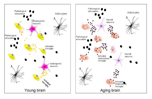
Microglia cells are the brain counterpart of macrophages and function as the first defense in the brain. Although they are neuroprotective in the young brain, microglia cells may be primed to react abnormally to stimuli in the aged brain and to become neurotoxic and destructive during neurodegeneration. Aging-induced immune senescence occurs in the brain as age-associated microglia senescence, which renders microglia to function abnormally and may eventually promote neurodegeneration. Microglia senescence is manifested by both morphological changes and alterations in immunophenotypic expression and inflammatory profile. These changes are likely caused by microinvironmental factors, but intrinsic factors cannot yet be completely excluded. Microglia senescence appears to underlie the switching of microglia from neuroprotective in the young brain to neurotoxic in the aged brain. The hypothesis of microglia senescence during aging offers a novel perspective on their roles in aging-related neurodegeneration. In Parkinson's disease and Alzheimer's disease, over-activation of microglia may play an active role in the pathogenesis because microglia senescence primes them to be neurotoxic during the development of the diseases.
Overview
The claims and statistics of this paper haven’t been checked yet.
Run the check — free, no account needed. It reads the open-access text and checks the sources it cites.
Integrity signals
Registry
Cites
Statistics
Figures
Verification
Claims the paper makes
The claims and statistics of this paper haven’t been checked yet.
Run the check — free, no account needed. It reads the open-access text and checks the sources it cites.
Claims other papers make about this one
Each is checked against this paper’s own text: does it say what the citing paper says it does?
In the citing paper
“The ageing brain is also characterised by structural and functional changes to microglial cells, which are the resident immune cell population of the central nervous system (CNS) and are the CNS equivalent of macrophages. They are activated by brain injury and local and systemic inflammation and become primed (hyper-responsive) to small stimuli with ageing, which can potentially cause damage and”
From Frailty in elderly people (2013) · cites this paper as [26]
Self-check
Percentages, sums and reported differences re-computed from the paper’s own numbers.
The self-check needs the paper’s full text.
Citations
Cites
Resting microglial cells are highly dynamic surveillants of brain parenchyma in vivo
Nimmerjahn, A., Kirchhoff, F., & Helmchen, F. (2005) · Science · doi:10.1126/science.1110647 · Cited by 5,827 · cited 2×
“By using in vivo two-photon imaging techniques, Nimmerjahn et al[37].”
BackgroundMethodInflamm‐aging: An Evolutionary Perspective on Immunosenescence
FRANCESCHI, C., BONAFÈ, M., VALENSIN, S., OLIVIERI, F., DE LUCA, M., OTTAVIANI, E., & DE BENEDICTIS, G. (2000) · Annals of the New York Academy of Sciences · doi:10.1111/j.1749-6632.2000.tb06651.x · Cited by 5,358 · cited 2×
“Extrinsic factors might include neurotransmitter, oxidative stress, neuroendocrine factors[10,68], hormones, and pathological factors such as abnormal protein aggregation [52] that exists in the microenvironment where microglia live.”
BackgroundMicroglia: a sensor for pathological events in the CNS
Kreutzberg, G. W. (1996) · Trends in Neurosciences · doi:10.1016/0166-2236(96)10049-7 · Cited by 4,396 · cited 1×
“When triggered by appropriate stimulation, microglia rapidly transformed from a resting state to an activated state with deramified shape and enhanced antigen presentation, which has been graded to a series of stages[18].”
BackgroundMicroglia: Active sensor and versatile effector cells in the normal and pathologic brain
Hanisch, U.-K., & Kettenmann, H. (2007) · Nature Neuroscience · doi:10.1038/nn1997 · Cited by 3,676 · cited 1×
“Microglia are distributed ubiquitously throughout the brain and function as resident macrophages and antigen-presenting cells in the CNS[35,36].”
BackgroundMicroglia and inflammation-mediated neurodegeneration: Multiple triggers with a common mechanism
Block, M. L., & Hong, J.-S. (2005) · Progress in Neurobiology · doi:10.1016/j.pneurobio.2005.06.004 · Cited by 1,557 · cited 1×
“These studies indicate that microglia are capable of being both neurotrophic and neurotoxic, depending on the specific stimulus, the severity of injury, and the environment [31-34].”
BackgroundMicroglial Dysfunction and Defective β-Amyloid Clearance Pathways in Aging Alzheimer's Disease Mice
Hickman, S. E., Allison, E. K., & El Khoury, J. (2008) · The Journal of Neuroscience · doi:10.1523/jneurosci.0616-08.2008 · Cited by 1,329 · cited 1×
“These genes include Abbinding scavenger receptors A (SRA), CD36, receptor for advanced glycosylation end products (RAGE), Abdegrading enzymes (insulysin, neprilysin) and MMP9 [74].”
BackgroundThe Endocrine Regulation of Aging by Insulin-like Signals
Tatar, M., Bartke, A., & Antebi, A. (2003) · Science · doi:10.1126/science.1081447 · Cited by 1,327 · cited 1×
“Insulin-like signaling pathway has been shown to regulate life span in worms, flies, and mice[ 72 ].”
BackgroundDown-Regulation of the Macrophage Lineage Through Interaction with OX2 (CD200)
Hoek, R. M., Ruuls, S. R., Murphy, C. A., Wright, G. J., Goddard, R., Zurawski, S. M., Blom, B., Homola, M. E., Streit, W. J., Brown, M. H., et al. (2000) · Science · doi:10.1126/science.290.5497.1768 · Cited by 1,022 · cited 1×
“Microglia activation is normally controlled under physiological conditions of the CNS through neuron-glia contact, CD200R-CD200 pathway[19], neuronal electrical activity[20], and/or some soluble neurotransmitters [21].”
BackgroundStreit, W. J., Braak, H., Xue, Q.-S., & Bechmann, I. (2009) · Acta Neuropathologica · doi:10.1007/s00401-009-0556-6 · Cited by 727
Dystrophic microglia in the aging human brain
Streit, W. J., Sammons, N. W., Kuhns, A. J., & Sparks, D. L. (2004) · Glia · doi:10.1002/glia.10319 · Cited by 705 · cited 2×
“”[50] Characteristics of “dystrophic” microglia that were observed in the aged brain include deramification (loss of finely branched cytoplasmic processes), cytoplasmic beading/spheroid formation, shortened and twisted cytoplasmic processes, and instances of partial or complete cytoplasmic fragmentation[50].”
BackgroundGlial reactions in Parkinson's disease
McGeer, P. L., & McGeer, E. G. (2008) · Movement Disorders · doi:10.1002/mds.21751 · Cited by 701 · cited 1×
“The considerable contribution of microglia activation to the pathogenesis of PD has long been proposed [45,46,75].”
BackgroundMicroglia derived from aging mice exhibit an altered inflammatory profile
Sierra, A., Gottfried‐Blackmore, A. C., McEwen, B. S., & Bulloch, K. (2007) · Glia · doi:10.1002/glia.20468 · Cited by 688 · cited 3×
“Isolated microglia from aged brains exhibit decreased process complexity, altered granularity, and increased basal cytokines as compared to those from the young brain, suggesting an elevated inflammatory state in the aged microglia[56].”
BackgroundKim, Y. S., & Joh, T. H. (2006) · Experimental & Molecular Medicine · doi:10.1038/emm.2006.40 · Cited by 655
Identification of normal and pathological aging in prospectively studied nondemented elderly humans
Dickson, D. W., Crystal, H. A., Mattiace, L. A., Masur, D. M., Blau, A. D., Davies, P., Yen, S.-H., & Aronson, M. K. (1992) · Neurobiology of Aging · doi:10.1016/0197-4580(92)90027-u · Cited by 643 · cited 3×
“It is still uncertain what triggers the microglial activation in the healthy aged brain; whether it is caused by degenerating neurons or by abnormal protein aggregation found in clinically normal elders is still unknown[22].”
BackgroundClinical Relevance of Age-Related Immune Dysfunction
Castle, S. C. (2000) · Clinical Infectious Diseases · doi:10.1086/313947 · Cited by 639
Cutting edge: impaired toll-like receptor expression and function in aging
Renshaw, M., Rockwell, J., Engleman, C., Gewirtz, A., Katz, J., & Sambhara, S. (2002) · The Journal of Immunology · doi:10.4049/jimmunol.169.9.4697 · Cited by 626 · cited 1×
“reported a reduced Toll-like receptor (TLR) expression in macrophages with advanced age[11], while Boehmer et al [12].”
BackgroundMicroglia Biology in Health and Disease
Garden, G. A., & Möller, T. (2006) · Journal of Neuroimmune Pharmacology · doi:10.1007/s11481-006-9015-5 · Cited by 511 · cited 1×
“Microglia are distributed ubiquitously throughout the brain and function as resident macrophages and antigen-presenting cells in the CNS[35,36].”
BackgroundDistinct Role for Microglia in Rotenone-Induced Degeneration of Dopaminergic Neurons
Gao, H., Hong, J., Zhang, W., & Liu, B. (2002) · Journal of Neuroscience · doi:10.1523/jneurosci.22-03-00782.2002 · Cited by 439 · cited 1×
“Most of the debate has arisen from in vitro studies [ 27-30 ], which usually give ambiguous effects of microglia on cultured neurons.”
BackgroundMicroglial phenotype: is the commitment reversible?
Schwartz, M., Butovsky, O., Brück, W., & Hanisch, U.-K. (2006) · Trends in Neurosciences · doi:10.1016/j.tins.2005.12.005 · Cited by 434 · cited 2×
“However, if the microglia activation in the brain oversteps the threshold of tolerability, it might contribute to pathology rather than have a sentinel or defensive role [ 38 ].”
BackgroundSimilar gene expression patterns characterize aging and oxidative stress in Drosophila melanogaster
Landis, G. N., Abdueva, D. A., Skvortsov, D., Yang, J., Rabin, B. E., Carrick, J., Tavaré, S., & Tower, J. (2004) · Proceedings of the National Academy of Sciences · doi:10.1073/pnas.0307605101 · Cited by 384 · cited 1×
“found upregulation of genes related to purine biosynthesis, heat-shock proteins, antioxidants, and innate immune responses in old flies and in oxygen-stressed flies, suggesting the specific gene expression profile during aging [71].”
BackgroundMicroglia Protect Neurons against Ischemia by Synthesis of Tumor Necrosis Factor
Lambertsen, K. L., Clausen, B. H., Babcock, A. A., Gregersen, R., Fenger, C., Nielsen, H. H., Haugaard, L. S., Wirenfeldt, M., Nielsen, M., Dagnæs‐Hansen, F., et al. (2009) · Journal of Neuroscience · doi:10.1523/jneurosci.5505-08.2009 · Cited by 374 · cited 1×
“Microglia have been demonstrated to protect neurons against ischemia either by synthesis of tumor necrosis factor[42] or by engulfment of harmful invading neutrophil granulocytes[43].”
BackgroundButovsky, O., Talpalar, A. E., Ben‐Yaakov, K., & Schwartz, M. (2005) · Molecular and Cellular Neuroscience · doi:10.1016/j.mcn.2005.03.005 · Cited by 372 · cited 1×
“Once activated, they are committed to various phenotypes[38,40,69].”
BackgroundCellular pathology of Parkinson?s disease: astrocytes, microglia and inflammation
Teismann, P., & Schulz, J. B. (2004) · Cell and Tissue Research · doi:10.1007/s00441-004-0944-0 · Cited by 371 · cited 1×
“In this slowly progressive procedure, activated microglia has been demonstrated to play important detrimental roles [45-47].”
BackgroundPlackett, T. P., Boehmer, E. D., Faunce, D. E., & Kovacs, E. J. (2004) · Journal of Leukocyte Biology · doi:10.1189/jlb.1103592 · Cited by 351 · cited 2×
“In rodents, the cytokine production (interleukin 1 (IL-1), tumor necrosis factor (TNF-a)) [6-9] as well as oxidative radicals and iNOS expression [9] were reported to decline in macrophages with aging.”
BackgroundControl of glial immune function by neurons
Neumann, H. (2001) · Glia · doi:10.1002/glia.1108 · Cited by 319 · cited 1×
“Microglia activation is normally controlled under physiological conditions of the CNS through neuron-glia contact, CD200R-CD200 pathway[19], neuronal electrical activity[20], and/or some soluble neurotransmitters [21].”
BackgroundEvidence That Aging And Amyloid Promote Microglial Cell Senescence
Flanary, B. E., Sammons, N. W., Nguyen, C., Walker, D., & Streit, W. J. (2007) · Rejuvenation Research · doi:10.1089/rej.2006.9096 · Cited by 272 · cited 2×
“who reported that microglial cells exhibit significant telomere shortening and reduction of telomerase activity during normal aging in rats[54].”
BackgroundBoehmer, E. D., Góral, J., Faunce, D. E., & Kovacs, E. J. (2003) · Journal of Leukocyte Biology · doi:10.1189/jlb.0803389 · Cited by 266 · cited 1×
“et al. reported a reduced Toll-like receptor (TLR) expression in macrophages with advanced age[11], while Boehmer et al [ 12 ].”
BackgroundNeumann, J. H. L., Sauerzweig, S., Rönicke, R., Gunzer, F., Dinkel, K., Ullrich, O., Gunzer, M., & Reymann, K. G. (2008) · Journal of Neuroscience · doi:10.1523/jneurosci.0060-08.2008 · Cited by 261 · cited 1×
“Microglia have been demonstrated to protect neurons against ischemia either by synthesis of tumor necrosis factor[42] or by engulfment of harmful invading neutrophil granulocytes[43].”
BackgroundRogers, J., Mastroeni, D., Leonard, B., Joyce, J., & Grover, A. (2007) · International Review of Neurobiology · doi:10.1016/s0074-7742(07)82012-5 · Cited by 260 · cited 2×
“The considerable contribution of microglia activation to the pathogenesis of PD has long been proposed [45,46,75].”
BackgroundRabchevsky, A. G., & Streit, W. J. (1997) · Journal of Neuroscience Research · doi:10.1002/(sici)1097-4547(19970101)47:1 · Cited by 256
Microglial Expression of MHC Class II Increases in Normal Aging of Nonhuman Primates
Sheffield, L. G., & Berman, N. E. J. (1998) · Neurobiology of Aging · doi:10.1016/s0197-4580(97)00168-1 · Cited by 234 · cited 3×
“During normal aging of the monkey brain, microglial expression of MHC class II increases with age[23], and the phagocytic activity of microglia increases age-dependently, leading to electron dense inclusions that have the appearance of myelin in microglia[24].”
BackgroundShaked, I., Tchoresh, D., Gersner, R., Meiri, G., Mordechai, S., Xiao, X., Hart, R. P., & Schwartz, M. (2005) · Journal of Neurochemistry · doi:10.1111/j.1471-4159.2004.02954.x · Cited by 210 · cited 2×
“Activated microglia can also clear glutamate without evoking inflammatory mediators after traumatic injury and thus reduce neurotoxicity[40].”
BackgroundYe, S.-M., & Johnson, R. W. (2001) · Neuroimmunomodulation · doi:10.1159/000049025 · Cited by 203 · cited 2×
“Additionally, increased pro-inflammatory cytokines and decreased anti-inflammatory cytokines have been demonstrated from lipopolysaccharide (LPS)-activated microglia explanted from aged mice[26], suggesting the abnormal immune state of microglia in the aged brain.”
BackgroundPeters, A., Josephson, K., & Vincent, S. L. (1991) · The Anatomical Record · doi:10.1002/ar.1092290311 · Cited by 196 · cited 2×
“During normal aging of the monkey brain, microglial expression of MHC class II increases with age[23], and the phagocytic activity of microglia increases age-dependently, leading to electron dense inclusions that have the appearance of myelin in microglia[24].”
BackgroundWilms, H., Zecca, L., Rosenstiel, P. C., Sievers, J., Deuschl, G., & Lucius, R. (2007) · Current Pharmaceutical Design · doi:10.2174/138161207780858429 · Cited by 195 · cited 1×
“Microglia activation in the aged brain It has been proposed that aberrant inflammatory responses play a role in the etiology of several neurodegenerative diseases, such as Parkinson’s disease (PD) or Alzheimer’s disease (AD)[13,14], in which aging is the most important risk factor.”
BackgroundTherapeutic approaches to inflammation in neurodegenerative disease
Klegeris, A., McGeer, E. G., & McGeer, P. L. (2007) · Current Opinion in Neurology · doi:10.1097/wco.0b013e3280adc943 · Cited by 189 · cited 1×
“Many studies have demonstrated coexisting neuro-inflammation and neurodegeneration[15,16].”
BackgroundExacerbated glial response in the aged mouse hippocampus following controlled cortical impact injury
Sandhir, R., Onyszchuk, G., & Berman, N. E. J. (2008) · Experimental Neurology · doi:10.1016/j.expneurol.2008.06.013 · Cited by 183 · cited 1×
“The distinct pattern of microglia response to injury in the aged brain has also been recorded in the 1-methyl-4-phenyl-1,2,3,6-tetrahydropyridine (MPTP)-induced neurotoxicity[59], the model of controlled cortical impact (CCI)[60], cortical stab injury [61], and transient retinal ischemia[62].”
BackgroundWalker, D. G., Link, J., Lue, L., Dalsing-Hernandez, J. E., & Boyes, B. E. (2005) · Journal of Leukocyte Biology · doi:10.1189/jlb.0705377 · Cited by 183 · cited 2×
“Walker et al[70] demonstrated that when compared with those from non-demented brains, large alterations in gene transcription were found in microglia derived from postmortem AD brain, including upregulation of many proinflammatory cytokines and chemokines such as IL1b, IL-8, and matrix metalloproteinases (MMP).”
BackgroundEffects of ageing on microglia in the normal rat brain: immunohistochemical observations
Ogura, K., Ogawa, M., & Yoshida, M. (1994) · Neuroreport · doi:10.1097/00001756-199406020-00016 · Cited by 181 · cited 2×
“Immunohistochemical studies by using activated microglia marker OX-6 staining also found microglial activation with age-related changes[25].”
BackgroundProtective effects of microglia in multiple sclerosis
Napoli, I., & Neumann, H. (2009) · Experimental Neurology · doi:10.1016/j.expneurol.2009.04.024 · Cited by 173 · cited 1×
“The protective roles of activated microglia have also been extensively discussed in multiple sclerosis[44].”
BackgroundProgressive telomere shortening occurs in cultured rat microglia, but not astrocytes
Flanary, B. E., & Streit, W. J. (2004) · Glia · doi:10.1002/glia.10301 · Cited by 167 · cited 3×
“For example, amyloid has been shown to accelerate microglia senescence[52].”
BackgroundStreit, W. J., & Graeber, M. B. (1993) · Glia · doi:10.1002/glia.440070112 · Cited by 159 · cited 1×
“However, the senescenced microglia are not universal in the aged brain since scattered dystrophic microglia are usually found alongside normal ramified microglial cells, suggesting that only a subset of microglia become dystrophic, and not all of them are of the same age and functional state[55].”
BackgroundSugama, S., Yang, L., Cho, B. P., DeGiorgio, L. A., Lorenzl, S., Albers, D. S., Beal, M. F., Volpe, B. T., & Joh, T. H. (2003) · Brain Research · doi:10.1016/s0006-8993(02)04085-4 · Cited by 153 · cited 1×
“The distinct pattern of microglia response to injury in the aged brain has also been recorded in the 1-methyl-4-phenyl-1,2,3,6-tetrahydropyridine (MPTP)-induced neurotoxicity[ 59 ], the model of controlled cortical impact (CCI)[60], cortical stab injury [61], and transient retinal ischemia[62].”
BackgroundJohnson, S. A., & Cambier, J. C. (2004) · Arthritis Research · doi:10.1186/ar1180 · Cited by 150 · cited 1×
“Although the compromise of adaptive immunity mediated by T and B lymphocytes has gained a significant amount of attention [3-5], the innate system also experiences significant changes with advanced age[2].”
BackgroundMicroglial degeneration in the aging brain - bad news for neurons?
Streit, W. (2008) · Frontiers in Bioscience · doi:10.2741/2937 · Cited by 150 · cited 1×
“Even though Streit et al[64] gave a suggestion in the difference between the two, describing that “hypertrophy and retraction and thickening of process” occurs in activation while “deramification, shortening and twisting of processes, cytoplasmic fragmentation” are features of aging, so far no definite biomarker or mo…”
BackgroundMicroglial activation in white matter lesions and nonlesional white matter of ageing brains
Simpson, J. E., Ince, P. G., Higham, C. E., Gelsthorpe, C., Fernando, M. S., Matthews, F. E., Forster, G., O'Brien, J. T., Barber, R., Kalaria, R. N., et al. (2007) · Neuropathology and Applied Neurobiology · doi:10.1111/j.1365-2990.2007.00890.x · Cited by 146 · cited 1×
“Ramified activated microglia are mainly associated with periventricular region, whereas innate, amoeboid phagocytic phenotype of microglia is seen within the deep subcortical region[67].”
BackgroundGlial cell dysregulation: a new perspective on Alzheimer disease
Rommy von Bernhardi, M. (2007) · Neurotoxicity Research · doi:10.1007/bf03033906 · Cited by 141
Influence of age on the production and regulation of interleukin-1 in mice.
Inamizu, T., Chang, M., & Makinodan, T. (1985) · PubMed · Cited by 135
Neutrophils and the blood–brain barrier dysfunction after trauma
Scholz, M., Cinatl, J. J., Schädel-Höpfner, M., & Windolf, J. (2006) · Medicinal Research Reviews · doi:10.1002/med.20064 · Cited by 134 · cited 1×
“The CNS is relatively immune-privileged, in which the activity of adaptive immunity mediated by lymphocytes is scarce[17].”
BackgroundThe frail elderly: role of dendritic cells in the susceptibility of infection
Uyemura, K., Castle, S. C., & Makinodan, T. (2002) · Mechanisms of Ageing and Development · doi:10.1016/s0047-6374(02)00033-7 · Cited by 127 · cited 1×
“Are the age-related alterations in microglia function and morphology resulting from intrinsic or extrinsic factors? Does microglia over-activation and malfunction in the aged brain come from their failure to respond correctly to their microenvironment, or from an overloaded toxic microenvironment? It has been demonstr…”
BackgroundWei, R., & Jonakait, G. M. (1999) · Journal of Neuroimmunology · doi:10.1016/s0165-5728(98)00248-3 · Cited by 121 · cited 1×
“Microglia activation is normally controlled under physiological conditions of the CNS through neuron-glia contact, CD200R-CD200 pathway[19], neuronal electrical activity[20], and/or some soluble neurotransmitters [21].”
BackgroundKyrkanides, S., O’Banion, M. K., Whiteley, P. E., Daeschner, J., & Olschowka, J. A. (2001) · Journal of Neuroimmunology · doi:10.1016/s0165-5728(01)00404-0 · Cited by 118 · cited 1×
“The distinct pattern of microglia response to injury in the aged brain has also been recorded in the 1-methyl-4-phenyl-1,2,3,6-tetrahydropyridine (MPTP)-induced neurotoxicity[59], the model of controlled cortical impact (CCI)[60], cortical stab injury [ 61 ], and transient retinal ischemia[62].”
BackgroundWasserman, J. K., Yang, H. H., & Schlichter, L. C. (2008) · European Journal of Neuroscience · doi:10.1111/j.1460-9568.2008.06442.x · Cited by 113 · cited 1×
“Such dystrophic microglia were prevalent and extensively distributed in the brain of older human subjects[50,51], whereas normal ramified microglial morphology with only rare instances of dystrophic microglia are seen in the young brain[49] These observations provide initial evidence of the age-associated changes in m…”
BackgroundLiu, X., Fan, X., Zhao, Y. G., Luo, G., Li, X., Li, R., & Le, W. (2005) · Journal of Neuroscience Research · doi:10.1002/jnr.20583 · Cited by 112 · cited 1×
“Most of the debate has arisen from in vitro studies [27-30], which usually give ambiguous effects of microglia on cultured neurons.”
BackgroundImmune response gene expression increases in the aging murine hippocampus
Terao, A., Apte-Deshpande, A. D., Dousman, L., Morairty, S. R., Eynon, B. P., Kilduff, T. S., & Freund, Y. R. (2002) · Journal of Neuroimmunology · doi:10.1016/s0165-5728(02)00317-x · Cited by 111 · cited 1×
“In the mouse hippocampus, the basal expression of 128 genes changes with aging[73].”
BackgroundBeach, T. G., Sue, L. I., Walker, D. G., Lue, L. F., Connor, D. J., Caviness, J. N., Sabbagh, M. N., & Adler, C. H. (2007) · Acta Neuropathologica · doi:10.1007/s00401-007-0250-5 · Cited by 104 · cited 2×
“Thus, increased microglia activation[78] and/or senescenced microglia in the aged brain has put the dopaminergic neurons in a more dangerous environment than that of the young brain.”
BackgroundLinton, P. (2001) · Frontiers in bioscience · doi:10.2741/linton · Cited by 97 · cited 1×
“Although the compromise of adaptive immunity mediated by T and B lymphocytes has gained a significant amount of attention [ 3-5 ], the innate system also experiences significant changes with advanced age[2].”
BackgroundWhite matter injury in young and aged rats after intracerebral hemorrhage
Wasserman, J. K., & Schlichter, L. C. (2008) · Experimental Neurology · doi:10.1016/j.expneurol.2008.08.010 · Cited by 96 · cited 1×
“In a study of intracerebral hemorrhage in young and aged rats, it was observed that microglia activation was significantly higher in young rats than in aged ones when examined three days after hemorrhage [ 58 ].”
BackgroundThe interplay between inflammation and neurodegeneration in CNS disease
Esiri, M. M. (2006) · Journal of Neuroimmunology · doi:10.1016/j.jneuroim.2006.11.013 · Cited by 87 · cited 1×
“Many studies have demonstrated coexisting neuro-inflammation and neurodegeneration[15,16].”
BackgroundEffect of aging on the microglial response to peripheral nerve injury
Conde, J. R., & Streit, W. J. (2006) · Neurobiology of Aging · doi:10.1016/j.neurobiolaging.2005.07.012 · Cited by 86 · cited 3×
“Microglia senescence renders microglia to function abnormally, to fail to respond correctly to stimuli[48,49] and, eventually, to promote neurodegeneration.”
BackgroundTelomeres Shorten with Age in Rat Cerebellum and Cortex in vivo
Flanary, B. E., & Streit, W. J. (2003) · Journal of Anti-Aging Medicine · doi:10.1089/109454503323028894 · Cited by 85 · cited 1×
“Previous studies have shown that telomere shortening, which is a sign of cell senescence, occurs in rat microglia overtime in vitro [52], as well as in vivo in rat cerebellum and cerebral cortex with age[53].”
BackgroundMitrasinovic, O. M., Grattan, A., Robinson, C. C., Lapustea, N., Poon, C., Ryan, H. S., Phong, C., & Murphy, G. M. (2005) · Journal of Neuroscience · doi:10.1523/jneurosci.0514-05.2005 · Cited by 83 · cited 1×
“Most of the debate has arisen from in vitro studies [ 27-30 ], which usually give ambiguous effects of microglia on cultured neurons.”
Backgroundβ-Amyloid peptides induce degeneration of cultured rat microglia
Korotzer, A. R., Pike, C. J., & Cotman, C. W. (1993) · Brain Research · doi:10.1016/0006-8993(93)90068-x · Cited by 82 · cited 1×
“However, both compromised amyloid-clearing ability of microglia[80] and the damage of microglia by amyloid[81] have been reported in AD brain.”
BackgroundImmunological changes in the elderly
Ginaldi, L., Martinis, M. D., D’Ostilio, A., Marini, L., Loreto, M. F., & Quaglino, D. (1999) · Aging Clinical and Experimental Research · doi:10.1007/bf03339801 · Cited by 81 · cited 2×
“Although the compromise of adaptive immunity mediated by T and B lymphocytes has gained a significant amount of attention [3-5], the innate system also experiences significant changes with advanced age[2].”
BackgroundBattisti, W. P., Wang, J., Bozek, K., & Murray, M. (1995) · The Journal of Comparative Neurology · doi:10.1002/cne.903540211 · Cited by 77 · cited 1×
“In axotomy of the optic nerve of amphibians, a rapid microglia response is exhibited with efficient clearance of myelin debris (which contains inhibiting molecules of axon growth) and finally successful axonal regeneration[39].”
BackgroundCorsini, E., Battaini, F., Lucchi, L., et al. (1999) · PubMed · Cited by 72
Sawada, M., Sawada, H., & Nagatsu, T. (2008) · Neurodegenerative Diseases · doi:10.1159/000113717 · Cited by 71 · cited 3×
“Microglia senescence renders microglia to function abnormally, to fail to respond correctly to stimuli[48,49] and, eventually, to promote neurodegeneration.”
BackgroundCorsini, E., Battaini, F., Lucchi, L., et al. (1999) · The Journal of Immunology · doi:10.4049/jimmunol.163.6.3468 · Cited by 69 · cited 1×
“In rodents, the cytokine production (interleukin 1 (IL-1), tumor necrosis factor (TNF-a)) [6-9] as well as oxidative radicals and iNOS expression [9] were reported to decline in macrophages with aging.”
BackgroundLiu, Fan, Xl, et al. (2006) · 中国生物学文摘 · Cited by 67
Age-Related Decrease in Accessory Cell Function of Human Alveolar Macrophages
Zissel, G., Schlaak, M. W., & Müller-Quernheim, J. (1999) · Journal of Investigative Medicine · doi:10.1177/108155899904700108 · Cited by 66 · cited 1×
“Are the age-related alterations in microglia function and morphology resulting from intrinsic or extrinsic factors? Does microglia over-activation and malfunction in the aged brain come from their failure to respond correctly to their microenvironment, or from an overloaded toxic microenvironment? It has been demonstr…”
BackgroundEffects of Enhanced Striatal Dopamine Turnover In Vivo on Glutathione Oxidation
Loeffler, D. A., DeMaggio, A. J., Juneau, P. L., Havaich, M. K., & LeWitt, P. A. (1994) · Clinical Neuropharmacology · doi:10.1097/00002826-199408000-00009 · Cited by 61 · cited 1×
“of neurons[79].”
Enhanced sensitivity of dopaminergic neurons to rotenone-induced toxicity with aging
Phinney, A. L., Andringa, G., Bol, J. G. J. M., Wolters, E. C., Muiswinkel, F. L. V., van Dam, A., & Drukarch, B. (2006) · Parkinsonism & Related Disorders · doi:10.1016/j.parkreldis.2005.12.002 · Cited by 57 · cited 1×
“Aging is the strongest risk factor for PD, which is true not only in the epidemiological studies, but also in the models of PD. A low systemic dose of rotenone had no effect on young rats, but led to a2 0- 30% reduction of dopaminergic neurons in the substantia nigra of older rats[ 76 ].”
BackgroundKim, K.-Y., Ju, W., & Neufeld, A. H. (2003) · Neurobiology of Aging · doi:10.1016/j.neurobiolaging.2003.07.005 · Cited by 56 · cited 1×
“The distinct pattern of microglia response to injury in the aged brain has also been recorded in the 1-methyl-4-phenyl-1,2,3,6-tetrahydropyridine (MPTP)-induced neurotoxicity[59], the model of controlled cortical impact (CCI)[60], cortical stab injury [61], and transient retinal ischemia[62].”
BackgroundParkinson disease IgG and C5a-induced synergistic dopaminergic neurotoxicity: Role of microglia
Wang, X., Yan, Z.-Q., Lü, G., Stuart, S., & Chen, S. (2006) · Neurochemistry International · doi:10.1016/j.neuint.2006.07.014 · Cited by 52 · cited 1×
“Most of the debate has arisen from in vitro studies [27-30], which usually give ambiguous effects of microglia on cultured neurons.”
BackgroundSelf-citationDecreases in macrophage mediated antitumor activity with aging
Wallace, P. K., Eisenstein, T. K., Meissler, J. J., & Morahan, P. S. (1995) · Mechanisms of Ageing and Development · doi:10.1016/0047-6374(94)01524-p · Cited by 48 · cited 1×
“In rodents, the cytokine production (interleukin 1 (IL-1), tumor necrosis factor (TNF-a)) [6-9] as well as oxidative radicals and iNOS expression [9] were reported to decline in macrophages with aging.”
BackgroundKumagai, N., Chiba, Y., Hosono, M., Fujii, M., Kawamura, N., Keino, H., Yoshikawa, K., Ishii, S., Saitoh, Y., Satoh, M., et al. (2007) · Brain Research · doi:10.1016/j.brainres.2007.09.021 · Cited by 47 · cited 1×
“These studies indicate that microglia are capable of being both neurotrophic and neurotoxic, depending on the specific stimulus, the severity of injury, and the environment [31-34].”
BackgroundThe Role of Microglial Cellular Senescence in the Aging and Alzheimer Diseased Brain
Flanary, B. (2005) · Rejuvenation Research · doi:10.1089/rej.2005.8.82 · Cited by 40 · cited 1×
“However, both compromised amyloid-clearing ability of microglia[80] and the damage of microglia by amyloid[81] have been reported in AD brain.”
BackgroundSawada, H., Hishida, R., Hirata, Y., Ono, K., Suzuki, H., Muramatsu, S., Nakano, I., Nagatsu, T., & Sawada, M. (2007) · Journal of Neuroscience Research · doi:10.1002/jnr.21241 · Cited by 40 · cited 2×
“By not responding to normal regulatory feedback mechanisms and/or having an impairment in their ability to clear Ab, microglia cells lose their ability to handle potentially toxic compounds and become cytotoxic due to their persistent activation[77].”
BackgroundIntracerebral Hemorrhage-Induced Brain Injury Is Aggravated in Senescence-Accelerated Prone Mice
Lee, J., Cho, G., Choi, B., Kim, H., Kim, Y., & Kim, W.-K. (2005) · Stroke · doi:10.1161/01.str.0000195151.46926.7b · Cited by 35 · cited 1×
“observed. In an intracerebral hemorrhage model of senescence-accelerated prone mice (SAMP8) and senescence-accelerated resistant mice (SAMR1), brain neutrophils and reactive astrocytes do not differ in number or distribution between SAMP8 and SAMR1 mice, but activated amoeboid microglia are distributed more abundantly…”
BackgroundFacial nerve axotomy in aged and young adult rats: analysis of the glial response
Hurley, S. D. (2003) · Neurobiology of Aging · doi:10.1016/s0197-4580(02)00097-0 · Cited by 30 · cited 2×
“Hurley SD, Coleman PD: Facial nerve axotomy in aged and young adult rats: analysis of the glial response.”
BackgroundInflammation versus neurodegeneration: Consequences for treatment
Palace, J. A. (2007) · Journal of the Neurological Sciences · doi:10.1016/j.jns.2006.05.072 · Cited by 30 · cited 1×
“Microglia activation in the aged brain It has been proposed that aberrant inflammatory responses play a role in the etiology of several neurodegenerative diseases, such as Parkinson’s disease (PD) or Alzheimer’s disease (AD)[13,14], in which aging is the most important risk factor.”
BackgroundHakim, F. T., & Gress, R. E. (2007) · Methods in molecular biology · doi:10.1007/978-1-59745-395-0_24 · Cited by 24
Imamura, N., Hida, H., Aihara, N., Ishida, K., Kanda, Y., Nishino, H., & Yamada, K. (2003) · Neuroscience Research · doi:10.1016/s0168-0102(03)00065-8 · Cited by 21 · cited 1×
“These studies indicate that microglia are capable of being both neurotrophic and neurotoxic, depending on the specific stimulus, the severity of injury, and the environment [31-34].”
BackgroundMicroglia and neurodegeneration
Rozemuller, & Muiswinkel, V. (2000) · European Journal of Clinical Investigation · doi:10.1046/j.1365-2362.2000.00665.x · Cited by 16 · cited 1×
“These studies indicate that microglia are capable of being both neurotrophic and neurotoxic, depending on the specific stimulus, the severity of injury, and the environment [31-34].”
BackgroundAging, immunity and neuroendocrine hormones
Mosley, R. L. (1996) · Advances in Neuroimmunology · doi:10.1016/s0960-5428(97)00031-9 · Cited by 16 · cited 1×
“Extrinsic factors might include neurotransmitter, oxidative stress, neuroendocrine factors[10,68], hormones, and pathological factors such as abnormal protein aggregation [52] that exists in the microenvironment where microglia live.”
Background
Cited by
Sorted by citations
Clegg, A., Young, J., Iliffe, S., Rikkert, M. O., & Rockwood, K. (2013) · The Lancet · doi:10.1016/s0140-6736(12)62167-9 · Cited by 9,503
Clearance of senescent glial cells prevents tau-dependent pathology and cognitive decline
Bussian, T. J., Aziz, A., Meyer, C. F., Swenson, B. L., van Deursen, J. M., & Baker, D. J. (2018) · Nature · doi:10.1038/s41586-018-0543-y · Cited by 1,289
Tay, T. L., Savage, J. C., Hui, C. W., Bisht, K., & Tremblay, M. (2017) · The Journal of Physiology · doi:10.1113/jp272134 · Cited by 562
“These parameters indicate that as humans and rodents age microglia take on a phenotype consistent with a pro-inflammatory response, even in the absence of overt pathological stimuli (Luo et al. 2010; Choi & Won, 2011; Wong, 2013; Sierra et al. 2014; Ojo et al. 2015; Barrientos et al. 2015a).”
BackgroundAge-related alterations in the dynamic behavior of microglia
Damani, M. R., Zhao, L., Fontainhas, A. M., Amaral, J., Fariss, R. N., & Wong, W. T. (2011) · Aging Cell · doi:10.1111/j.1474-9726.2010.00660.x · Cited by 504
“…aging changes in microglia have been hypothesized to drive the pathogenic progression through a diminution of neuroprotective functions, direct increases in neurotoxicity, and dysregulated responses to signals and perturbations (Flanary et al., 2007; Streit et al., 2008; Luo et al., 2010).”
BackgroundMicroglia during development and aging
Harry, G. J. (2013) · Pharmacology & Therapeutics · doi:10.1016/j.pharmthera.2013.04.013 · Cited by 524
“…hypothesized that changes in the aging microglia drive pathogenic progression of diseases or injury through a diminution of neuroprotective functions, increase in neurotoxicity, and dysregulation of responses to signals and perturbations (Flanary et al., 2007; Lopes et al., 2008; Luo et al., 2010).”
Background
Metadata
- Authors
- Luo, X.-G., Ding, J.-Q., & Chen, S.-D.
- Venue
- Molecular Neurodegeneration
- Published
- 2010-03-24
- Volume
- 5
- Issue
- 1
- Pages
- 12-12
- ISSN
- 1750-1326 (ISSN-L)
- PMC
- PMC2852379
- OpenAlex
- W2156427445
- Cited by
- 236
- References
- 85
- Citations by year
- 2026: 7 · 2025: 12 · 2024: 10 · 2023: 11 · 2022: 17 · 2021: 14 · 2020: 15 · 2019: 11 · 2018: 20 · 2017: 13 · 2016: 18 · 2015: 19 · 2014: 12 · 2013: 22 · 2012: 15
- FWCI
- 10.86
- Type
- journal article
- Language
- en
- Open access
- gold
- doi.org
- Licence
- cc-by
- Locations
- Molecular Neurodegeneration (publishedVersion, cc-by)
- PubMed Central (publishedVersion, cc-by)
- PubMed (publishedVersion)
- DOAJ (DOAJ: Directory of Open Access Journals) (submittedVersion, cc-by-sa)
- Topics
- Life Sciences › Neuroscience › Neurology › Neuroinflammation and Neurodegeneration Mechanisms
- Life Sciences › Biochemistry, Genetics and Molecular Biology › Molecular Biology › Inflammasome and immune disorders
- Life Sciences › Neuroscience › Neurology › Neurological Disease Mechanisms and Treatments
- Keywords
- microglia, brain aging, neurodegeneration, microglial senescence, immunosenescence, neuroinflammation, neuroprotection, Parkinson's disease, Alzheimer's disease, microglial activation, neurotoxicity
About these checks
Registry, retraction, Cites and statistics results are computed, not judged, with one exception: a printed reference that no index lists is read once by a model before it counts as a citation error. Claims are extracted by a model and kept only when their quote appears verbatim in the text; each cited source is then checked, with a model’s help, for whether it says what the claim relies on. Those verdicts can be wrong — read the claim’s quote and the cited source before relying on one.
Statistics and arithmetic are recomputed, not judged.
Confidence bands are shown only for fields calibrated on our annotated test set. None is calibrated yet, so no band is shown.
What counts as a claim ›How we measure confidence ›How we check this ›
Are you an author, or do you see a problem with this page? Request a review
