

The DOI matches its Crossref record; 129 of the 129 references it cites were found in a registry or an index.
Aberrant repair and fibrosis development in skeletal muscle
Open accessAbstract
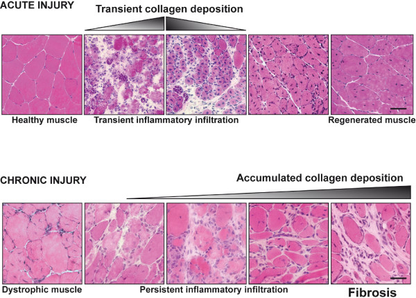
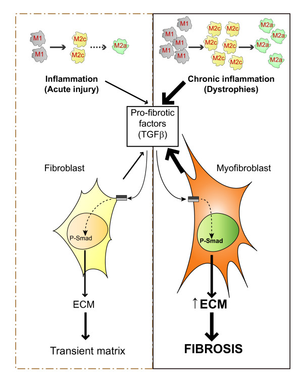
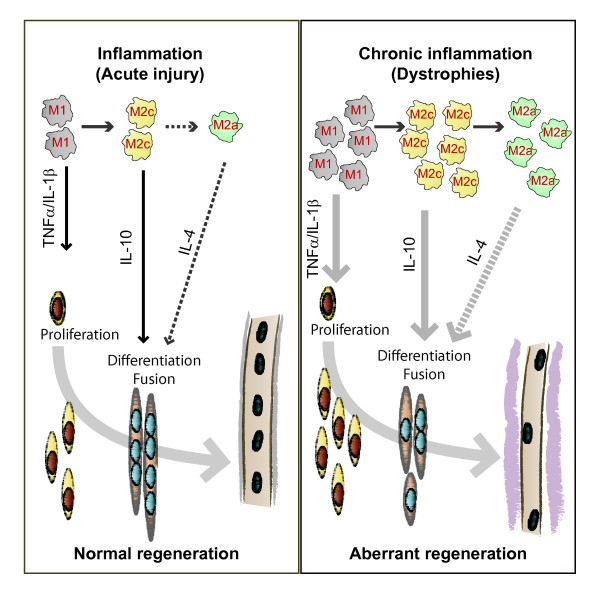
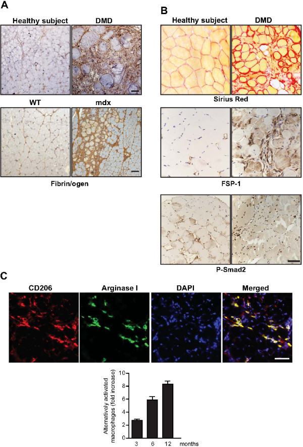
The repair process of damaged tissue involves the coordinated activities of several cell types in response to local and systemic signals. Following acute tissue injury, infiltrating inflammatory cells and resident stem cells orchestrate their activities to restore tissue homeostasis. However, during chronic tissue damage, such as in muscular dystrophies, the inflammatory-cell infiltration and fibroblast activation persists, while the reparative capacity of stem cells (satellite cells) is attenuated. Abnormal dystrophic muscle repair and its end stage, fibrosis, represent the final common pathway of virtually all chronic neurodegenerative muscular diseases. As our understanding of the pathogenesis of muscle fibrosis has progressed, it has become evident that the muscle provides a useful model for the regulation of tissue repair by the local microenvironment, showing interplay among muscle-specific stem cells, inflammatory cells, fibroblasts and extracellular matrix components of the mammalian wound-healing response. This article reviews the emerging findings of the mechanisms that underlie normal versus aberrant muscle-tissue repair.
Overview
The claims and statistics of this paper haven’t been checked yet.
Run the check — free, no account needed. It reads the open-access text and checks the sources it cites.
Integrity signals
Registry
Cites
Statistics
Figures
Verification
Claims the paper makes
The claims and statistics of this paper haven’t been checked yet.
Run the check — free, no account needed. It reads the open-access text and checks the sources it cites.
Claims other papers make about this one
Each is checked against this paper’s own text: does it say what the citing paper says it does?
In the citing paper
“Previous work has shown that the accumulation of inflammatory macrophages and age-related declines in interferon-gamma signaling delay muscle repair following injury”
From Skeletal muscle aging, cellular senescence, and senotherapeutics: current knowledge and future directions (2021) · cites this paper as Mann et al., 2011
Self-check
Percentages, sums and reported differences re-computed from the paper’s own numbers.
The self-check needs the paper’s full text.
Citations
Cites
1 retracted
RETRACTED ARTICLE: The role of nitric oxide during healing of trauma to the skeletal muscle
Filippin, L. I., Cuevas, M. J., Lima‐Cabello, E., Marroni, N. P., González‐Gallego, J., & Xavier, R. M. (2010) · Inflammation Research · doi:10.1007/s00011-010-0277-2 · Cited by 34 · cited 4×
“In addition, NO, which is released by local and invading cells during injury, can act as a pro-inflammatory molecule, and is able to activate MMP-2, which in turn releases HGF. Importantly, blockers of NOS function, such as nitro- Larginine methyl ester (L-NAME), have been shown to decrease the acute inflammatory reac…”
MethodInfluentialRetractedThe chemokine system in diverse forms of macrophage activation and polarization
MANTOVANI, A., SICA, A., SOZZANI, S., ALLAVENA, P., VECCHI, A., & LOCATI, M. (2004) · Trends in Immunology · doi:10.1016/j.it.2004.09.015 · Cited by 6,534 · cited 2×
“A nomenclature for polarized macrophages has been proposed [24,25] and they are now referred to as classically and alternatively activated macrophages, or M1 and M2 macrophages, respectively (Figure 2, Figure 3).”
BackgroundMechanisms of TGF-β Signaling from Cell Membrane to the Nucleus
Shi, Y., & Massagué, J. (2003) · Cell · doi:10.1016/s0092-8674(03)00432-x · Cited by 5,860
Visse, R., & Nagase, H. (2003) · Circulation Research · doi:10.1161/01.res.0000070112.80711.3d · Cited by 4,575 · cited 1×
“Activation requires proteolytic cleavage of the inactive precursor by either membrane-type (MT)1MMP [105] or plasmin, and cleavage of their corresponding inhibitors [106].”
Cellular and molecular mechanisms of fibrosis
Wynn, T. (2008) · The Journal of Pathology · doi:10.1002/path.2277 · Cited by 4,495
SATELLITE CELL OF SKELETAL MUSCLE FIBERS
Mauro, A. (1961) · The Journal of Cell Biology · doi:10.1083/jcb.9.2.493 · Cited by 3,683
Hinz, B., Phan, S. H., Thannickal, V. J., Galli, A., Bochaton-Piallat, M.-L., & Gabbiani, G. (2007) · The American Journal of Pathology · doi:10.2353/ajpath.2007.070112 · Cited by 2,068 · cited 1×
“These cells are associated with tissue repair and fibrosis in many tissues and organs, including muscle, skin, liver, lung, bone and cartilage [56].”
BackgroundArnold, L., Henry, A., Poron, F., Baba-Amer, Y., van Rooijen, N., Plonquet, A., Gherardi, R. K., & Chazaud, B. (2007) · The Journal of Experimental Medicine · doi:10.1084/jem.20070075 · Cited by 2,002 · cited 3×
“An important development in our understanding of muscle repair and fibrosis was the demonstration that a heterogeneous population of macrophages exists in regenerating muscle after injury, exhibiting opposing activities (either pro-inflammatory or anti-inflammatory) and different kinetics [23].”
BackgroundInfluentialFibrotic disease and the TH1/TH2 paradigm
Wynn, T. A. (2004) · Nature Reviews Immunology · doi:10.1038/nri1412 · Cited by 1,765
Muscle injury activates resident fibro/adipogenic progenitors that facilitate myogenesis
Joe, A. W. B., Yi, L., Natarajan, A., Le Grand, F., So, L., Wang, J., Rudnicki, M. A., & Rossi, F. M. V. (2010) · Nature Cell Biology · doi:10.1038/ncb2015 · Cited by 1,690 · cited 7×
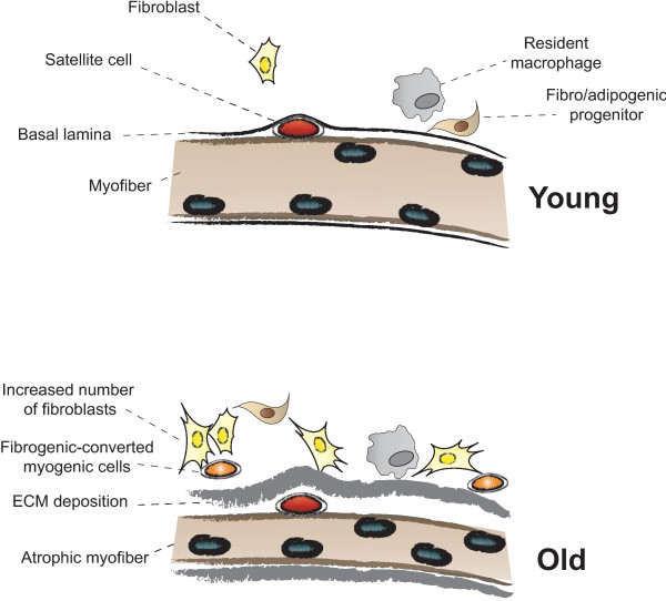
“Whether FAP conversion also occurs during aging, and whether Basal lamina ECM deposition Satellite cell Fibro/adipogenic progenitor Resident macrophage Fibroblast Increased number of fibroblasts Fibrogenic-converted myogenic cells Myofiber Atrophic myofiber Young Old Figure 5 Myofiber growth and extracellular matrix (…”
BackgroundInfluential
Cited by
Sorted by citations
Skeletal muscle performance and ageing
Tieland, M., Trouwborst, I., & Clark, B. C. (2018) · Journal of Cachexia, Sarcopenia and Muscle · doi:10.1002/jcsm.12238 · Cited by 904
“84 There is no direct evidence of muscle fi brosis with age in humans due to the dif fi culty in assessing fi brosis in population studies, but animal work indicated > 17% increase in fi brotic tissue in older rats as compared with younger rats.”
Lemos, D. R., Babaeijandaghi, F., Low, M., Chang, C.-K., Lee, S. T., Fiore, D., Zhang, R.-H., Natarajan, A., Nedospasov, S. A., & Rossi, F. M. V. (2015) · Nature Medicine · doi:10.1038/nm.3869 · Cited by 740
Macrophages in inflammation, repair and regeneration
Oishi, Y., & Manabe, I. (2018) · International Immunology · doi:10.1093/intimm/dxy054 · Cited by 770
Skeletal muscle: A review of molecular structure and function, in health and disease
Mukund, K., & Subramaniam, S. (2019) · WIREs Systems Biology and Medicine · doi:10.1002/wsbm.1462 · Cited by 805
“This uncontrolled and irreversible ECM expansion accompanied by an accumulation of ECM due to inhibited degradation (turnover), results in a fibrotic phenotype within muscle (Mann et al., 2011), especially in chronic diseases such as dystrophinopathies (Serrano & Muñoz-Cánoves, 2017).”
BackgroundMcGregor, R. A., Cameron-Smith, D., & Poppitt, S. D. (2014) · Longevity & Healthspan · doi:10.1186/2046-2395-3-9 · Cited by 532
Metadata
- Authors
- Mann, C. J., Perdiguero, E., Kharraz, Y., Aguilar, S., Pessina, P., Serrano, A. L., & Muñoz-Cánoves, P.
- Affiliations
- Universitat Pompeu Fabra (ES)
- Venue
- Skeletal Muscle
- Published
- 2011-05-04
- Volume
- 1
- Issue
- 1
- Pages
- 21-21
- ISSN
- 2044-5040 (ISSN-L)
- PMC
- PMC3156644
- OpenAlex
- W2076787014
- Cited by
- 853
- References
- 129
- Citations by year
- 2026: 37 · 2025: 59 · 2024: 55 · 2023: 53 · 2022: 59 · 2021: 71 · 2020: 81 · 2019: 85 · 2018: 76 · 2017: 51 · 2016: 68 · 2015: 48 · 2014: 49 · 2013: 33 · 2012: 27
- FWCI
- 14.30
- Type
- journal article
- Language
- en
- Open access
- gold
- doi.org
- Licence
- cc-by
- Locations
- Skeletal Muscle (publishedVersion, cc-by)
- Europe PMC (PubMed Central) (publishedVersion, cc-by)
- PubMed Central (publishedVersion, cc-by)
- PubMed (publishedVersion)
- RECERCAT (Consorci de Serveis Universitaris de Catalunya) (publishedVersion, other-oa)
- DOAJ (DOAJ: Directory of Open Access Journals) (submittedVersion, cc-by-sa)
- Repositori digital de la UPF (Universitat Pompeu Fabra) (submittedVersion, cc-by)
- Funders
- Ministerio de Ciencia e Innovación (PLE2009)
- Centro de Investigación Biomédica en Red sobre Enfermedades Neurodegenerativas
- Topics
- Life Sciences › Biochemistry, Genetics and Molecular Biology › Molecular Biology › Muscle Physiology and Disorders
- Health Sciences › Medicine › Cardiology and Cardiovascular Medicine › Cardiac Fibrosis and Remodeling
- Health Sciences › Medicine › Surgery › Wound Healing and Treatments
- Keywords
- muscle fibrosis, muscle repair, muscular dystrophy, satellite cells, muscle stem cells, fibroblast activation, inflammatory cell infiltration, extracellular matrix, wound healing, microenvironment
About these checks
Registry, retraction, Cites and statistics results are computed, not judged, with one exception: a printed reference that no index lists is read once by a model before it counts as a citation error. Claims are extracted by a model and kept only when their quote appears verbatim in the text; each cited source is then checked, with a model’s help, for whether it says what the claim relies on. Those verdicts can be wrong — read the claim’s quote and the cited source before relying on one.
Statistics and arithmetic are recomputed, not judged.
Confidence bands are shown only for fields calibrated on our annotated test set. None is calibrated yet, so no band is shown.
What counts as a claim ›How we measure confidence ›How we check this ›
Are you an author, or do you see a problem with this page? Request a review
