

The DOI matches its Crossref record; 129 of the 129 references it cites were found in a registry or an index.
Aberrant repair and fibrosis development in skeletal muscle
Open accessAbstract
The repair process of damaged tissue involves the coordinated activities of several cell types in response to local and systemic signals. Following acute tissue injury, infiltrating inflammatory cells and resident stem cells orchestrate their activities to restore tissue homeostasis. However, during chronic tissue damage, such as in muscular dystrophies, the inflammatory-cell infiltration and fibroblast activation persists, while the reparative capacity of stem cells (satellite cells) is attenuated. Abnormal dystrophic muscle repair and its end stage, fibrosis, represent the final common pathway of virtually all chronic neurodegenerative muscular diseases. As our understanding of the pathogenesis of muscle fibrosis has progressed, it has become evident that the muscle provides a useful model for the regulation of tissue repair by the local microenvironment, showing interplay among muscle-specific stem cells, inflammatory cells, fibroblasts and extracellular matrix components of the mammalian wound-healing response. This article reviews the emerging findings of the mechanisms that underlie normal versus aberrant muscle-tissue repair.
Overview
The claims and statistics of this paper haven’t been checked yet.
Run the check — free, no account needed. It reads the open-access text and checks the sources it cites.
Integrity signals
Registry
Cites
Statistics
Figures
Verification
Claims the paper makes
The claims and statistics of this paper haven’t been checked yet.
Run the check — free, no account needed. It reads the open-access text and checks the sources it cites.
Claims other papers make about this one
Each is checked against this paper’s own text: does it say what the citing paper says it does?
In the citing paper
“Previous work has shown that the accumulation of inflammatory macrophages and age-related declines in interferon-gamma signaling delay muscle repair following injury”
From Skeletal muscle aging, cellular senescence, and senotherapeutics: current knowledge and future directions (2021) · cites this paper as Mann et al., 2011
Self-check
Percentages, sums and reported differences re-computed from the paper’s own numbers.
The self-check needs the paper’s full text.
Citations
Cites
1 retracted
RETRACTED ARTICLE: The role of nitric oxide during healing of trauma to the skeletal muscle
Filippin, L. I., Cuevas, M. J., Lima‐Cabello, E., Marroni, N. P., González‐Gallego, J., & Xavier, R. M. (2010) · Inflammation Research · doi:10.1007/s00011-010-0277-2 · Cited by 34 · cited 4×
“In addition, NO, which is released by local and invading cells during injury, can act as a pro-inflammatory molecule, and is able to activate MMP-2, which in turn releases HGF. Importantly, blockers of NOS function, such as nitro- Larginine methyl ester (L-NAME), have been shown to decrease the acute inflammatory reac…”
MethodInfluentialRetractedThe chemokine system in diverse forms of macrophage activation and polarization
MANTOVANI, A., SICA, A., SOZZANI, S., ALLAVENA, P., VECCHI, A., & LOCATI, M. (2004) · Trends in Immunology · doi:10.1016/j.it.2004.09.015 · Cited by 6,534 · cited 2×
“A nomenclature for polarized macrophages has been proposed [24,25] and they are now referred to as classically and alternatively activated macrophages, or M1 and M2 macrophages, respectively (Figure 2, Figure 3).”
BackgroundMechanisms of TGF-β Signaling from Cell Membrane to the Nucleus
Shi, Y., & Massagué, J. (2003) · Cell · doi:10.1016/s0092-8674(03)00432-x · Cited by 5,860
Visse, R., & Nagase, H. (2003) · Circulation Research · doi:10.1161/01.res.0000070112.80711.3d · Cited by 4,575 · cited 1×
“Activation requires proteolytic cleavage of the inactive precursor by either membrane-type (MT)1MMP [105] or plasmin, and cleavage of their corresponding inhibitors [106].”
Cellular and molecular mechanisms of fibrosis
Wynn, T. (2008) · The Journal of Pathology · doi:10.1002/path.2277 · Cited by 4,495
SATELLITE CELL OF SKELETAL MUSCLE FIBERS
Mauro, A. (1961) · The Journal of Cell Biology · doi:10.1083/jcb.9.2.493 · Cited by 3,683
Hinz, B., Phan, S. H., Thannickal, V. J., Galli, A., Bochaton-Piallat, M.-L., & Gabbiani, G. (2007) · The American Journal of Pathology · doi:10.2353/ajpath.2007.070112 · Cited by 2,068 · cited 1×
“These cells are associated with tissue repair and fibrosis in many tissues and organs, including muscle, skin, liver, lung, bone and cartilage [56].”
BackgroundArnold, L., Henry, A., Poron, F., Baba-Amer, Y., van Rooijen, N., Plonquet, A., Gherardi, R. K., & Chazaud, B. (2007) · The Journal of Experimental Medicine · doi:10.1084/jem.20070075 · Cited by 2,002 · cited 3×
“An important development in our understanding of muscle repair and fibrosis was the demonstration that a heterogeneous population of macrophages exists in regenerating muscle after injury, exhibiting opposing activities (either pro-inflammatory or anti-inflammatory) and different kinetics [23].”
BackgroundInfluentialFibrotic disease and the TH1/TH2 paradigm
Wynn, T. A. (2004) · Nature Reviews Immunology · doi:10.1038/nri1412 · Cited by 1,765
Muscle injury activates resident fibro/adipogenic progenitors that facilitate myogenesis
Joe, A. W. B., Yi, L., Natarajan, A., Le Grand, F., So, L., Wang, J., Rudnicki, M. A., & Rossi, F. M. V. (2010) · Nature Cell Biology · doi:10.1038/ncb2015 · Cited by 1,690 · cited 7×
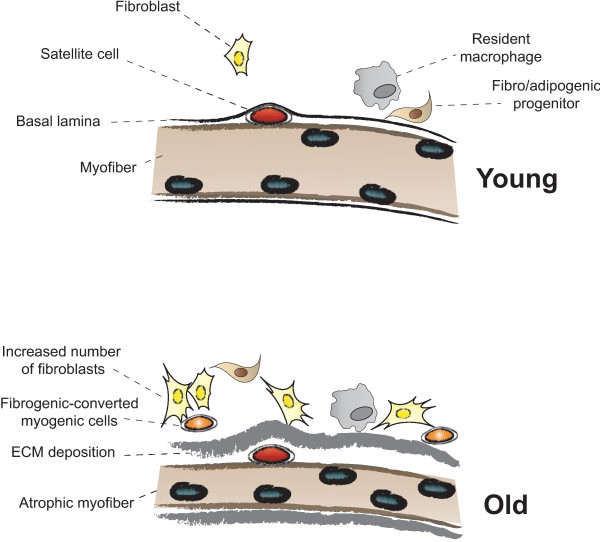
“Whether FAP conversion also occurs during aging, and whether Basal lamina ECM deposition Satellite cell Fibro/adipogenic progenitor Resident macrophage Fibroblast Increased number of fibroblasts Fibrogenic-converted myogenic cells Myofiber Atrophic myofiber Young Old Figure 5 Myofiber growth and extracellular matrix (…”
BackgroundInfluentialEmery, A. E. (2002) · The Lancet · doi:10.1016/s0140-6736(02)07815-7 · Cited by 1,667
Increased Wnt signaling during aging alters muscle stem cell fate and increases fibrosis
Brack, A. S., Conboy, M. J., Roy, S., Lee, M., Kuo, C. J., Keller, C., & Rando, T. A. (2007) · Science · doi:10.1126/science.1144090 · Cited by 1,564 · cited 1×
“An earlier study by the same group had already highlighted the role of the canonical Wnt pathway in ageassociated fibrosis, linking increased collagen deposition in aged regenerating muscles to a greater percentage of fibrogenic cells arising from the conversion of myogenic into non-myogenic cells [65].”
BackgroundInflammatory processes in muscle injury and repair
Tidball, J. G. (2005) · American Journal of Physiology-Regulatory, Integrative and Comparative Physiology · doi:10.1152/ajpregu.00454.2004 · Cited by 1,416 · cited 3×
“outcome of muscle repair, or alternatively, fibrosis [12,14,17,18].”
BackgroundMacrophages: master regulators of inflammation and fibrosis
Wynn, T., & Barron, L. (2010) · Seminars in Liver Disease · doi:10.1055/s-0030-1255354 · Cited by 1,392 · cited 2×
“A nomenclature for polarized macrophages has been proposed [24,25] and they are now referred to as classically and alternatively activated macrophages, or M1 and M2 macrophages, respectively (Figure 2, Figure 3).”
BackgroundUezumi, A., Fukada, S., Yamamoto, N., Takeda, S., & Tsuchida, K. (2010) · Nature Cell Biology · doi:10.1038/ncb2014 · Cited by 1,315 · cited 8×
“Whether FAP conversion also occurs during aging, and whether Basal lamina ECM deposition Satellite cell Fibro/adipogenic progenitor Resident macrophage Fibroblast Increased number of fibroblasts Fibrogenic-converted myogenic cells Myofiber Atrophic myofiber Young Old Figure 5 Myofiber growth and extracellular matrix (…”
BackgroundInfluentialFunction and Genetics of Dystrophin and Dystrophin-Related Proteins in Muscle
Blake, D. J., Weir, A., Newey, S. E., & Davies, K. E. (2002) · Physiological Reviews · doi:10.1152/physrev.00028.2001 · Cited by 1,270
Immunogenic and tolerogenic cell death
Green, D. R., Ferguson, T., Zitvogel, L., & Kroemer, G. (2009) · Nature Reviews Immunology · doi:10.1038/nri2545 · Cited by 1,176 · cited 1×
“The earliest phases of tissue repair are generally characterized by local activation of the innate immune system, even though the original immunogenic stimuli are not always known (see below) [20].”
BackgroundRegulatory interactions between muscle and the immune system during muscle regeneration
Tidball, J. G., & Villalta, S. A. (2010) · American Journal of Physiology-Regulatory, Integrative and Comparative Physiology · doi:10.1152/ajpregu.00735.2009 · Cited by 1,105 · cited 4×
“Immediately after skeletal-muscle injury, cytokines and growth factors are released from both the injured blood vessels and from infiltrating inflammatory cells [13,14].”
BackgroundInfluentialArginase-1–Expressing Macrophages Suppress Th2 Cytokine–Driven Inflammation and Fibrosis
Pesce, J. T., Ramalingam, T. R., Mentink-Kane, M. M., Wilson, M. S., El Kasmi, K. C., Smith, A. M., Thompson, R. W., Cheever, A. W., Murray, P. J., & Wynn, T. A. (2009) · PLoS Pathogens · doi:10.1371/journal.ppat.1000371 · Cited by 816
Regulation of PDGF and its receptors in fibrotic diseases
Bonner, J. C. (2004) · Cytokine & Growth Factor Reviews · doi:10.1016/j.cytogfr.2004.03.006 · Cited by 768 · cited 1×
“In vitro, PDGF production can be induced by TGFb, which also stimulates fibroblasts to contract collagen matrices and differentiate into myofibroblasts [88].”
BackgroundFrazier, K. S., Williams, S. J., Kothapalli, D., Klapper, H., & Grotendorst, G. R. (1996) · Journal of Investigative Dermatology · doi:10.1111/1523-1747.ep12363389 · Cited by 750 · cited 2×
“This fibrotic response is specific to TGFb and CTGF, and is not mimicked by EGF, PDGF or FGF2 [87].”
BackgroundCohn, R. D., van Erp, C., Habashi, J. P., Soleimani, A. A., Klein, E. C., Lisi, M. T., Gamradt, M., Rhys, C. M. A., Holm, T. M., Loeys, B., et al. (2007) · Nature Medicine · doi:10.1038/nm1536 · Cited by 672
Ruffell, D., Mourkioti, F., Gambardella, A., Kirstetter, P., Lopez, R. G., Rosenthal, N., & Nerlov, C. (2009) · Proceedings of the National Academy of Sciences · doi:10.1073/pnas.0908641106 · Cited by 626 · cited 1×
“Mutant mice were able to remove necrotic tissue from injured muscle, but they exhibited severe defects in myofiber regeneration [43].”
BackgroundIL-4 Acts as a Myoblast Recruitment Factor during Mammalian Muscle Growth
Horsley, V., Jansen, K. M., Mills, S. T., & Pavlath, G. K. (2003) · Cell · doi:10.1016/s0092-8674(03)00319-2 · Cited by 532 · cited 1×
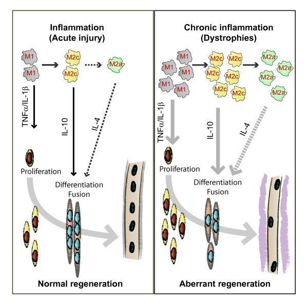
“interleukin (IL)-4 was shown to regulate fusion of myoblasts in vitro and in vivo [129].”
BackgroundA nitric oxide synthase transgene ameliorates muscular dystrophy in mdx mice
Wehling, M., Spencer, M. J., & Tidball, J. G. (2001) · The Journal of Cell Biology · doi:10.1083/jcb.200105110 · Cited by 530 · cited 1×
“Several studies of macrophage depletion or impaired macrophage recruitment have revealed crucial functions of macrophages in the regulation of fibrogenesis in dystrophic muscle, and the potential for therapeutic intervention [19,35,37,38].”
BackgroundVillalta, S. A., Nguyen, H. X., Deng, B., Gotoh, T., & Tidball, J. G. (2008) · Human Molecular Genetics · doi:10.1093/hmg/ddn376 · Cited by 512 · cited 2×
“Indeed, similar shifts in macrophage polarization and macrophage competition for arginine metabolism were seen to influence the severity of the muscle pathology in mdx dystrophic mice [28].”
BackgroundImbalance between pSmad3 and Notch induces CDK inhibitors in old muscle stem cells
Carlson, M. E., Hsu, M., & Conboy, I. M. (2008) · Nature · doi:10.1038/nature07034 · Cited by 498 · cited 1×
“For example, analysis of the microniche of aged murine muscle stem cells found high levels of TGFb and its activated effector Smad3 in both differentiated muscle fibers and satellite cells, which was reciprocal to the levels of active Notch, which is more abundant in the young microniche [62].”
BackgroundLi, Y., Foster, W. H., Deasy, B. M., Chan, Y., Prisk, V. R., Tang, Y., Cummins, J. H., & Huard, J. (2004) · American Journal Of Pathology · doi:10.1016/s0002-9440(10)63188-4 · Cited by 462
Porter, J. D. (2002) · Human Molecular Genetics · doi:10.1093/hmg/11.3.263 · Cited by 442 · cited 1×
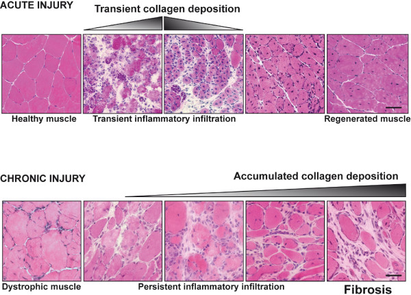
“However, in chronic human diseases such as DMD and many other dystrophies, newly generated fibers are also prone to degeneration because they retain the underlying molecular defect, producing constant cycles of fiber degeneration associated with chronic inflammation (Figure 1) [4].”
BackgroundDurbeej, M., & Campbell, K. P. (2002) · Current Opinion in Genetics & Development · doi:10.1016/s0959-437x(02)00309-x · Cited by 434 · cited 1×
“Furthermore, the ECM sequesters and presents heparinbinding growth factors to the fibers, such as hepatocyte growth factor (HGF) and fibroblast growth factor (FGF), and signals to the differentiated fibers through dystroglycan and sarcoglycan complexes [102,103].”
BackgroundPlattner, R., Kadlec, L., DeMali, K. A., Kazlauskas, A., & Pendergast, A. M. (1999) · Genes & Development · doi:10.1101/gad.13.18.2400 · Cited by 428 · cited 1×
“In addition, PDGF can induce c-abl kinase activity, similar to TGFb, in a non-canonical manner [89].”
BackgroundCellular and molecular mechanisms underlying age-related skeletal muscle wasting and weakness
Ryall, J. G., Schertzer, J. D., & Lynch, G. S. (2008) · Biogerontology · doi:10.1007/s10522-008-9131-0 · Cited by 423 · cited 1×
“In addition, there is infiltration of fat and other non-contractile material, which causes a reduction in muscle ‘quality’ [8].”
BackgroundLoss of myostatin attenuates severity of muscular dystrophy in mdx mice
Wagner, K. R., McPherron, A. C., Winik, N., & Lee, S. (2002) · Annals of Neurology · doi:10.1002/ana.10385 · Cited by 378 · cited 1×
“Similarly, mdx mice lacking myostatin displayed less fibrosis in the diaphragm, and were also stronger and more muscular than their normal mdx counterparts [97].”
BackgroundVascular endothelial growth factor stimulates skeletal muscle regeneration in Vivo
Arsic, N., Zacchigna, S., Zentilin, L., Ramirez‐Correa, G. A., Pattarini, L., Salvi, A., Sinagra, G., & Giacca, M. (2004) · Molecular Therapy · doi:10.1016/j.ymthe.2004.08.007 · Cited by 360 · cited 1×
“Interestingly, there is the suggestion that the type of injury in acute models may drive the repair response towards more fibrotic or more adipogenic repair, for example after glycerol injury and acute ischemia, respectively [68,69], although the mechanisms have not been extensively investigated.”
BackgroundSignaling pathways perturbing muscle mass.
Glass, D. J. (2010) · Current Opinion in Clinical Nutrition and Metabolic Care · doi:10.1097/mco.0b013e32833862df · Cited by 359 · cited 1×
“Deficiency of myostatin increases skeletal muscle mass, myofiber diameter and strength [93,94].”
BackgroundConnective tissue fibroblasts and Tcf4 regulate myogenesis
Mathew, S. J., Hansen, J. M., Merrell, A. J., Murphy, M. M., Lawson, J. A., Hutcheson, D. A., Hansen, M. S. T., Angus-Hill, M. L., & Kardon, G. (2010) · Development · doi:10.1242/dev.057463 · Cited by 323
Transforming growth factor-β and myostatin signaling in skeletal muscle
Kollias, H. D., & McDermott, J. C. (2007) · Journal of Applied Physiology · doi:10.1152/japplphysiol.01091.2007 · Cited by 317
Kherif, S. A. A. B., Lafuma, C., Dehaupas, M., Lachkar, S., Fournier, J. G., Verdière‐Sahuqué, M., Fardeau, M., & Alameddine, H. S. (1999) · Developmental Biology · doi:10.1006/dbio.1998.9107 · Cited by 312 · cited 1×
“Perhaps more importantly, the skeletal muscle of adult mdx mice expresses high levels of latent and active MMP-2 and MMP-9, and inhibiting MMP-9 activity was shown to significantly improve regeneration and contractile functions by reducing the structural deterioration of muscle, necrosis, inflammation and fibrosis [16…”
BackgroundRegulation and dysregulation of fibrosis in skeletal muscle
Serrano, A. L., & Muñoz‐Cánoves, P. (2010) · Experimental Cell Research · doi:10.1016/j.yexcr.2010.05.035 · Cited by 302
The COX-2 pathway is essential during early stages of skeletal muscle regeneration
Bondesen, B. A., Mills, S. T., Kegley, K. M., & Pavlath, G. K. (2004) · American Journal of Physiology-Cell Physiology · doi:10.1152/ajpcell.00088.2004 · Cited by 300 · cited 1×
“PGs have been shown to play various roles in many stages of myogenesis, and are secreted from regenerating muscle [48,49].”
BackgroundVillalta, S. A., Rinaldi, C., Deng, B., Liu, G., Fedor, B., & Tidball, J. G. (2011) · Human Molecular Genetics · doi:10.1093/hmg/ddq523 · Cited by 299 · cited 1×
“One example of this was the deletion of IL-10 in mdx mice, which increased muscle damage and reduced muscle strength, due to an imbalance between M1 and M2 macrophages [29].”
BackgroundVetrone, S., Montecino-Rodriguez, E., Kudryashova, E., Kramerova, I., Hoffman, E. P., Liu, S., Miceli, M., & Spencer, M. J. (2009) · Journal of Clinical Investigation · doi:10.1172/jci37662 · Cited by 297 · cited 2×
“These results correlated well with improvements in muscle strength and reduced diaphragmatic and cardiac fibrosis [53].”
BackgroundResultRelationships between Transforming Growth Factor-β1, Myostatin, and Decorin
Zhu, J., Li, Y., Shen, W., Qiao, C., Ambrosio, F., Lavasani, M., Nozaki, M., Branca, M. F., & Huard, J. (2007) · Journal of Biological Chemistry · doi:10.1074/jbc.m704146200 · Cited by 270 · cited 3×
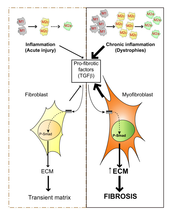
“Direct injection of recombinant TGFb into skeletal muscle in vivo stimulates TGFb expression in myogenic cells in an autocrine fashion and induces connective-tissue formation in the area of the injection [73,74].”
BackgroundDual and Beneficial Roles of Macrophages During Skeletal Muscle Regeneration
Chazaud, B., Brigitte, M., Yacoub‐Youssef, H., Arnold, L., Gherardi, R. K., Sonnet, C., Lafuste, P., & Chrétien, F. (2008) · Exercise and Sport Sciences Reviews · doi:10.1097/jes.0b013e318190ebdb · Cited by 270 · cited 2×
“Immediately after skeletal-muscle injury, cytokines and growth factors are released from both the injured blood vessels and from infiltrating inflammatory cells [13,14].”
BackgroundHelper (CD4+) and Cytotoxic (CD8+) T Cells Promote the Pathology of Dystrophin-Deficient Muscle
Spencer, M. J., Montecino‐Rodriguez, E., Dorshkind, K. A., & Tidball, J. G. (2001) · Clinical Immunology · doi:10.1006/clim.2000.4966 · Cited by 268 · cited 1×
“when followed by anti-CD4 and/or antiCD8 antibody treatment, failed to improve diaphragm fibrosis at 6 months of age in mdx mice [51,54,55].”
BackgroundPelosi, L., Giacinti, C., Nardis, C., Borsellino, G., Rizzuto, E., Nicoletti, C., Wannenes, F., Battistini, L., Rosenthal, N. A., Molinaro, M., et al. (2007) · The FASEB Journal · doi:10.1096/fj.06-7690com · Cited by 268 · cited 2×
“Local expression of a muscle-restricted IGF-1 transgene accelerates the regenerative process in injured skeletal muscle, modulating the inflammatory response and limiting fibrosis [39].”
BackgroundMyostatin Directly Regulates Skeletal Muscle Fibrosis
Li, Z. B., Kollias, H. D., & Wagner, K. R. (2008) · Journal of Biological Chemistry · doi:10.1074/jbc.m802585200 · Cited by 267 · cited 1×
“However, myostatin not only regulates the growth of muscle cells, but also regulates muscle fibroblast activation and hence the progression of fibrosis [96].”
BackgroundFibrinogen drives dystrophic muscle fibrosis via a TGFβ/alternative macrophage activation pathwayPreprint
Vidal, B., Serrano, A. L., Tjwa, M., Suelves, M., Ardite, E., Mori, R. D., Baeza-Raja, B., de Lagrán, M. M., Lafuste, P., Ruiz‐Bonilla, V., et al. (2008) · Genes & Development · doi:10.1101/gad.465908 · Cited by 257 · cited 8×
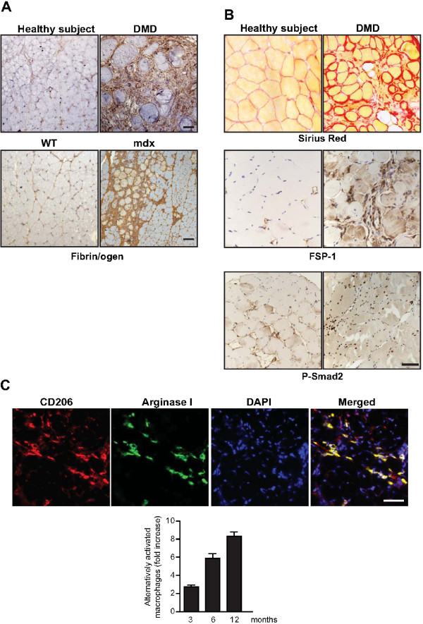
“Fibrin(ogen) deposits may serve as important triggers for the inflammatory responses, because of their capacity to bind to the integrin receptor Mac-1 on classically activated M1 macrophages [27] (see above).”
BackgroundInfluentialThe impact of sarcopenia and exercise training on skeletal muscle satellite cells
Snijders, T., Verdijk, L. B., & van Loon, L. J. C. (2009) · Ageing Research Reviews · doi:10.1016/j.arr.2009.05.003 · Cited by 254 · cited 1×
“The net consequence of these alterations firstly involves progressive atrophy and loss of individual muscle fibers, associated with concomitant loss of motor units [7].”
BackgroundTGF-β and Smad3 Signaling Link Inflammation to Chronic Fibrogenesis
Bonniaud, P., Margetts, P. J., Ask, K., Flanders, K., Gauldie, J., & Kolb, M. R. J. (2005) · The Journal of Immunology · doi:10.4049/jimmunol.175.8.5390 · Cited by 252
Aging and microRNA expression in human skeletal muscle: a microarray and bioinformatics analysis
Drummond, M. J., McCarthy, J. J., Sinha, M., Spratt, H. M., Volpi, E., Esser, K. A., & Rasmussen, B. B. (2010) · Physiological Genomics · doi:10.1152/physiolgenomics.00148.2010 · Cited by 248 · cited 1×
“Because Let-7 family members been related to cell-cycle control, they may function to downregulate other genes involved in muscle-cell proliferation, which in turn might negatively affect the reduced regenerative capacity of muscle seen with aging [126].”
BackgroundRole of matrix metalloproteinases in skeletal muscle
Chen, X., & Li, Y. (2009) · Cell Adhesion & Migration · doi:10.4161/cam.3.4.9338 · Cited by 241 · cited 6×
“The large family of MMPs includes various collagenases (MMP-1, MMP-8 and MMP-13), gelatinases (MMP-2 and MMP-9), stromelysins (MMP-3, MMP-7, MMP-10 and MMP-11), membrane-type metalloproteinases (MMP-14, MMP-15, MMP-16, MMP-17, MMP24 and MMP-25), and the metalloelastase MMP-12 [16].”
BackgroundInfluentialBrigitte, M., Schilte, C., Plonquet, A., Baba‐Amer, Y., Henry, A., Charlier, C., Tajbakhsh, S., Albert, M. L., Gherardi, R. K., & Chrétien, F. (2009) · Arthritis & Rheumatism · doi:10.1002/art.27183 · Cited by 222 · cited 1×
“Recent studies have shown that resident macrophages in the muscle epimysium/perimysium orchestrate the innate immune response to injury, which is linked to adaptive immunity through inflammatory dendritic cells [21].”
BackgroundSuppression of macrophage functions impairs skeletal muscle regeneration with severe fibrosis
Segawa, M., Fukada, S., Yamamoto, Y., Yahagi, H., Kanematsu, M., SATO, M., Ito, T., Uezumi, A., HAYASHI, S., & Miyagoe‐Suzuki, Y. (2008) · Experimental Cell Research · doi:10.1016/j.yexcr.2008.08.008 · Cited by 221 · cited 2×
“Several studies of macrophage depletion or impaired macrophage recruitment have revealed crucial functions of macrophages in the regulation of fibrogenesis in dystrophic muscle, and the potential for therapeutic intervention [19,35,37,38].”
BackgroundImproved muscle healing through enhanced regeneration and reduced fibrosis in myostatin-null mice
McCroskery, S., Thomas, M., Platt, L., Hennebry, A., Nishimura, T., McLeay, L. M., Sharma, M., & Kambadur, R. (2005) · Journal of Cell Science · doi:10.1242/jcs.02482 · Cited by 219 · cited 1×
“As a potential therapeutic approach, immunological neutralization of myostatin in mdx mice was shown to attenuate dystrophy by improving muscle regeneration and reducing ECM accumulation [98].”
BackgroundLatent TGF-β–binding protein 4 modifies muscular dystrophy in mice
Heydemann, A., Ceco, E., Lim, J. E., Hadhazy, M., Ryder, P. V., Moran, J. L., Beier, D. R., Palmer, A. A., & McNally, E. M. (2009) · Journal of Clinical Investigation · doi:10.1172/jci39845 · Cited by 212 · cited 1×
“Finally, latent TGFb-binding protein (LTBP)4 regulates the release and bioavailability of TGFb from the ECM, where it has been shown to modulate fibrosis in the context of muscular dystrophy in mice [77] Taken together, these studies highlight the crucial role of TGFb in the initiation of fibrotic processes, and in th…”
BackgroundBedair, H. S., Karthikeyan, T., Quintero, A., Li, Y., & Huard, J. (2008) · The American Journal of Sports Medicine · doi:10.1177/0363546508315470 · Cited by 199 · cited 1×
“Reduced tissue fibrosis after injury was also detected in mice treated with both decorin- and angiotensin receptor antagonists, with decorin also preventing TGFb-induced differentiation of myogenic cells into fibrotic cells [80].”
BackgroundDesguerre, I., Christov, C., Mayer, M., Zeller, R., Bécane, H. M., Bastuji‐Garin, S., Leturcq, F., Chiron, C., Chelly, J., & Gherardi, R. K. (2009) · PLoS ONE · doi:10.1371/journal.pone.0004347 · Cited by 194 · cited 1×
“In patients with DMD, fibrosis has also been associated with increased numbers of alternatively activated macrophages [42].”
BackgroundInhibition of myostatin with emphasis on follistatin as a therapy for muscle disease
Rodino‐Klapac, L. R., Haidet, A. M., Kota, J., Handy, C., Kaspar, B. K., & Mendell, J. R. (2009) · Muscle & Nerve · doi:10.1002/mus.21244 · Cited by 194 · cited 1×
“Normal myostatin signaling can be counter-regulated by the extracellular protein follistatin [95].”
BackgroundThe role of corticosteroids in muscular dystrophy: A critical appraisal
Angelini, C. I. (2007) · Muscle & Nerve · doi:10.1002/mus.20812 · Cited by 191
Hematopoietic stem cell and progenitor defects in Sca-1/Ly-6A–null mice
Ito, C. Y., Li, C. Y. J., Bernstein, A., Dick, J. E., & Stanford, W. L. (2002) · Blood · doi:10.1182/blood-2002-06-1918 · Cited by 184 · cited 1×
“A more recent follow-up study [118] related the increased fibrosis of Sca-1-deficient mice to a reduced recruitment of IgM and C3 to injured muscle, due to a specific reduction in peritoneal IgM-producing B-1a cells.”
BackgroundRelation between myofibers and connective tissue during muscle injury repair
Kääriäinen, M., Järvinen, T. A. H., Järvinen, M., Rantanen, J., & Kalimo, H. (2000) · Scandinavian Journal of Medicine and Science in Sports · doi:10.1034/j.1600-0838.2000.010006332.x · Cited by 184 · cited 2×
“In normal muscle, the external lamina is composed of collagen IV, laminin, and heparan sulfate proteoglycans, while the interstitial matrix that surrounds the basal lamina contains collagens I, III and V, fibronectin and perlecan [10,11,102].”
BackgroundAlexakis, C., Partridge, T. A., & Bou–Gharios, G. (2007) · American Journal of Physiology-Cell Physiology · doi:10.1152/ajpcell.00061.2007 · Cited by 182 · cited 2×
“Although fibroblasts are the major collagen-producing cells, myofiber-associated satellite cells and C2C12 myoblasts have also been shown to express significant levels of interstitial collagens I and III, which diminish during the process of differentiation [58].”
BackgroundLi, H., Mittal, A., Makonchuk, D. Y., Bhatnagar, S., & Kumar, A. (2009) · Human Molecular Genetics · doi:10.1093/hmg/ddp191 · Cited by 179 · cited 1×
“Perhaps more importantly, the skeletal muscle of adult mdx mice expresses high levels of latent and active MMP-2 and MMP-9, and inhibiting MMP-9 activity was shown to significantly improve regeneration and contractile functions by reducing the structural deterioration of muscle, necrosis, inflammation and fibrosis [16…”
BackgroundStrength at the extracellular matrix–muscle interface
Grounds, M. D., Sorokin, L. M., & White, J. D. (2005) · Scandinavian Journal of Medicine and Science in Sports · doi:10.1111/j.1600-0838.2005.00467.x · Cited by 175
Markiewski, M. M., Mastellos, D. C., Tudoran, R., DeAngelis, R. A., Strey, C. W., Franchini, S., Wetsel, R. A., Erdei, A., & Lambris, J. D. (2004) · The Journal of Immunology · doi:10.4049/jimmunol.173.2.747 · Cited by 168 · cited 1×
“Because Sca-1 also plays a role in the maintenance of progenitor cells [119], it was hypothesized that the reduction in B-1a cells was due to lineage-specific defects in self-renewal.”
BackgroundMartinez, C. O., McHale, M. J., Wells, J. T., Ochoa, O., Michalek, J. E., McManus, L. M., & Shireman, P. K. (2010) · American Journal of Physiology-Regulatory, Integrative and Comparative Physiology · doi:10.1152/ajpregu.00797.2009 · Cited by 167 · cited 1×
“Conversely, BM transplantation experiments have revealed an important role for the CC chemokine receptor 2/monocyte chemotactic protein-1 (CCR2/MCP-1) ligand, and for the proteolytic activity of urokinase plasminogen activator (uPA)/plasmin in skeletal-muscle repair by regulating the recruitment of BMderived macrophag…”
BackgroundCencetti, F., Bernacchioni, C., Nincheri, P., Donati, C., & Bruni, P. (2010) · Molecular Biology of the Cell · doi:10.1091/mbc.e09-09-0812 · Cited by 166 · cited 2×
“Rho/ Rho kinase signaling also appeared to be implicated in the TGFb-mediated transition of myoblasts into myofibroblasts downstream of SK1 activation [60].”
BackgroundDecorin Gene Transfer Promotes Muscle Cell Differentiation and Muscle Regeneration
Li, Y., Li, J., Zhu, J., Sun, B., Branca, M. F., Tang, Y., Foster, W. H., Xiao, X., & Huard, J. (2007) · Molecular Therapy · doi:10.1038/sj.mt.6300250 · Cited by 162 · cited 2×
“whereas loss of biglycan in transgenic mice does not affect regenerative capacity, despite inducing delays in new myofiber growth [100], decorin delivery to regenerating muscle via adenoassociated viral (AAV) vectors prevents TGFb activation and fibrosis [76].”
BackgroundMarco, S. D., Mazrouï, R., Dallaire, P., Chittur, S. V., Tenenbaum, S. A., Radzioch, D., Marette, A., & Gallouzi, I. (2005) · Molecular and Cellular Biology · doi:10.1128/mcb.25.15.6533-6545.2005 · Cited by 155
Regulation of GDF-8 signaling by the p38 MAPK
Philip, B., Lu, Z., & Gao, Y. (2004) · Cellular Signalling · doi:10.1016/j.cellsig.2004.08.003 · Cited by 153 · cited 1×
“In fibroblasts, myostatin induces activation of the Smad, p38 MAPK and AKT pathways to promote ECM synthesis [99].”
BackgroundAntifibrotic effects of suramin in injured skeletal muscle after laceration
Chan, Y., Li, Y., Foster, W. H., Horaguchi, T., Somogyi, G. T., Fu, F. H., & Huard, J. (2003) · Journal of Applied Physiology · doi:10.1152/japplphysiol.00915.2002 · Cited by 142 · cited 1×
“Similar results were reported in animals treated with suramin, which acts by competing with TGFb for receptor binding [82].”
ResultMechanisms of Muscle Atrophy Induced by Glucocorticoids
Schakman, O., Gilson, H., Kalista, S., & Thissen, J. (2009) · Hormone Research in Paediatrics · doi:10.1159/000229762 · Cited by 139 · cited 1×
“drugs has been their ability to promote fiber atrophy [47], in addition to their negative effect on prostaglandin (PG) synthesis via interference with cyclooxygenase (COX) gene functions.”
BackgroundAndreetta, F., Bernasconi, P., Baggi, F., Ferro, P., Oliva, L., Arnoldi, E., Cornelio, F., Mantegazza, R., & Confalonieri, P. A. (2006) · Journal of Neuroimmunology · doi:10.1016/j.jneuroim.2006.03.005 · Cited by 126 · cited 1×
“For example, direct immunomodulation of TGFb inhibited connective-tissue accumulation and the progression of fibrosis in the diaphragm of mdx mice, although inflammation increased [79].”
BackgroundGargioli, C., Coletta, M., Grandis, F. D., Cannata, S. M., & Cossu, G. (2008) · Nature Medicine · doi:10.1038/nm.1852 · Cited by 125 · cited 1×
“These cells restored a vascular network and diminished collagen deposition, raising the exciting possibility of using similar cell-based or gene therapies to improve muscle function in patients with currently untreatable advanced-stage dystrophies [114].”
BackgroundT-Cell-Dependent Fibrosis in the mdx Dystrophic Mouse
Morrison, J. I., Lu, Q. L., Pastoret, C., Partridge, T. A., & Bou–Gharios, G. (2000) · Laboratory Investigation · doi:10.1038/labinvest.3780092 · Cited by 125 · cited 3×
“In nu/nu/ mdx mice, (immunodeficient nude mice in the mdx background) the lack of functional T cells alone was associated with less diaphragm fibrosis at 3 months, supporting the pathogenic role for T cells in mdx muscle, and revealing this lymphocyte subclass to be an important source of TGFb1 [52].”
BackgroundOhtake, Y., Tojo, H., & Seiki, M. (2006) · Journal of Cell Science · doi:10.1242/jcs.03158 · Cited by 125 · cited 2×
“For example, MT1-MMP is presumed to help maintain myofiber integrity, as transgenic mice deficient in MT1-MMP develop centrally nucleated myofibers that are smaller than normal [105].”
BackgroundVial, C., Zúñiga, L. M., Cabello‐Verrugio, C., Cañón, P., Fadić, R., & Brandan, E. (2007) · Journal of Cellular Physiology · doi:10.1002/jcp.21324 · Cited by 122 · cited 2×
“These results suggest a role for both TGFb and CTGF in skeletal-muscle remodeling by inducing fibrosis, inhibiting myogenesis and promoting dedifferentiation of myoblasts into myofibroblast-like cells [86].”
BackgroundBone marrow‐derived cell regulation of skeletal muscle regeneration
Sun, D., Martinez, C. O., Ochoa, O., Ruiz‐Willhite, L., Bonilla, J. R., Centonze, V. E., Waite, L. L., Michalek, J. E., McManus, L. M., & Shireman, P. K. (2008) · The FASEB Journal · doi:10.1096/fj.07-095901 · Cited by 117 · cited 1×
“Conversely, BM transplantation experiments have revealed an important role for the CC chemokine receptor 2/monocyte chemotactic protein-1 (CCR2/MCP-1) ligand, and for the proteolytic activity of urokinase plasminogen activator (uPA)/plasmin in skeletal-muscle repair by regulating the recruitment of BMderived macrophag…”
BackgroundConnective tissue growth factor is overexpressed in muscles of human muscular dystrophy
Sun, G., Haginoya, K., Wu, Y., Chiba, Y., Nakanishi, T., Onuma, A., Sato, Y., Takigawa, M., Iinuma, K., & Tsuchiya, S. (2007) · Journal of the Neurological Sciences · doi:10.1016/j.jns.2007.09.043 · Cited by 112 · cited 1×
“Increased levels of CTGF have been reported in skeletal muscle from patients with DMD, dystrophic dogs and mdx mice [85,86].”
BackgroundLluı́s, F., Roma, J. M., Suelves, M., Parra, M., Aniorte, G., Gallardo, E., Illa, I., Rodrı́guez, L., Hughes, S. M., Carmeliet, P., et al. (2001) · Blood · doi:10.1182/blood.v97.6.1703 · Cited by 111 · cited 4×
“Whereas it has been shown that both uPA and plasmin activity are necessary for skeletal-muscle regeneration, tPA activity is dispensable, indicating that no redundancy exists in muscle [33,35].”
BackgroundInfluentialPlasmin activity is required for myogenesis in vitro and skeletal muscle regeneration in vivo
Suelves, M., López‐Alemany, R., Lluı́s, F., Aniorte, G., Serrano, E., Parra, M., Carmeliet, P., & Muñoz‐Cánoves, P. (2002) · Blood · doi:10.1182/blood.v99.8.2835 · Cited by 109 · cited 4×
“Several studies of macrophage depletion or impaired macrophage recruitment have revealed crucial functions of macrophages in the regulation of fibrogenesis in dystrophic muscle, and the potential for therapeutic intervention [19,35,37,38].”
BackgroundInfluentialBrack, A. S., Murphy-Seiler, F., Hanifi, J. M., Deka, J., Eyckerman, S., Keller, C., Aguet, M., & Rando, T. A. (2009) · Developmental Biology · doi:10.1016/j.ydbio.2009.08.014 · Cited by 107 · cited 2×
“In additional studies of aged muscle, the fate of muscle stem-cell progeny was reported to be controlled by an interaction between the Wnt and Notch pathways in which glycogen synthase kinase (GSK)3b plays an important role [64].”
BackgroundThe Effect of Relaxin Treatment on Skeletal Muscle Injuries
Negishi, S., Li, Y., Ūsas, A., Fu, F. H., & Huard, J. (2005) · The American Journal of Sports Medicine · doi:10.1177/0363546505278701 · Cited by 106 · cited 1×
“Another recent study identified an increase in a muscle-resident stromal cell (mrSC) population in the muscles of mdx mice [122].”
BackgroundFarini, A., Meregalli, M., Belicchi, M., Battistelli, M., Parolini, D., D’Antona, G., Gavina, M., Ottoboni, L., Constantin, G., Bottinelli, R., et al. (2007) · The Journal of Pathology · doi:10.1002/path.2213 · Cited by 104 · cited 2×
“when followed by anti-CD4 and/or antiCD8 antibody treatment, failed to improve diaphragm fibrosis at 6 months of age in mdx mice [51,54,55].”
BackgroundImatinib attenuates skeletal muscle dystrophy in mdx mice
Huang, P. C., Zhao, X. S., Fields, M., Ransohoff, R. M., & Zhou, L. (2009) · The FASEB Journal · doi:10.1096/fj.09-129833 · Cited by 104 · cited 1×
“Consequently, administering imatinib to mdx mice reduced fibrosis in the diaphragm, and attenuated skeletal-muscle necrosis and inflammation, with concomitant improvements in muscle function [91].”
BackgroundShen, W., Li, Y., Zhu, J., Schwendener, R. A., & Huard, J. (2007) · Journal of Cellular Physiology · doi:10.1002/jcp.21212 · Cited by 102 · cited 2×
“PGs have been shown to play various roles in many stages of myogenesis, and are secreted from regenerating muscle [48,49].”
BackgroundCasar, J. C., McKechnie, B. A., Fallon, J. R., Young, M. F., & Brandan, E. (2004) · Developmental Biology · doi:10.1016/j.ydbio.2003.12.025 · Cited by 101 · cited 2×
“whereas loss of biglycan in transgenic mice does not affect regenerative capacity, despite inducing delays in new myofiber growth [100], decorin delivery to regenerating muscle via adenoassociated viral (AAV) vectors prevents TGFb activation and fibrosis [76].”
BackgroundContext matters: In vivo and in vitro influences on muscle satellite cell activity
Cornelison, D. (2008) · Journal of Cellular Biochemistry · doi:10.1002/jcb.21892 · Cited by 101 · cited 2×
“In normal muscle, the external lamina is composed of collagen IV, laminin, and heparan sulfate proteoglycans, while the interstitial matrix that surrounds the basal lamina contains collagens I, III and V, fibronectin and perlecan [10,11,102].”
BackgroundThalacker‐Mercer, A. E., Dell’Italia, L. J., Cui, X., Cross, J. M., & Bamman, M. M. (2009) · Physiological Genomics · doi:10.1152/physiolgenomics.00151.2009 · Cited by 99 · cited 1×
“Aged human muscle also behaves differently from young muscle after a similar modest level of damage, with dysregulation of a particular set of transcripts including transcripts related to fibrosis and connectivetissue function [127].”
BackgroundZhou, L., Porter, J. D., Cheng, G., Gong, B., Hatala, D. A., Merriam, A. P., Zhou, X., Rafael, J. A., & Kaminski, H. J. (2005) · Neuromuscular Disorders · doi:10.1016/j.nmd.2005.09.009 · Cited by 98 · cited 2×
“TGFb is expressed in normal muscle after injury and in the dystrophic muscle of patients with DMD and mdx mice [70], where it has the potential to induce fibrosis around myofibers, probably by stimulating fibroblasts to produce ECM proteins such as collagen and fibronectin.”
Functional resolution of fibrosis in mdx mouse dystrophic heart and skeletal muscle by halofuginone
Huebner, K. D., Jassal, D. S., Halevy, O., Pines, M., & Anderson, J. E. (2008) · American Journal of Physiology-Heart and Circulatory Physiology · doi:10.1152/ajpheart.01253.2007 · Cited by 96 · cited 1×
“Indeed, halofuginone treatment was also shown to enhance myogenesis, a feature that may be relevant to improve muscle regeneration and function in muscular dystrophy [84].”
BackgroundBrandan, E., Cabello‐Verrugio, C., & Vial, C. (2008) · Matrix Biology · doi:10.1016/j.matbio.2008.07.004 · Cited by 95 · cited 2×
“Direct injection of recombinant TGFb into skeletal muscle in vivo stimulates TGFb expression in myogenic cells in an autocrine fashion and induces connective-tissue formation in the area of the injection [73,74].”
BackgroundPremature Aging in Skeletal Muscle Lacking Serum Response Factor
Lahoute, C., Sotiropoulos, A., Favier, M., Guillet-Deniau, I., Charvet, C., Ferry, A., Butler-Browne, G. S., METZGER, D., Tuil, D. G., & Daegelen, D. (2008) · PLoS ONE · doi:10.1371/journal.pone.0003910 · Cited by 94 · cited 1×
“For example, a major role for myofibers themselves in controlling the homeostasis of the ECM during age-related sarcopenia has been proposed [125].”
BackgroundNitric oxide regulates the repair of injured skeletal muscle
Filippin, L. I., Cuevas, M. J., Lima‐Cabello, E., Marroni, N. P., González‐Gallego, J., & Xavier, R. M. (2010) · Nitric Oxide · doi:10.1016/j.niox.2010.11.003 · Cited by 94 · cited 4×
“In addition, NO, which is released by local and invading cells during injury, can act as a pro-inflammatory molecule, and is able to activate MMP-2, which in turn releases HGF. Importantly, blockers of NOS function, such as nitro- Larginine methyl ester (L-NAME), have been shown to decrease the acute inflammatory reac…”
MethodInfluentialDecorin and biglycan expression is differentially altered in several muscular dystrophies
Zanotti, S., Negri, T., Cappelletti, C., Bernasconi, P., Canioni, E., Blasi, C. D., Pegoraro, E., Angelini, C. I., Ciscato, P., Prelle, A. C., et al. (2005) · Brain · doi:10.1093/brain/awh635 · Cited by 93 · cited 2×
“Based on all of the emerging data, it is tempting to speculate that the pronounced fibrosis in seen in patients with DMD is related to altered net proteolytic activity in the dystrophic muscles, due to imbalances in expression and activity of PA/MMP system components [101,116].”
BackgroundFukushima, K., Nakamura, A., Ueda, H., Yuasa, K., Yoshida, K., Takeda, S., & Ikeda, S. (2007) · BMC Musculoskeletal Disorders · doi:10.1186/1471-2474-8-54 · Cited by 92 · cited 1×
“Perhaps more importantly, the skeletal muscle of adult mdx mice expresses high levels of latent and active MMP-2 and MMP-9, and inhibiting MMP-9 activity was shown to significantly improve regeneration and contractile functions by reducing the structural deterioration of muscle, necrosis, inflammation and fibrosis [16…”
BackgroundYogo, Y., Fujishima, S., Inoue, T., Saito, F., Shiomi, T., Yamaguchi, K., & Ishizaka, A. (2009) · Respiratory Research · doi:10.1186/1465-9921-10-80 · Cited by 91 · cited 1×
“CCL17 in particular has been shown to enhance fibrosis in several mouse models of pulmonary disease by binding to CC chemokine receptor (CCR)4 [30].”
BackgroundRadley‐Crabb, H. G., Davies, M. J., & Grounds, M. D. (2008) · Neuromuscular Disorders · doi:10.1016/j.nmd.2007.11.002 · Cited by 91 · cited 1×
“Similar benefits on the dystrophic phenotype in mdx mice were reported after using a variety of anti-inflammatory agents acting on cytokines such as TNFa and on their cellular receptors, or on other major pro-inflammatory mediators such as nuclear factor (NF)- B [39-41].”
BackgroundTranscriptional and posttranscriptional regulation of the plasminogen activator system
Medcalf, R. L., Muñoz‐Cánoves, P., & Nagamine, Y. (2005) · Thrombosis and Haemostasis · doi:10.1160/th04-12-0814 · Cited by 90 · cited 3×
“The main function of the plasminogen activation system main function is to degrade fibrin [104], but it also fulfills an important role in muscle ECM remodeling by cleaving ECM-associated molecules, liberating and/or activating latent forms of bioactive molecules such as growth factors, angiogenic factors and cytokine…”
BackgroundFibro/adipogenic progenitors: A double-edged sword in skeletal muscle regeneration
Natarajan, A., Lemos, D. R., & Rossi, F. M. V. (2010) · Cell Cycle · doi:10.4161/cc.9.11.11854 · Cited by 88
Trensz, F., Haroun, S., Cloutier, A., Richter, M. V., & Grenier, G. (2010) · American Journal of Physiology-Cell Physiology · doi:10.1152/ajpcell.00253.2010 · Cited by 87 · cited 1×
“In aged mice, a Sca-1+, non-myogenic (MyoD-) and non-immunohematopoietic (CD45-) cell population has been proposed to favor fibrosis [121].”
BackgroundCan muscle regeneration fail in chronic inflammation: a weakness in inflammatory myopathies?
Loell, I., & Lundberg, I. E. (2010) · Journal of Internal Medicine · doi:10.1111/j.1365-2796.2010.02334.x · Cited by 81
Friedrich, O., Both, M., Weber, C., Schürmann, S., Teichmann, M. D. H., von Wegner, F., Fink, R., Vogel, M., Chamberlain, J. S., & Garbe, C. S. (2010) · Biophysical Journal · doi:10.1016/j.bpj.2009.11.005 · Cited by 81 · cited 1×
“At the ultrastructural level, aging has also been associated with myofibril disarrangements in a dystrophic animal model, and drops in force without alterations in motor protein function as measured by in vitro motility assays [9].”
BackgroundPeterson, J. M., & Guttridge, D. C. (2008) · International Reviews of Immunology · doi:10.1080/08830180802302389 · Cited by 79 · cited 1×
“Similar benefits on the dystrophic phenotype in mdx mice were reported after using a variety of anti-inflammatory agents acting on cytokines such as TNFa and on their cellular receptors, or on other major pro-inflammatory mediators such as nuclear factor (NF)- B [39-41].”
BackgroundEmerging strategies for cell and gene therapy of the muscular dystrophies
Muir, L. A., & Chamberlain, J. S. (2009) · Expert Reviews in Molecular Medicine · doi:10.1017/s1462399409001100 · Cited by 75 · cited 1×
“intervention, or impairs the efficiency of these therapies [5].”
BackgroundAlterations in the TGFβ signaling pathway in myogenic progenitors with age
Beggs, M. L., Nagarajan, R., Taylor‐Jones, J. M., Nolen, G., MacNicol, M., & Peterson, C. A. (2004) · Aging Cell · doi:10.1111/j.1474-9728.2004.00135.x · Cited by 74 · cited 1×
“Microarray analyses of myogenic cells from aged animals also revealed major alterations in the expression of many genes dependent upon the activation of TGFb signaling [78].”
BackgroundHaus, J. M., Carrithers, J. A., Carroll, C. C., Tesch, P. A., & Trappe, T. A. (2007) · American Journal of Physiology-Regulatory, Integrative and Comparative Physiology · doi:10.1152/ajpregu.00292.2007 · Cited by 72 · cited 1×
“Collagen modifications, such as non-enzymatically regulated crosslinking to produce advanced glycation end (AGE) products, also increase the stiffness of muscle connective tissue, thereby contributing to impaired muscle function in the older person [59].”
BackgroundRoffe, S., Hagai, Y., Pines, M., & Halevy, O. (2010) · Experimental Cell Research · doi:10.1016/j.yexcr.2010.01.003 · Cited by 71
Matrix metalloproteinase-1 treatment of muscle fibrosis
Kaar, J. L., Li, Y., Blair, H. C., Asche, G., Koepsel, R. R., Huard, J., & Russell, A. J. (2008) · Acta Biomaterialia · doi:10.1016/j.actbio.2008.03.010 · Cited by 71 · cited 1×
“MMP-13 and MMP-1 also participate in ECM remodeling during muscle repair [16,107,108], while gelatinase MMP-2 affects new fiber formation by promoting collagen IV degradation in the basement membrane during myoblast proliferation, migration and fusion.”
BackgrounduPA deficiency exacerbates muscular dystrophy in MDX mice
Suelves, M., Vidal, B., Serrano, A. L., Tjwa, M., Roma, J. M., López‐Alemany, R., Luttun, A., de Lagrán, M. M., Díaz, M. À., Jardı́, M., et al. (2007) · The Journal of Cell Biology · doi:10.1083/jcb.200705127 · Cited by 70
Mu, X., Urso, M. L., Murray, K., Fu, F. H., & Li, Y. (2010) · American Journal Of Pathology · doi:10.2353/ajpath.2010.091121 · Cited by 65
Notch2 negatively regulates myofibroblastic differentiation of myoblasts
Ono, Y., Sensui, H., Okutsu, S., & Nagatomi, R. (2006) · Journal of Cellular Physiology · doi:10.1002/jcp.20838 · Cited by 63 · cited 1×
“Finally, Notch2 can inhibit the differentiation of myoblasts into myofibroblasts by directly counter-regulating Notch3 and limiting its expression [61].”
BackgroundKafadar, K. A., Yi, L., Ahmad, Y., So, L., Rossi, F. M. V., & Pavlath, G. K. (2008) · Developmental Biology · doi:10.1016/j.ydbio.2008.10.036 · Cited by 61 · cited 2×
“Based on all of the emerging data, it is tempting to speculate that the pronounced fibrosis in seen in patients with DMD is related to altered net proteolytic activity in the dystrophic muscles, due to imbalances in expression and activity of PA/MMP system components [101,116].”
BackgroundWu, N., Jansen, E. D., & Davidson, J. M. (2003) · Journal of Investigative Dermatology · doi:10.1046/j.1523-1747.2003.12497.x · Cited by 60 · cited 1×
“MMP-13 and MMP-1 also participate in ECM remodeling during muscle repair [16,107,108], while gelatinase MMP-2 affects new fiber formation by promoting collagen IV degradation in the basement membrane during myoblast proliferation, migration and fusion.”
BackgroundExercise-Induced Activation of STAT3 Signaling Is Increased with Age
Caldow, M. K., Carey, K. A., Ward, A. C., Farnfield, M. M., & Cameron‐Smith, D. (2008) · Rejuvenation Research · doi:10.1089/rej.2007.0643 · Cited by 57 · cited 1×
“Increased STAT3-dependent responses in aged versus young muscles have also been reported in humans after resistance exercise, which has being hypothesized to be related to the efficiency of muscle repair and regeneration [128].”
BackgroundEffects of T-Lymphocyte Depletion on Muscle Fibrosis in the mdx Mouse
Morrison, J. I., Palmer, D. B., Cobbold, S. P., Partridge, T. A., & Bou–Gharios, G. (2005) · American Journal Of Pathology · doi:10.1016/s0002-9440(10)62480-7 · Cited by 55 · cited 1×
“when followed by anti-CD4 and/or antiCD8 antibody treatment, failed to improve diaphragm fibrosis at 6 months of age in mdx mice [51,54,55].”
BackgroundFibbi, G., Barletta, E., Dini, G., Rosso, A. D., Pucci, M., Cerletti, M., & Rosso, M. D. (2001) · Laboratory Investigation · doi:10.1038/labinvest.3780209 · Cited by 49 · cited 2×
“Similar effects were also seen in aged muscle [124].”
BackgroundResultLekic, P., Rubbino, I., Krasnoshtein, F., Cheifetz, S., McCulloch, C. A., & Tenenbaum, H. C. (1997) · The Anatomical Record · doi:10.1002/(sici)1097-0185(199703)247:3 · Cited by 42
Sca-1-Expressing Nonmyogenic Cells Contribute to Fibrosis in Aged Skeletal Muscle
Hidestrand, M., Richards-Malcolm, S. A., Gurley, C. M., Nolen, G. T., Grimes, B. A., Waterstrat, A., Zant, G. E. V., & Peterson, C. A. (2008) · The Journals of Gerontology Series A · doi:10.1093/gerona/63.6.566 · Cited by 41 · cited 1×
“Although more detailed studies of the role of IgM and C3 in muscle regeneration and fibrosis are necessary, C3 has already been shown to influence the development of liver fibrosis in other animal models [120].”
BackgroundSuelves, M. (2005) · Frontiers in bioscience · doi:10.2741/1754 · Cited by 40 · cited 2×
“repair seen after injury in PAI-1-deficient animals [36].”
BackgroundLluri, G., Langlois, G. D., McClellan, B., Soloway, P. D., & Jaworski, D. M. (2006) · Journal of Neurobiology · doi:10.1002/neu.20315 · Cited by 37 · cited 1×
“Basement membranes and temporary ECM components are also crucial for guiding the formation of neuromuscular junctions (NMJs) [15].”
BackgroundRole of MKP-1 in Osteoclasts and Bone Homeostasis
Carlson, J., Cui, W., Zhang, Q. H., Xu, X., Mercan, F., Bennett, A. M., & Vignery, A. (2009) · American Journal Of Pathology · doi:10.2353/ajpath.2009.090035 · Cited by 37 · cited 1×
“Similar scenarios of reduced Notch activation and increased TGFb/pSmad3 signaling have been reported recently in aged human muscles [63].”
BackgroundJørgensen, L. H., Jensen, C. H., Wewer, U. M., & Schrøder, H. D. (2007) · American Journal Of Pathology · doi:10.2353/ajpath.2007.070435 · Cited by 35 · cited 1×
“The protein ADAM12 (a disintegrin and metalloproteinase 12) has been shown to produce benefits in the context of muscle dystrophy and aging by upregulating utrophin and stabilizing the plasma membrane [115].”
BackgroundImatinib mesylate ameliorates the dystrophic phenotype in exercised mdx mice
Bizário, J. C. D. S., Cerri, D. G., Rodrigues, L. C., de Oliveira, G. L. V., Nomizo, A., de Araujo, D. D., Fukuhara, P. S., Ribeiro, J. C., de Castro, F. A., & Costa, M. C. R. (2009) · Journal of Neuroimmunology · doi:10.1016/j.jneuroim.2009.05.006 · Cited by 28 · cited 1×
“Complementary studies showed that imatinib ameliorated dystrophy in mdx mice after exercise, although treated mice also exhibited significant weight loss [92].”
BackgroundImatinib and the Treatment of Fibrosis: Recent Trials and Tribulations
Gordon, J., & Spiera, R. (2010) · Current Rheumatology Reports · doi:10.1007/s11926-010-0146-6 · Cited by 25 · cited 1×
“For example, the anti-neoplastic agent imatinib selectively and competitively blocks the ATP binding site of c-abl and several other tyrosine kinases, including PDGF receptors and c-kit, and it diminishes tissue fibrosis in many experimental mouse models of fibrotic disorders [90].”
BackgroundLoell, I., Helmers, S. B., Dastmalchi, M., Alexanderson, H., Munters, L. A., Nennesmo, I., Lindroos, E., Borg, K., Lundberg, I. E., & Esbjörnsson, M. (2010) · Clinical Physiology and Functional Imaging · doi:10.1111/j.1475-097x.2010.00973.x · Cited by 24 · cited 1×
“One other limitation of the drugs used to treat IIMs is that they do not produce complete recovery, nor do they address the underlying defects, which in most cases are multifactorial and not fully defined [50].”
BackgroundClinical Applications of Creatine Supplementation on Paediatrics
Evangeliou, A., Vasilaki, K., Karagianni, P., & Nikolaidis, N. (2009) · Current Pharmaceutical Biotechnology · doi:10.2174/138920109789542075 · Cited by 16 · cited 1×
“More importantly from the clinical perspective, long-term dietary supplementation with arginine increased skeletal and cardiac muscle fibrosis in dystrophic mice, in contrast to the reported benefits from short-term supplementation, thus suggesting caution is needed regarding dietary arginine supplementation for patie…”
BackgroundSca-1 influences the innate immune response during skeletal muscle regeneration
Long, K. K., Pavlath, G. K., & Montano, M. A. (2010) · American Journal of Physiology-Cell Physiology · doi:10.1152/ajpcell.00319.2010 · Cited by 13 · cited 1×
“It is still not clear if Sca-1 acts directly or indirectly to upregulate MMP activity, which could in turn increase matrix breakdown and efficient muscle regeneration, while halting fibrosis [117].”
Background
Cited by
Sorted by citations
Skeletal muscle performance and ageing
Tieland, M., Trouwborst, I., & Clark, B. C. (2018) · Journal of Cachexia, Sarcopenia and Muscle · doi:10.1002/jcsm.12238 · Cited by 904
“84 There is no direct evidence of muscle fi brosis with age in humans due to the dif fi culty in assessing fi brosis in population studies, but animal work indicated > 17% increase in fi brotic tissue in older rats as compared with younger rats.”
Lemos, D. R., Babaeijandaghi, F., Low, M., Chang, C.-K., Lee, S. T., Fiore, D., Zhang, R.-H., Natarajan, A., Nedospasov, S. A., & Rossi, F. M. V. (2015) · Nature Medicine · doi:10.1038/nm.3869 · Cited by 740
Macrophages in inflammation, repair and regeneration
Oishi, Y., & Manabe, I. (2018) · International Immunology · doi:10.1093/intimm/dxy054 · Cited by 770
Skeletal muscle: A review of molecular structure and function, in health and disease
Mukund, K., & Subramaniam, S. (2019) · WIREs Systems Biology and Medicine · doi:10.1002/wsbm.1462 · Cited by 805
“This uncontrolled and irreversible ECM expansion accompanied by an accumulation of ECM due to inhibited degradation (turnover), results in a fibrotic phenotype within muscle (Mann et al., 2011), especially in chronic diseases such as dystrophinopathies (Serrano & Muñoz-Cánoves, 2017).”
BackgroundMcGregor, R. A., Cameron-Smith, D., & Poppitt, S. D. (2014) · Longevity & Healthspan · doi:10.1186/2046-2395-3-9 · Cited by 532
Metadata
- Authors
- Mann, C. J., Perdiguero, E., Kharraz, Y., Aguilar, S., Pessina, P., Serrano, A. L., & Muñoz-Cánoves, P.
- Affiliations
- Universitat Pompeu Fabra (ES)
- Venue
- Skeletal Muscle
- Published
- 2011-05-04
- Volume
- 1
- Issue
- 1
- Pages
- 21-21
- ISSN
- 2044-5040 (ISSN-L)
- PMC
- PMC3156644
- OpenAlex
- W2076787014
- Cited by
- 853
- References
- 129
- Citations by year
- 2026: 37 · 2025: 59 · 2024: 55 · 2023: 53 · 2022: 59 · 2021: 71 · 2020: 81 · 2019: 85 · 2018: 76 · 2017: 51 · 2016: 68 · 2015: 48 · 2014: 49 · 2013: 33 · 2012: 27
- FWCI
- 14.30
- Type
- journal article
- Language
- en
- Open access
- gold
- doi.org
- Licence
- cc-by
- Locations
- Skeletal Muscle (publishedVersion, cc-by)
- Europe PMC (PubMed Central) (publishedVersion, cc-by)
- PubMed Central (publishedVersion, cc-by)
- PubMed (publishedVersion)
- RECERCAT (Consorci de Serveis Universitaris de Catalunya) (publishedVersion, other-oa)
- DOAJ (DOAJ: Directory of Open Access Journals) (submittedVersion, cc-by-sa)
- Repositori digital de la UPF (Universitat Pompeu Fabra) (submittedVersion, cc-by)
- Funders
- Ministerio de Ciencia e Innovación (PLE2009)
- Centro de Investigación Biomédica en Red sobre Enfermedades Neurodegenerativas
- Topics
- Life Sciences › Biochemistry, Genetics and Molecular Biology › Molecular Biology › Muscle Physiology and Disorders
- Health Sciences › Medicine › Cardiology and Cardiovascular Medicine › Cardiac Fibrosis and Remodeling
- Health Sciences › Medicine › Surgery › Wound Healing and Treatments
- Keywords
- muscle fibrosis, muscle repair, muscular dystrophy, satellite cells, muscle stem cells, fibroblast activation, inflammatory cell infiltration, extracellular matrix, wound healing, microenvironment
About these checks
Registry, retraction, Cites and statistics results are computed, not judged, with one exception: a printed reference that no index lists is read once by a model before it counts as a citation error. Claims are extracted by a model and kept only when their quote appears verbatim in the text; each cited source is then checked, with a model’s help, for whether it says what the claim relies on. Those verdicts can be wrong — read the claim’s quote and the cited source before relying on one.
Statistics and arithmetic are recomputed, not judged.
Confidence bands are shown only for fields calibrated on our annotated test set. None is calibrated yet, so no band is shown.
What counts as a claim ›How we measure confidence ›How we check this ›
Are you an author, or do you see a problem with this page? Request a review
