

The DOI matches its Crossref record; 129 of the 129 references it cites were found in a registry or an index.
Aberrant repair and fibrosis development in skeletal muscle
Open accessAbstract
The repair process of damaged tissue involves the coordinated activities of several cell types in response to local and systemic signals. Following acute tissue injury, infiltrating inflammatory cells and resident stem cells orchestrate their activities to restore tissue homeostasis. However, during chronic tissue damage, such as in muscular dystrophies, the inflammatory-cell infiltration and fibroblast activation persists, while the reparative capacity of stem cells (satellite cells) is attenuated. Abnormal dystrophic muscle repair and its end stage, fibrosis, represent the final common pathway of virtually all chronic neurodegenerative muscular diseases. As our understanding of the pathogenesis of muscle fibrosis has progressed, it has become evident that the muscle provides a useful model for the regulation of tissue repair by the local microenvironment, showing interplay among muscle-specific stem cells, inflammatory cells, fibroblasts and extracellular matrix components of the mammalian wound-healing response. This article reviews the emerging findings of the mechanisms that underlie normal versus aberrant muscle-tissue repair.
Overview
The claims and statistics of this paper haven’t been checked yet.
Run the check — free, no account needed. It reads the open-access text and checks the sources it cites.
Integrity signals
Registry
Cites
Statistics
Figures
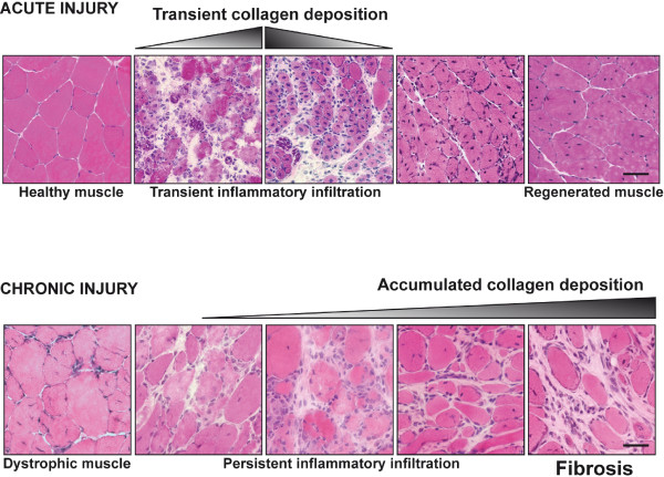
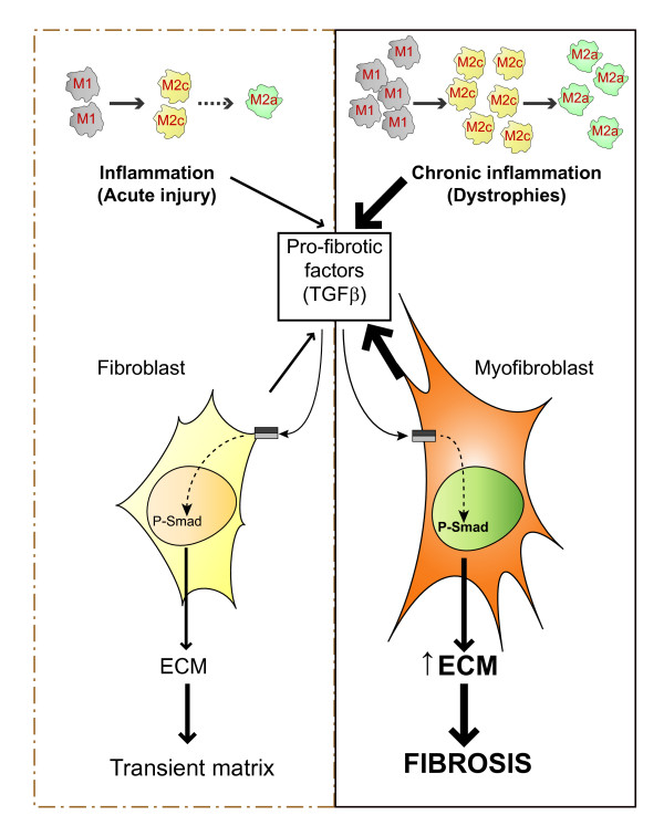
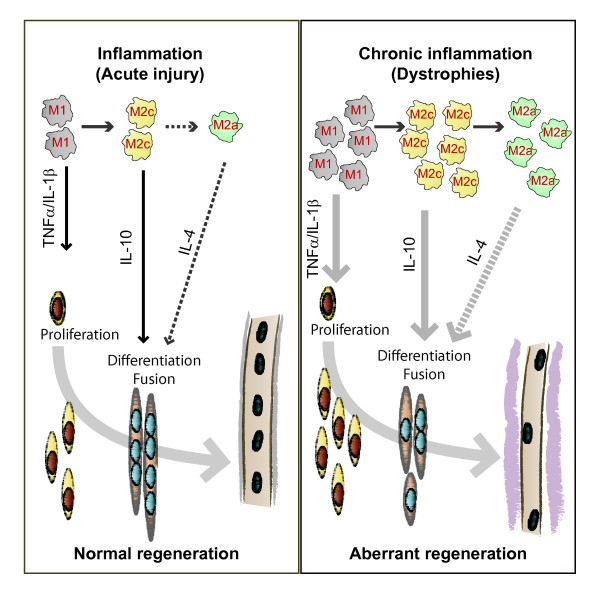
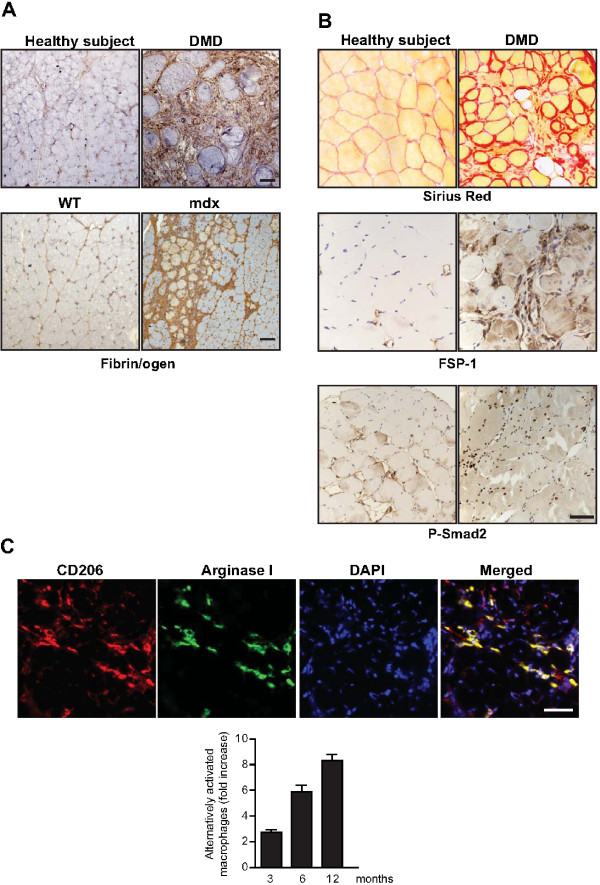
Verification
Claims the paper makes
The claims and statistics of this paper haven’t been checked yet.
Run the check — free, no account needed. It reads the open-access text and checks the sources it cites.
Claims other papers make about this one
Each is checked against this paper’s own text: does it say what the citing paper says it does?
In the citing paper
“Previous work has shown that the accumulation of inflammatory macrophages and age-related declines in interferon-gamma signaling delay muscle repair following injury”
From Skeletal muscle aging, cellular senescence, and senotherapeutics: current knowledge and future directions (2021) · cites this paper as Mann et al., 2011
Self-check
Percentages, sums and reported differences re-computed from the paper’s own numbers.
The self-check needs the paper’s full text.
Citations
Cites
1 retracted
RETRACTED ARTICLE: The role of nitric oxide during healing of trauma to the skeletal muscle
Filippin, L. I., Cuevas, M. J., Lima‐Cabello, E., Marroni, N. P., González‐Gallego, J., & Xavier, R. M. (2010) · Inflammation Research · doi:10.1007/s00011-010-0277-2 · Cited by 34 · cited 4×
“In addition, NO, which is released by local and invading cells during injury, can act as a pro-inflammatory molecule, and is able to activate MMP-2, which in turn releases HGF. Importantly, blockers of NOS function, such as nitro- Larginine methyl ester (L-NAME), have been shown to decrease the acute inflammatory reac…”
MethodInfluentialRetractedThe chemokine system in diverse forms of macrophage activation and polarization
MANTOVANI, A., SICA, A., SOZZANI, S., ALLAVENA, P., VECCHI, A., & LOCATI, M. (2004) · Trends in Immunology · doi:10.1016/j.it.2004.09.015 · Cited by 6,534 · cited 2×
“A nomenclature for polarized macrophages has been proposed [24,25] and they are now referred to as classically and alternatively activated macrophages, or M1 and M2 macrophages, respectively (Figure 2, Figure 3).”
BackgroundMechanisms of TGF-β Signaling from Cell Membrane to the Nucleus
Shi, Y., & Massagué, J. (2003) · Cell · doi:10.1016/s0092-8674(03)00432-x · Cited by 5,860
Visse, R., & Nagase, H. (2003) · Circulation Research · doi:10.1161/01.res.0000070112.80711.3d · Cited by 4,575 · cited 1×
“Activation requires proteolytic cleavage of the inactive precursor by either membrane-type (MT)1MMP [105] or plasmin, and cleavage of their corresponding inhibitors [106].”
Cellular and molecular mechanisms of fibrosis
Wynn, T. (2008) · The Journal of Pathology · doi:10.1002/path.2277 · Cited by 4,495
SATELLITE CELL OF SKELETAL MUSCLE FIBERS
Mauro, A. (1961) · The Journal of Cell Biology · doi:10.1083/jcb.9.2.493 · Cited by 3,683
Hinz, B., Phan, S. H., Thannickal, V. J., Galli, A., Bochaton-Piallat, M.-L., & Gabbiani, G. (2007) · The American Journal of Pathology · doi:10.2353/ajpath.2007.070112 · Cited by 2,068 · cited 1×
“These cells are associated with tissue repair and fibrosis in many tissues and organs, including muscle, skin, liver, lung, bone and cartilage [56].”
BackgroundArnold, L., Henry, A., Poron, F., Baba-Amer, Y., van Rooijen, N., Plonquet, A., Gherardi, R. K., & Chazaud, B. (2007) · The Journal of Experimental Medicine · doi:10.1084/jem.20070075 · Cited by 2,002 · cited 3×
“An important development in our understanding of muscle repair and fibrosis was the demonstration that a heterogeneous population of macrophages exists in regenerating muscle after injury, exhibiting opposing activities (either pro-inflammatory or anti-inflammatory) and different kinetics [23].”
BackgroundInfluentialFibrotic disease and the TH1/TH2 paradigm
Wynn, T. A. (2004) · Nature Reviews Immunology · doi:10.1038/nri1412 · Cited by 1,765
Muscle injury activates resident fibro/adipogenic progenitors that facilitate myogenesis
Joe, A. W. B., Yi, L., Natarajan, A., Le Grand, F., So, L., Wang, J., Rudnicki, M. A., & Rossi, F. M. V. (2010) · Nature Cell Biology · doi:10.1038/ncb2015 · Cited by 1,690 · cited 7×
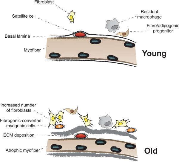
“Whether FAP conversion also occurs during aging, and whether Basal lamina ECM deposition Satellite cell Fibro/adipogenic progenitor Resident macrophage Fibroblast Increased number of fibroblasts Fibrogenic-converted myogenic cells Myofiber Atrophic myofiber Young Old Figure 5 Myofiber growth and extracellular matrix (…”
BackgroundInfluential
Cited by
Sorted by citations
Skeletal muscle performance and ageing
Tieland, M., Trouwborst, I., & Clark, B. C. (2018) · Journal of Cachexia, Sarcopenia and Muscle · doi:10.1002/jcsm.12238 · Cited by 904
“84 There is no direct evidence of muscle fi brosis with age in humans due to the dif fi culty in assessing fi brosis in population studies, but animal work indicated > 17% increase in fi brotic tissue in older rats as compared with younger rats.”
Lemos, D. R., Babaeijandaghi, F., Low, M., Chang, C.-K., Lee, S. T., Fiore, D., Zhang, R.-H., Natarajan, A., Nedospasov, S. A., & Rossi, F. M. V. (2015) · Nature Medicine · doi:10.1038/nm.3869 · Cited by 740
Macrophages in inflammation, repair and regeneration
Oishi, Y., & Manabe, I. (2018) · International Immunology · doi:10.1093/intimm/dxy054 · Cited by 770
Skeletal muscle: A review of molecular structure and function, in health and disease
Mukund, K., & Subramaniam, S. (2019) · WIREs Systems Biology and Medicine · doi:10.1002/wsbm.1462 · Cited by 805
“This uncontrolled and irreversible ECM expansion accompanied by an accumulation of ECM due to inhibited degradation (turnover), results in a fibrotic phenotype within muscle (Mann et al., 2011), especially in chronic diseases such as dystrophinopathies (Serrano & Muñoz-Cánoves, 2017).”
BackgroundMcGregor, R. A., Cameron-Smith, D., & Poppitt, S. D. (2014) · Longevity & Healthspan · doi:10.1186/2046-2395-3-9 · Cited by 532
Collagen VI regulates satellite cell self-renewal and muscle regeneration
Urciuolo, A., Quarta, M., Morbidoni, V., Gattazzo, F., Molon, S., Grumati, P., Montemurro, F., Tedesco, F. S., Blaauw, B., Cossu, G., et al. (2013) · Nature Communications · doi:10.1038/ncomms2964 · Cited by 511
“Muscle substitution by fibrotic connective tissue is a common feature of several conditions, such as aging and Duchenne muscular dystrophy, pointing at a negative effect for excessive production of fibrillar collagens and interstitial ECM components 32 .”
Allen, D. G., Whitehead, N. P., & Froehner, S. C. (2015) · Physiological Reviews · doi:10.1152/physrev.00007.2015 · Cited by 446
Cellular dynamics in the muscle satellite cell niche
Bentzinger, C. F., Wang, Y. X., Dumont, N. A., & Rudnicki, M. A. (2013) · The EMBO Reports · doi:10.1038/embor.2013.182 · Cited by 398
“Notably, the attenuation of muscle repair in most forms of muscular dystrophy is correlated with a build-up of fibrotic scarring, adipose tissue and immune infiltrations [97].”
von Maltzahn, J., Chang, N. C., Bentzinger, C. F., & Rudnicki, M. A. (2012) · Trends in Cell Biology · doi:10.1016/j.tcb.2012.07.008 · Cited by 408
“Fibrosis is often associated with muscular dystrophies, which is thought to contribute to disease pathology [51].”
Skeletal Muscle as an Endocrine Organ: The Role of Myokines in Exercise Adaptations
Hoffmann, C., & Weigert, C. (2017) · Cold Spring Harbor Perspectives in Medicine · doi:10.1101/cshperspect.a029793 · Cited by 393
“TGF-b is an inhibitor of myogenic differentiation (Massague et al. 1986) and plays an important role in inflammatory process after muscle injury and fibrosis (Mann et al. 2011).”
Fibronectin Regulates Wnt7a Signaling and Satellite Cell Expansion
Bentzinger, C. F., Wang, Y. X., von Maltzahn, J., Soleimani, V. D., Yin, H., & Rudnicki, M. A. (2013) · Cell Stem Cell · doi:10.1016/j.stem.2012.09.015 · Cited by 386
“Fibrosis is generally considered to impede satellite cell function and muscle repair (Mann et al., 2011).”
BackgroundBao, H., Cao, J., Chen, M., Min, C., Chen, W., Chen, X., Chen, Y., Chen, Y., Chen, Y., Chen, Z., et al. (2023) · Science China Life Sciences · doi:10.1007/s11427-023-2305-0 · Cited by 402
Fry, C. S., Kirby, T. J., Kosmac, K., McCarthy, J. J., & Peterson, C. A. (2017) · Cell stem cell · doi:10.1016/j.stem.2016.09.010 · Cited by 372
“Under clinical conditions characterized by reduced satellite cell activity, such as muscular dystrophy and aging, excessive fibrosis occurs, which, in turn, impairs muscle adaptation (Alnaqeeb et al., 1984; Brack et al., 2007; Mann et al., 2011; Stedman et al., 1991).”
Lukjanenko, L., Karaz, S., Stuelsatz, P., Gurriaran-Rodriguez, U., Michaud, J., Dammone, G., Sizzano, F., Mashinchian, O., Ancel, S., Migliavacca, E., et al. (2019) · Cell stem cell · doi:10.1016/j.stem.2018.12.014 · Cited by 374
“Several inflammatory processes have been implicated in fibrosis in skeletal muscle (Mann et al., 2011).”
BackgroundNew twist on artificial muscles
Haines, C. S., Li, N., Spinks, G. M., Aliev, A. E., Di, J., & Baughman, R. H. (2016) · Proceedings of the National Academy of Sciences · doi:10.1073/pnas.1605273113 · Cited by 365
Skeletal muscle fibrosis: an overview
Mahdy, M. A. A. (2018) · Cell and Tissue Research · doi:10.1007/s00441-018-2955-2 · Cited by 320
“The quiescent satellite cells (SCs) activate, proliferate and differentiate forming new myotubes with production of new ECM, blood vessels and nerves (Laumonier and Menetrey 2016; Mann et al. 2011; Saclier et al. 2013; Yin et al. 2013).”
BackgroundInfluentialA Muscle Stem Cell Support Group: Coordinated Cellular Responses in Muscle Regeneration
Wosczyna, M. N., & Rando, T. A. (2018) · Developmental Cell · doi:10.1016/j.devcel.2018.06.018 · Cited by 363
Lukjanenko, L., Jung, M. J., Hegde, N., Perruisseau-Carrier, C., Migliavacca, E., Rozo, M., Karaz, S., Jacot, G. E., Schmidt, M., Li, L., et al. (2016) · Nature Medicine · doi:10.1038/nm.4126 · Cited by 347
Collins, K. H., Herzog, W., MacDonald, G. Z., Reimer, R. A., Rios, J. L., Smith, I. C. P., Zernicke, R. F., & Hart, D. A. (2018) · Frontiers in Physiology · doi:10.3389/fphys.2018.00112 · Cited by 327
“the presence of low-level inflammation, which may result in increased lipid and fibrosis deposition, andmay limit reversibility of fibrosis (Mann et al., 2011).”
BackgroundInfluentialLieber, R. L., & Ward, S. R. (2013) · American Journal of Physiology-Cell Physiology · doi:10.1152/ajpcell.00173.2013 · Cited by 315
“TGF 1 is released from injured muscle fibers or, in an autocrine fashion, from fibers exposed to TGF 1 (71) and accompanies muscle inflammation (78), whereby inflammatory cells can deposit this cytokine.”
BackgroundMacrophage Plasticity and the Role of Inflammation in Skeletal Muscle Repair
Kharraz, Y., Guerra, J., Mann, C. J., Serrano, A. L., & Muñoz‐Cánoves, P. (2013) · Mediators of Inflammation · doi:10.1155/2013/491497 · Cited by 325
“Although several studies have shown that satellite cell-derived myoblasts may synthesize many components of the ECM, the major matrix-producing cell is the �broblast [9].”
BackgroundMuscle injuries and strategies for improving their repair
Laumonier, T., & Ménétrey, J. (2016) · Journal of Experimental Orthopaedics · doi:10.1186/s40634-016-0051-7 · Cited by 297
“TGF-β1, by stimulating fibroblasts/myofibroblasts to produce extracellular proteins such as fibronectin and type I/III collagen, has been identified as the key element in this process (Mann et al. 2011),.”
BackgroundHaemers, P., Hamdi, H., Guedj, K., Suffee, N., Farahmand, P., Popović, N., Claus, P., Leprince, P. N., Nicoletti, A., Jalife, J., et al. (2015) · European Heart Journal · doi:10.1093/eurheartj/ehv625 · Cited by 267
“–22 For instance, after chronic injury, skeletal muscle is replaced by a mix of fibrous tissue and white adipocytes.(21,23) In obese patients, fibrosis of the adipose tissue is a hallmark of disease progression.”
BackgroundForcina, L., Cosentino, M., & Musarò, A. (2020) · Cells · doi:10.3390/cells9051297 · Cited by 282
Understanding the Process of Fibrosis in Duchenne Muscular Dystrophy
Kharraz, Y., Guerra, J., Pessina, P., Serrano, A. L., & Muñoz‐Cánoves, P. (2014) · BioMed Research International · doi:10.1155/2014/965631 · Cited by 270
“cells and factors in the development of not only fibrosis arising during aberrant regeneration and DMD progression, but in other diseases such as diabetes and in aging (reviewed in [8]).”
BackgroundThe effects of obesity on skeletal muscle regeneration
Akhmedov, D., & Berdeaux, R. (2013) · Frontiers in Physiology · doi:10.3389/fphys.2013.00371 · Cited by 241
“In skeletal muscle, inflammation is activated after injury and is coordinated with myogenic differentiation to achieve efficient muscle regeneration (reviewed in Mann et al., 2011; Kharraz et al., 2013).”
BackgroundCosta, A., Toschi, A., Murfuni, I., Pelosi, L., Sica, G., Adamo, S., & Scicchitano, B. M. (2014) · BioMed Research International · doi:10.1155/2014/235426 · Cited by 248
“However, if inflammatory cell infiltration and fibroblast activation persist, an aberrant tissue repair response will produce a nonfunctionalmass of fibrotic tissue [41].”
BackgroundInfiltration of intramuscular adipose tissue impairs skeletal muscle contraction
Biltz, N. K., Collins, K. H., Shen, K. C., Schwartz, K., Harris, C. A., & Meyer, G. A. (2020) · The Journal of Physiology · doi:10.1113/jp279595 · Cited by 214
“This may reflect the complex role of the extracellular matrix in muscle contraction, as it has both an important physiological function in force transmission and healing and a pathological role in the development of stiffness and contracture (Mann et al. 2011).”
BackgroundThe genetic basis of pectoralis major myopathies in modern broiler chicken lines
Bailey, R. A., Watson, K., Bilgili, S. F., & Avendaño, S. (2015) · Poultry Science · doi:10.3382/ps/pev304 · Cited by 194
“Satellite cell activity is greatest immediately after hatch, but declines as the birds age, which potentially could have an impact on their ability to support growth of muscle fibers (Harthan et al., 2013) and/or their repair (Mann et al., 2011).”
BackgroundSharples, A. P., Stewart, C. E., & Seaborne, R. A. (2016) · Aging Cell · doi:10.1111/acel.12486 · Cited by 180
“…vs. skeletal muscle tissue accretion in the foetal skeletal muscle (Tong et al., 2009), a hall mark of sarcopenia, and one of the fundamental causes of reduced muscle force and quality (force per cross-sectional area of muscle) (reviewed in Serrano & Munoz-Canoves, 2010; Mann et al., 2011).”
BackgroundChin, L., Xia, Y., Discher, D. E., & Janmey, P. A. (2016) · Current Opinion in Chemical Engineering · doi:10.1016/j.coche.2016.01.011 · Cited by 181
Principles of Cell Circuits for Tissue Repair and Fibrosis
Adler, M., Mayo, A., Zhou, X., Franklin, R. A., Meizlish, M. L., Medzhitov, R., Kallenberger, S. M., & Alon, U. (2020) · iScience · doi:10.1016/j.isci.2020.100841 · Cited by 177
Liu, L., Charville, G. W., Cheung, T. H., Yoo, B., Santos, P. J., Schroeder, M., & Rando, T. A. (2018) · Cell Stem Cell · doi:10.1016/j.stem.2018.08.019 · Cited by 156
“In aging animals, muscle regeneration is often delayed and accompanied by increased fibrosis and adipogenesis (Conboy et al., 2005; Mann et al., 2011).”
Renin‐Angiotensin System: An Old Player with Novel Functions in Skeletal Muscle
Cabello‐Verrugio, C., Morales, M. G., Rivera, J. C. A., Cabrera, D., & Simón, F. (2015) · Medicinal Research Reviews · doi:10.1002/med.21343 · Cited by 170
Acuña, M. J., Pessina, P., Olguín, H. C., Cabrera, D., Vio, C. P., Bäder, M., Muñoz‐Cánoves, P., Santos, R. A. S., Cabello‐Verrugio, C., & Brandan, E. (2013) · Human Molecular Genetics · doi:10.1093/hmg/ddt514 · Cited by 162
“Acting via the Mas receptor, angiotensin-1-7 [Ang-(1-7)], is part of the renin–angiotensin system, with the opposite effect to that of angiotensin II.”
BackgroundInfluentialPerandini, L. A., Chimin, P., Lutkemeyer, D. D. S., & Câmara, N. O. S. (2018) · FEBS Journal · doi:10.1111/febs.14417 · Cited by 167
Therapeutic strategies for preventing skeletal muscle fibrosis after injury
Garg, K., Corona, B. T., & Walters, T. (2015) · Frontiers in Pharmacology · doi:10.3389/fphar.2015.00087 · Cited by 151
“…and FGF-2 released from neutrophils, macrophages, fibroblasts and myogenic precursors can promote fibrosis, the most pro-fibrogenic growth factor identified in the literature is TGF-β1 (Sheppard, 2006; Serrano andMunoz-Canoves, 2010; Burks and Cohn, 2011; Mann et al., 2011; Serrano et al., 2011).”
BackgroundPanduro, M., Benoist, C. O., & Mathis, D. J. (2018) · Proceedings of the National Academy of Sciences · doi:10.1073/pnas.1800618115 · Cited by 164
“7G) on day 7 of regeneration, which are indicators of ineffective muscle repair (26).”
BackgroundIncreased Stiffness in Aged Skeletal Muscle Impairs Muscle Progenitor Cell Proliferative Activity
Lacraz, G., Rouleau, A.-J., Couture, V., Söllrald, T., Drouin, G., Veillette, N., Grandbois, M., & Grenier, G. (2015) · PLoS ONE · doi:10.1371/journal.pone.0136217 · Cited by 156
“Notably, excessive collagen deposition in muscle leads to fibrosis, a major feature of aging and dystrophic/reparative disorders [44–47].”
BackgroundOwens, D. J., Sharples, A. P., Polydorou, I., Alwan, N., Donovan, T. F., TANG, J. C. Y., Fraser, W. D., Cooper, R. G., Morton, J. P., Stewart, C. E., et al. (2015) · American Journal of Physiology-Endocrinology and Metabolism · doi:10.1152/ajpendo.00375.2015 · Cited by 156
“Effective repair usually leads to full functional recovery of the damaged tissue, whereas ineffective repair leads to fibrosis and suboptimal rescue of function, such as in severe muscular dystrophies (32).”
BackgroundSanta, F. D., Vitiello, L., Torcinaro, A., & Ferraro, E. (2018) · Antioxidants and Redox Signaling · doi:10.1089/ars.2017.7420 · Cited by 157
“…promote phagocytosis of necrotic fibers and debris, sustain inflammation, and support activation and proliferation of SCs during early regeneration; whereas M2 promote the resolution of inflammation and enhance muscle regeneration by inducing muscle progenitor cells (MPCs) differentiation (194, 291).”
Coulis, G., Jaime, D., Guerrero-Juarez, C., Kastenschmidt, J. M., Farahat, P. K., Nguyen, Q., Pervolarakis, N., McLinden, K., Thurlow, L., Movahedi, S., et al. (2023) · Science Advances · doi:10.1126/sciadv.add9984 · Cited by 150
Contreras, O., Rebolledo, D. L., Oyarzún, J. E., Olguín, H. C., & Brandan, E. (2016) · Cell and Tissue Research · doi:10.1007/s00441-015-2343-0 · Cited by 154
Intramuscular mechanisms of overtraining
Cheng, A. J., Jude, B., & Lanner, J. T. (2020) · Redox Biology · doi:10.1016/j.redox.2020.101480 · Cited by 176
“TGF-β1 can be secreted by macrophages and is known to induce pathological fibrosis [110,111], but other cells e.”
BackgroundWasting mechanisms in muscular dystrophy
Shin, J. S., Tajrishi, M. M., Ogura, Y., & Kumar, A. (2013) · The International Journal of Biochemistry & Cell Biology · doi:10.1016/j.biocel.2013.05.001 · Cited by 151
Reducing CTGF/CCN2 slows down mdx muscle dystrophy and improves cell therapy
Morales, M. G., Gutiérrez, J., Cabello‐Verrugio, C., Cabrera, D., Lipson, K. E., Goldschmeding, R., & Brandan, E. (2013) · Human Molecular Genetics · doi:10.1093/hmg/ddt352 · Cited by 152
Role of Transforming Growth Factor-β in Skeletal Muscle Fibrosis: A Review
Ismaeel, A., Kim, J., Kirk, J. S., SMITH, R. S., Bohannon, W. T., & Koutakis, P. (2019) · International Journal of Molecular Sciences · doi:10.3390/ijms20102446 · Cited by 153
Sugg, K. B., Lubardic, J., Gumucio, J. P., & Mendias, C. L. (2014) · Journal of Orthopaedic Research® · doi:10.1002/jor.22624 · Cited by 143
“Previous work in damaged skeletal muscle has demonstrated that suppression of either M1 or M2 macrophage function severely impairs tissue regeneration, indicating the important balance between inflammation and regeneration for proper tissue repair.(11) Fibroblasts are the predominant cell type in tendons and are respo…”
BackgroundMuire, P. J., Mangum, L. H., & Wenke, J. C. (2020) · Frontiers in Immunology · doi:10.3389/fimmu.2020.01056 · Cited by 152
“Instead, there is elevated deposition of ECM components at the injury site, which inhibits myofiber formation and leads to replacement of muscle with fibrotic/scar tissue, a hallmark of fibrosis (38).”
BackgroundThéret, M., Rossi, F. M. V., & Contreras, O. (2021) · Frontiers in Physiology · doi:10.3389/fphys.2021.673404 · Cited by 154
Pericytes: multitasking cells in the regeneration of injured, diseased, and aged skeletal muscle
Birbrair, A., Zhang, T., Wang, Z., Messi, M. L., Mintz, A., & Delbono, O. (2014) · Frontiers in Aging Neuroscience · doi:10.3389/fnagi.2014.00245 · Cited by 145
“However, the analyzed samples were unstained, so the technique could not precisely distinguish pericytes from other surrounding cells.”
BackgroundBier, A., Berenstein, P., Kronfeld, N., Morgoulis, D., Ziv-Av, A., Goldstein, H., Kazimirsky, G., Cazacu, S., Meir, R., Popovtzer, R., et al. (2018) · Biomaterials · doi:10.1016/j.biomaterials.2018.04.055 · Cited by 141
The Basis of Muscle Regeneration
Musarò, A. (2014) · Advances in Biology · doi:10.1155/2014/612471 · Cited by 149
“The remodeling of connective tissue and angiogenesis defines the fourth stage of the regenerative process [44, 150, 152, 153] (Figure 2).”
BackgroundSkeletal muscle fibroblasts in health and disease
Chapman, M., Meza, R. C., & Lieber, R. L. (2016) · Differentiation · doi:10.1016/j.diff.2016.05.007 · Cited by 143
“It has even been suggested that myofibroblasts may not exist at all in skeletal muscle, instead they could be mature fibroblasts expressing ECM proteins (Mann et al., 2011).”
BackgroundInfluentialUnwavering Pathobiology of Volumetric Muscle Loss Injury
Greising, S. M., Rivera, J. C., Goldman, S. M., Watts, A., Aguilar, C. A., & Corona, B. T. (2017) · Scientific Reports · doi:10.1038/s41598-017-13306-2 · Cited by 136
Multiscale analysis of a regenerative therapy for treatment of volumetric muscle loss injury
Aguilar, C. A., Greising, S. M., Watts, A., Goldman, S. M., Peragallo, C., Zook, C., Larouche, J. A., & Corona, B. T. (2018) · Cell Death Discovery · doi:10.1038/s41420-018-0027-8 · Cited by 136
The Muscle Stem Cell Niche in Health and Disease
Mashinchian, O., Pisconti, A., Moal, E. L., & Bentzinger, C. F. (2017) · Current topics in developmental biology/Current Topics in Developmental Biology · doi:10.1016/bs.ctdb.2017.08.003 · Cited by 140
Contreras, O., Cruz-Soca, M., Théret, M., Soliman, H. S., Tung, L. W., Groppa, E., Rossi, F. M. V., & Brandan, E. (2019) · Journal of Cell Science · doi:10.1242/jcs.232157 · Cited by 143
“Both TGF-β and PDGF signaling pathways are known regulators of mesenchymal cell migration and invasion (Hoch and Soriano, 2003; Mann et al., 2011).”
BackgroundIntronic polyadenylation of PDGFRα in resident stem cells attenuates muscle fibrosis
Mueller, A. A., van Velthoven, C. T. J., Fukumoto, K. D., Cheung, T. H., & Rando, T. A. (2016) · Nature · doi:10.1038/nature20160 · Cited by 130
Smith, L., & Barton, E. R. (2014) · American Journal of Physiology-Cell Physiology · doi:10.1152/ajpcell.00383.2013 · Cited by 127
González, D., Contreras, O., Rebolledo, D. L., Espinoza, J. P., van Zundert, B., & Brandan, E. (2017) · PLoS ONE · doi:10.1371/journal.pone.0177649 · Cited by 128
Human skeletal muscle fibroblasts stimulate in vitro myogenesis and in vivo muscle regeneration
Mackey, A. L., Magnan, M., Chazaud, B., & Kjær, M. (2017) · The Journal of Physiology · doi:10.1113/jp273997 · Cited by 127
“The timely resolution of this transient matrix is important for successful restoration of the muscle and for avoiding chronic injury situations where fibrotic and adipogenic activity dominate (Mann et al. 2011).”
BackgroundCollins, K. H., Paul, H. A., Hart, D. A., Reimer, R. A., Smith, I. C., Rios, J. L., Seerattan, R. A., & Herzog, W. (2016) · Scientific Reports · doi:10.1038/srep37278 · Cited by 125
Wang, Y. X., Dumont, N. A., & Rudnicki, M. A. (2014) · Journal of Cell Science · doi:10.1242/jcs.151209 · Cited by 129
“Most notably, the use of anti-inflammatory and anti-fibrosis drugs in muscular dystrophy normalises the muscle stem cell niche and stimulates regeneration (Mann et al., 2011; Manzur et al., 1996).”
BackgroundClinical Relevance of Fascial Tissue and Dysfunctions
Klingler, W., Velders, M., Hoppe, K., Pedro, M., & Schleip, R. (2014) · Current Pain and Headache Reports · doi:10.1007/s11916-014-0439-y · Cited by 122
Dingal, P. C. D. P., Bradshaw, A. M., Cho, S., Raab, M., Buxboim, A., Swift, J., & Discher, D. E. (2015) · Nature Materials · doi:10.1038/nmat4350 · Cited by 127
“34 .”
Capote, J. C., Kramerova, I. A., Martinez, L., Vetrone, S. A., Barton, E. R., Sweeney, H. L., Miceli, M. C., & Spencer, M. J. (2016) · The Journal of Cell Biology · doi:10.1083/jcb.201510086 · Cited by 124
“Macrophages that infiltrate dystrophic muscles are a heterogeneous mix of classically (M1) and alternatively (M2) activated types (Villalta et al., 2009; Mann et al., 2011).”
BackgroundEngineered skeletal muscles for disease modeling and drug discovery
Wang, J., Khodabukus, A., Rao, L., VanDusen, K. W., Abutaleb, N. O., & Bursac, N. (2019) · Biomaterials · doi:10.1016/j.biomaterials.2019.119416 · Cited by 125
Molecular mechanisms of skeletal muscle development, regeneration, and osteogenic conversion
Endo, T. (2015) · Bone · doi:10.1016/j.bone.2015.02.028 · Cited by 117
“In skeletal muscle of chronic degenerative muscle diseases such as muscular dystrophies, fibrosis and adipogenesis (fatty degeneration) as well asmyofiber destruction become prominent, and consequentlymuscle functions deteriorate [149,150].”
BackgroundPAI-1–regulated miR-21 defines a novel age-associated fibrogenic pathway in muscular dystrophy
Ardite, E., Perdiguero, E., Vidal, B., Gutarra, S., Serrano, A. L., & Muñoz‐Cánoves, P. (2012) · The Journal of Cell Biology · doi:10.1083/jcb.201105013 · Cited by 118
“Fibroblasts were detected using the antibodies rabbit polyclonal fibroblast-specific protein–1 (FSP-1; Abcam) and rabbit polyclonal TCF-4 (Cell Signaling Technology; Okada et al., 1997; Zeng and Cullen, 2003; Mathew et al., 2011; Murphy et al., 2011).”
MethodAlameddine, H. S., & Morgan, J. E. (2016) · Journal of Neuromuscular Diseases · doi:10.3233/jnd-160183 · Cited by 122
Six1 regulates stem cell repair potential and self-renewal during skeletal muscle regeneration
Grand, F. L., Grifone, R., Mourikis, P., Houbron, C., Gigaud, C., Pujol, J., Maillet, M., Pagès, G., Rudnicki, M. A., Tajbakhsh, S., et al. (2012) · The Journal of Cell Biology · doi:10.1083/jcb.201201050 · Cited by 113
“Unsuccessful repair after tissue damage leads to the derangement of normal structure, and an increased development of fibroblasts during the regeneration process was previously linked with defects in the myogenic process (Mann et al., 2011).”
Satellite cells from dystrophic muscle retain regenerative capacity
Boldrin, L., Zammit, P. S., & Morgan, J. E. (2014) · Stem Cell Research · doi:10.1016/j.scr.2014.10.007 · Cited by 112
Li, H., Hicks, J. J., Wang, L., Oyster, N., Philippon, M. J., Hurwitz, S. R., Hogan, M. V., & Huard, J. (2016) · Biomaterials · doi:10.1016/j.biomaterials.2016.02.017 · Cited by 111
αv integrins on mesenchymal cells regulate skeletal and cardiac muscle fibrosis
Murray, I. R., Gonzalez-Galofre, Z. N., Baily, J., Dobie, R., Wallace, R. J., MacKinnon, A. C., Smith, J. R., Greenhalgh, S. N., Thompson, A., Conroy, K. P., et al. (2017) · Nature Communications · doi:10.1038/s41467-017-01097-z · Cited by 108
Effects of Low-Level Laser Therapy on Skeletal Muscle Repair
Alves, A. N., Fernandes, K. P. S., Deana, A. M., Bussadori, S. K., & Mesquita‐Ferrari, R. A. (2014) · American Journal of Physical Medicine & Rehabilitation · doi:10.1097/phm.0000000000000158 · Cited by 109
“Effect of LLLT on Inflammation The inflammatory phase is characterized by the degeneration of muscle fibers, the recruitment of neutrophils and macrophages to the injury site, as well as the production of inflammatory cytokines (tumor necrosis factor alpha [TNF->], interleukin 1 beta [IL-1A], IL-6, and IL-8) and prote…”
BackgroundInfluentialMuscle injury: review of experimental models.
de Souza, J. B., & Gottfried, C. (2013) · Journal of Electromyography & Kinesiology · doi:10.1016/j.jelekin.2013.07.009 · Cited by 115
“Fibroblasts play an important role in muscle tissue repair by secreting extracellular matrix proteins including: collagen types I and III, fibronectin, elastin, proteoglycans, laminin and growth factors (Li et al., 2008; Mann et al., 2011).”
BackgroundHydrogel biomaterials and their therapeutic potential for muscle injuries and muscular dystrophies
Lev, R., & Seliktar, D. (2018) · Journal of The Royal Society Interface · doi:10.1098/rsif.2017.0380 · Cited by 105
Skeletal and cardiac muscle pericytes: Functions and therapeutic potential
Murray, I. R., Baily, J. E., Chen, W. C. W., Dar, A., Gonzalez-Galofre, Z. N., Jensen, A. R., Petrigliano, F. A., Deb, A., & Henderson, N. C. (2016) · Pharmacology & Therapeutics · doi:10.1016/j.pharmthera.2016.09.005 · Cited by 107
“Fibrosis secondary to acute skeletal muscle injury results in significant functional impairment and predisposes to further injury (Mann et al., 2011; Uezumi et al., 2014).”
BackgroundLoreti, M., & Sacco, A. (2022) · npj Regenerative Medicine · doi:10.1038/s41536-022-00204-z · Cited by 111
Crosstalk Between Skeletal Muscle and Immune System: Which Roles Do IL-6 and Glutamine Play?
Rogeri, P. S., Gasparini, S. O., Martins, G. L., Costa, L. K. F., Araujo, C. C., Lugaresi, R., Kopfler, M., & Lancha, A. H. (2020) · Frontiers in Physiology · doi:10.3389/fphys.2020.582258 · Cited by 108
“This initial increase on the FAPs is essential for the production of the extracellular matrix components in order to stabilize the tissue, acting as a scaffolding for new fibers, being used by the satellite cells as a basal membrane to assure that the myofibers will remain aligned (Chen and Li, 2009; Mann et al., 2011…”
BackgroundInfluentialFibrogenic Cell Plasticity Blunts Tissue Regeneration and Aggravates Muscular Dystrophy
Pessina, P., Kharraz, Y., Jardı́, M., Fukada, S., Serrano, A. L., Perdiguero, E., & Muñoz‐Cánoves, P. (2015) · Stem Cell Reports · doi:10.1016/j.stemcr.2015.04.007 · Cited by 101
“…dystrophin, successive cycles of tissue degeneration and regeneration lead to an eventual muscle regenerative failure and replacement of dystrophic muscle by fibrotic tissue, resulting in respiratory failure and early death (Mann et al., 2011; Stedman et al., 1991; Wallace and McNally, 2009).”
InfluentialEffect of Aging on Tendon Biology, Biomechanics and Implications for Treatment Approaches
Kwan, K. Y. C., Ng, K. W., Rao, Y., Zhu, C., Qi, S., Tuan, R. S., Ker, D. F. E., & Wang, D. M. (2023) · International Journal of Molecular Sciences · doi:10.3390/ijms242015183 · Cited by 101
“Yet, its expansion has also been associated with excessive tissue scarring [141].”
BackgroundNovel and optimized strategies for inducing fibrosis in vivo: focus on Duchenne Muscular Dystrophy
Pessina, P., Cabrera, D., Morales, M. G., Riquelme, C. A., Gutiérrez, J., Serrano, A. L., Brandan, E., & Muñoz‐Cánoves, P. (2014) · Skeletal Muscle · doi:10.1186/2044-5040-4-7 · Cited by 100
On the anisotropy of skeletal muscle tissue under compression
Böl, M., Ehret, A. E., Leichsenring, K., Weichert, C., & Kruse, R. (2014) · Acta Biomaterialia · doi:10.1016/j.actbio.2014.03.003 · Cited by 99
Garg, K., Corona, B. T., & Walters, J. (2014) · Journal of Applied Physiology · doi:10.1152/japplphysiol.00689.2014 · Cited by 101
Induction of Murine Macrophage M2 Polarization by Cigarette Smoke Extract via the JAK2/STAT3 Pathway
Yuan, F., Fu, X., Shi, H., Chen, G., Dong, P., & Zhang, W. (2014) · PLoS ONE · doi:10.1371/journal.pone.0107063 · Cited by 97
“Macrophages also produce inflammatory factors that participate in various physiological and pathological processes [7].”
BackgroundIeronimakis, N. M., Hays, A. L., Prasad, A., Janebodin, K., Duffield, J. S., & Reyes, M. (2016) · The Journal of Pathology · doi:10.1002/path.4801 · Cited by 96
“Therefore, collagen-producing cells are not easily distinguishable from other cell types, such as inflammatory cells, that also occupy damaged tissue and influence fibrosis in disease or injury [23].”
BackgroundEnglund, D. A., Zhang, X., Aversa, Z., & LeBrasseur, N. K. (2021) · Mechanisms of Ageing and Development · doi:10.1016/j.mad.2021.111595 · Cited by 94
Fibronectin is a serum biomarker for D uchenne muscular dystrophy
Martin, F. C., Hiller, M., Spitali, P., Oonk, S., Dalebout, H., Palmblad, M., Chaouch, A., Guglieri, M., Straub, V. W., Lochmüller, H., et al. (2014) · PROTEOMICS - CLINICAL APPLICATIONS · doi:10.1002/prca.201300072 · Cited by 96
“Several studies showed that excessive deposition of fibronectin precedes collagen deposition [40, 41].”
BackgroundGe, J., Liu, K., Niu, W., Chen, M., Wang, M., Xue, Y., Gao, C., Ma, P. X., & Lei, B. (2018) · Biomaterials · doi:10.1016/j.biomaterials.2018.05.027 · Cited by 94
Striated muscle function, regeneration, and repair
Shadrin, I. Y., Khodabukus, A., & Bursac, N. (2016) · Cellular and Molecular Life Sciences · doi:10.1007/s00018-016-2285-z · Cited by 106
“Thus, dystrophic muscles are damaged more easily, resulting in a repetitive degeneration/regeneration cycles, eventual exhaustion of the SC pool, muscle loss, and fibrosis [9, 84].”
BackgroundSkeletal muscle fibrosis in pancreatic Cancer patients with respect to survival
Judge, S. M., Nosacka, R. L., Delitto, D., Gerber, M. H., Cameron, M. E., Trevino, J. G., & Judge, A. R. (2018) · JNCI Cancer Spectrum · doi:10.1093/jncics/pky043 · Cited by 91
“The long-term consequences of chronic muscle damage and impaired regeneration are well established and include non-resolute inflammation, proliferation of FAP cells, and progressive replacement of muscle fibers with both fat and fibrotic tissue (25,26).”
BackgroundSciorati, C., Clementi, E., Manfredi, A. A., & Rovere, P. Q. (2015) · Cellular and Molecular Life Sciences · doi:10.1007/s00018-015-1857-7 · Cited by 99
“SC Satellite cells MRF Myogenic regulatory factors MHC Myosin heavy chain MEF Myogenic enhancer factors EMC Extracellular matrix components MMP Matrix metalloproteinases PPARc2 Peroxisome proliferator-activated receptor c2 UCP1 Uncoupling protein 1 DMD Duchenne muscular dystrophy MRI Magnetic resonance imaging sca-1 S…”
BackgroundInfluentialInterstitial Cell Remodeling Promotes Aberrant Adipogenesis in Dystrophic Muscles
Camps, J., Breuls, N., Sifrim, A., Giarratana, N., Corvelyn, M., Danti, L., Grosemans, H., Vanuytven, S., Thiry, I., Belicchi, M., et al. (2020) · Cell Reports · doi:10.1016/j.celrep.2020.107597 · Cited by 90
“In chronic injury, regeneration deteriorates, leading to increased adipogenesis and fibrosis at the expense of the muscle fibers (Hamrick et al., 2016; Mann et al., 2011).”
BackgroundWhole‐body magnetic resonance imaging evaluation of facioscapulohumeral muscular dystrophy
Leung, D. G., Carrino, J. A., Wagner, K. R., & Jacobs, M. A. (2015) · Muscle & Nerve · doi:10.1002/mus.24569 · Cited by 89
Mitochondrial Function in Muscle Stem Cell Fates
Bhattacharya, D., & Scimè, A. (2020) · Frontiers in Cell and Developmental Biology · doi:10.3389/fcell.2020.00480 · Cited by 95
Effects of aging, exercise, and disease on force transfer in skeletal muscle
Hughes, D. C., Wallace, M. A., & Baar, K. (2015) · American Journal of Physiology-Endocrinology and Metabolism · doi:10.1152/ajpendo.00095.2015 · Cited by 84
Delineating the roles of neutrophils and macrophages in zebrafish regeneration models
Keightley, M.-C., Wang, C., Pazhakh, V., & Lieschke, G. J. (2014) · The International Journal of Biochemistry & Cell Biology · doi:10.1016/j.biocel.2014.07.010 · Cited by 82
Cellular senescence-mediated exacerbation of Duchenne muscular dystrophy
Sugihara, H., Teramoto, N., Nakamura, K., Shiga, T., Shirakawa, T., Matsuo, M., Ogasawara, M., Nishino, I., Matsuwaki, T., Nishihara, M., et al. (2020) · Scientific Reports · doi:10.1038/s41598-020-73315-6 · Cited by 85
Metadata
- Authors
- Mann, C. J., Perdiguero, E., Kharraz, Y., Aguilar, S., Pessina, P., Serrano, A. L., & Muñoz-Cánoves, P.
- Affiliations
- Universitat Pompeu Fabra (ES)
- Venue
- Skeletal Muscle
- Published
- 2011-05-04
- Volume
- 1
- Issue
- 1
- Pages
- 21-21
- ISSN
- 2044-5040 (ISSN-L)
- PMC
- PMC3156644
- OpenAlex
- W2076787014
- Cited by
- 853
- References
- 129
- Citations by year
- 2026: 37 · 2025: 59 · 2024: 55 · 2023: 53 · 2022: 59 · 2021: 71 · 2020: 81 · 2019: 85 · 2018: 76 · 2017: 51 · 2016: 68 · 2015: 48 · 2014: 49 · 2013: 33 · 2012: 27
- FWCI
- 14.30
- Type
- journal article
- Language
- en
- Open access
- gold
- doi.org
- Licence
- cc-by
- Locations
- Skeletal Muscle (publishedVersion, cc-by)
- Europe PMC (PubMed Central) (publishedVersion, cc-by)
- PubMed Central (publishedVersion, cc-by)
- PubMed (publishedVersion)
- RECERCAT (Consorci de Serveis Universitaris de Catalunya) (publishedVersion, other-oa)
- DOAJ (DOAJ: Directory of Open Access Journals) (submittedVersion, cc-by-sa)
- Repositori digital de la UPF (Universitat Pompeu Fabra) (submittedVersion, cc-by)
- Funders
- Ministerio de Ciencia e Innovación (PLE2009)
- Centro de Investigación Biomédica en Red sobre Enfermedades Neurodegenerativas
- Topics
- Life Sciences › Biochemistry, Genetics and Molecular Biology › Molecular Biology › Muscle Physiology and Disorders
- Health Sciences › Medicine › Cardiology and Cardiovascular Medicine › Cardiac Fibrosis and Remodeling
- Health Sciences › Medicine › Surgery › Wound Healing and Treatments
- Keywords
- muscle fibrosis, muscle repair, muscular dystrophy, satellite cells, muscle stem cells, fibroblast activation, inflammatory cell infiltration, extracellular matrix, wound healing, microenvironment
About these checks
Registry, retraction, Cites and statistics results are computed, not judged, with one exception: a printed reference that no index lists is read once by a model before it counts as a citation error. Claims are extracted by a model and kept only when their quote appears verbatim in the text; each cited source is then checked, with a model’s help, for whether it says what the claim relies on. Those verdicts can be wrong — read the claim’s quote and the cited source before relying on one.
Statistics and arithmetic are recomputed, not judged.
Confidence bands are shown only for fields calibrated on our annotated test set. None is calibrated yet, so no band is shown.
What counts as a claim ›How we measure confidence ›How we check this ›
Are you an author, or do you see a problem with this page? Request a review
