

The DOI matches its Crossref record; 215 of the 215 references it cites were found in a registry or an index.
Mechanisms of fluid movement into, through and out of the brain: evaluation of the evidence
Open accessAbstract
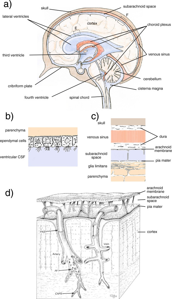
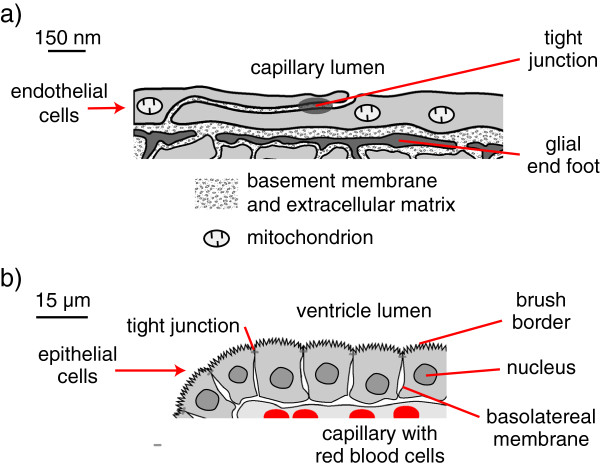
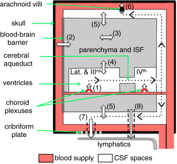
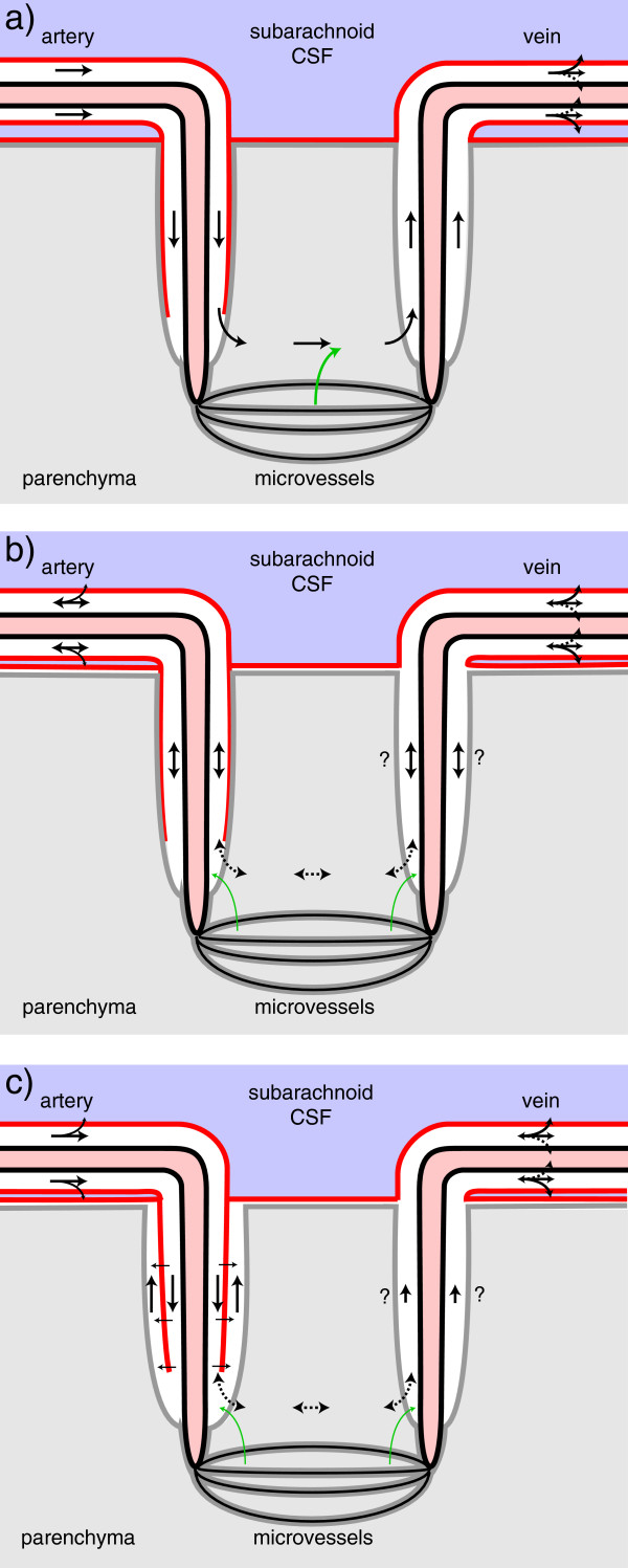
Interstitial fluid (ISF) surrounds the parenchymal cells of the brain and spinal cord while cerebrospinal fluid (CSF) fills the larger spaces within and around the CNS. Regulation of the composition and volume of these fluids is important for effective functioning of brain cells and is achieved by barriers that prevent free exchange between CNS and blood and by mechanisms that secrete fluid of controlled composition into the brain and distribute and reabsorb it. Structures associated with this regular fluid turnover include the choroid plexuses, brain capillaries comprising the blood-brain barrier, arachnoid villi and perineural spaces penetrating the cribriform plate. ISF flow, estimated from rates of removal of markers from the brain, has been thought to reflect rates of fluid secretion across the blood-brain barrier, although this has been questioned because measurements were made under barbiturate anaesthesia possibly affecting secretion and flow and because CSF influx to the parenchyma via perivascular routes may deliver fluid independently of blood-brain barrier secretion. Fluid secretion at the blood-brain barrier is provided by specific transporters that generate solute fluxes so creating osmotic gradients that force water to follow. Any flow due to hydrostatic pressures driving water across the barrier soon ceases unless accompanied by solute transport because water movements modify solute concentrations. CSF is thought to be derived primarily from secretion by the choroid plexuses. Flow rates measured using phase contrast magnetic resonance imaging reveal CSF movements to be more rapid and variable than previously supposed, even implying that under some circumstances net flow through the cerebral aqueduct may be reversed with net flow into the third and lateral ventricles. Such reversed flow requires there to be alternative sites for both generation and removal of CSF. Fluorescent tracer analysis has shown that fluid flow can occur from CSF into parenchyma along periarterial spaces. Whether this represents net fluid flow and whether there is subsequent flow through the interstitium and net flow out of the cortex via perivenous routes, described as glymphatic circulation, remains to be established. Modern techniques have revealed complex fluid movements within the brain. This review provides a critical evaluation of the data.
Overview
The claims and statistics of this paper haven’t been checked yet.
Run the check — free, no account needed. It reads the open-access text and checks the sources it cites.
Integrity signals
Registry
Cites
Statistics
Figures
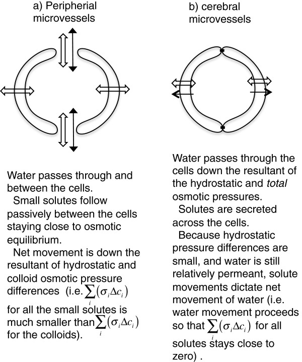
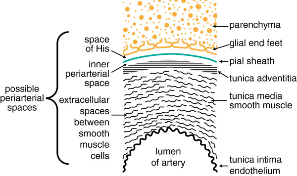
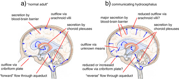
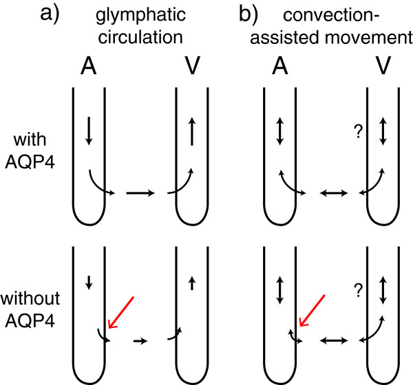
Verification
Claims the paper makes
The claims and statistics of this paper haven’t been checked yet.
Run the check — free, no account needed. It reads the open-access text and checks the sources it cites.
Claims other papers make about this one
Each is checked against this paper’s own text: does it say what the citing paper says it does?
The cited source says what the claim attributes to it.
In the citing paper
“Other recent studies (252, 255, 547, 555), however, did not support the proposed glymphatic mechanism, nor the convective, pressure-driven fluid flow of CSF from para-arterial to paravenous spaces throughout the parenchymal ECS (31, 30, 255, 296, 547, 255).”
From Blood-Brain Barrier: From Physiology to Disease and Back (2019) · cites this paper as [252]
Self-check
Percentages, sums and reported differences re-computed from the paper’s own numbers.
The self-check needs the paper’s full text.
Citations
Cites
Intravenous Fluids and Acute Kidney Injury
Ding, X., Cheng, Z., & Qian, Q. (2017) · Blood Purification · doi:10.1159/000452702 · Cited by 6,868
Iliff, J. J., Wang, M., Liao, Y., Plogg, B. A., Peng, W., Gundersen, G. A., Benveniste, H., Vates, G. E., Deane, R., Goldman, S. A., et al. (2012) · Science Translational Medicine · doi:10.1126/scitranslmed.3003748 · Cited by 5,824
Sleep Drives Metabolite Clearance from the Adult Brain
Xie, L., Kang, H., Xu, Q., Chen, M. J., Liao, Y., Thiyagarajan, M., O’Donnell, J., Christensen, D. J., Nicholson, C., Iliff, J. J., et al. (2013) · Science · doi:10.1126/science.1241224 · Cited by 5,411
FINE STRUCTURAL LOCALIZATION OF A BLOOD-BRAIN BARRIER TO EXOGENOUS PEROXIDASE
Reese, T. S., & Karnovsky, M. J. (1967) · The Journal of Cell Biology · doi:10.1083/jcb.34.1.207 · Cited by 2,629
JUNCTIONS BETWEEN INTIMATELY APPOSED CELL MEMBRANES IN THE VERTEBRATE BRAIN
Brightman, M., & Reese, T. S. (1969) · The Journal of Cell Biology · doi:10.1083/jcb.40.3.648 · Cited by 2,457
Thermodynamic analysis of the permeability of biological membranes to non-electrolytes
Kedem, O., & Katchalsky, A. (1958) · Biochimica et Biophysica Acta · doi:10.1016/0006-3002(58)90330-5 · Cited by 2,214
On the Absorption of Fluids from the Connective Tissue Spaces
Starling, E. H. (1896) · The Journal of Physiology · doi:10.1113/jphysiol.1896.sp000596 · Cited by 2,126
Diffusion in Brain Extracellular Space
Syková, E., & Nicholson, C. (2008) · Physiological Reviews · doi:10.1152/physrev.00027.2007 · Cited by 1,481
Convection-enhanced delivery of macromolecules in the brain.
Bobo, R. H., Laske, D. W., Akbaşak, A., Morrison, P. F., Dedrick, R. L., & Oldfield, E. H. (1994) · Proceedings of the National Academy of Sciences · doi:10.1073/pnas.91.6.2076 · Cited by 1,442
Effect of anoxia on ion distribution in the brain
Hansen, A. J. (1985) · Physiological Reviews · doi:10.1152/physrev.1985.65.1.101 · Cited by 1,342
Cited by
Sorted by citations
Items 201–300 of 636
Date, P., Ackermann, P., Furey, C. G., Fink, I. B., Jonas, S., Khokha, M. K., Kahle, K. T., & Deniz, E. (2019) · Scientific Reports · doi:10.1038/s41598-019-42549-4 · Cited by 40
Nedelcovych, M. T., Kim, B.-H., Zhu, X., Lovell, L. E., Manning, A. A., Kelschenbach, J., Hadas, E., Chao, W., Prchalová, E., Dash, R. P., et al. (2019) · Journal of Neuroimmune Pharmacology · doi:10.1007/s11481-019-09859-w · Cited by 41
Magnetic Resonance Imaging and Modeling of the Glymphatic System
Kaur, J., Davoodi‐Bojd, E., Fahmy, L. M., Zhang, L., Ding, G., Hu, J., Zhang, Z., Chopp, M., & Jiang, Q. (2020) · Diagnostics · doi:10.3390/diagnostics10060344 · Cited by 40
Sensitivity analysis on a network model of glymphatic flow
Stevens, K. A., Tithof, J., Cook, D. D., Thomas, J. H., & Kelley, D. H. (2022) · Journal of The Royal Society Interface · doi:10.1098/rsif.2022.0257 · Cited by 39
Choroid plexus perfusion and intracranial cerebrospinal fluid changes after angiogenesis
Johnson, S. E., McKnight, C. D., Lants, S. K., Juttukonda, M. R., Fusco, M. R., Chitale, R. V., Donahue, P. M. C., Claassen, D. O., & Donahue, M. J. (2019) · Journal of Cerebral Blood Flow & Metabolism · doi:10.1177/0271678x19872563 · Cited by 39
The glymphatic system and multiple sclerosis: An evolving connection
Alghanimy, A., Work, L. M., & Holmes, W. M. (2024) · Multiple Sclerosis and Related Disorders · doi:10.1016/j.msard.2024.105456 · Cited by 39
Enhanced in vitro model of the CSF dynamics
Benninghaus, A., Balédent, O., Lokossou, A., Wembers, C. C., Leonhardt, S. K., & Radermacher, K. (2019) · Fluids and Barriers of the CNS · doi:10.1186/s12987-019-0131-z · Cited by 38
Nizari, S., Wells, J. A., Carare, R. O., Romero, I. A., & Hawkes, C. A. (2021) · Acta Neuropathologica Communications · doi:10.1186/s40478-020-01108-z · Cited by 40
Tang, Y., & Cao, Y. (2021) · Pharmaceutics · doi:10.3390/pharmaceutics13030422 · Cited by 42
Coles, J. A., Stewart-Hutchinson, P. J., Myburgh, E., & Brewer, J. M. (2017) · Methods · doi:10.1016/j.ymeth.2017.03.020 · Cited by 40
Porro, G., Basile, M., Xie, Z., Tuveri, G. M., Battaglia, G., & Lopes, C. D. F. (2025) · Advanced Drug Delivery Reviews · doi:10.1016/j.addr.2025.115637 · Cited by 39
Transependymal Cerebrospinal Fluid Flow: Opportunity for Drug Delivery?
Casaca-Carreira, J., Temel, Y., Hescham, S., & Jahanshahi, A. (2017) · Molecular Neurobiology · doi:10.1007/s12035-017-0501-y · Cited by 39
Mechanisms Underlying Aquaporin-4 Subcellular Mislocalization in Epilepsy
Szu, J. I., & Binder, D. K. (2022) · Frontiers in Cellular Neuroscience · doi:10.3389/fncel.2022.900588 · Cited by 38
Enhanced interstitial fluid drainage in the hippocampus of spontaneously hypertensive rats
Bedussi, B., Naessens, D. M. P., de Vos, J., Engberink, R. O., Wilhelmus, M. M. M., Richard, E., ten Hove, M., VanBavel, E., & Bakker, E. N. T. P. (2017) · Scientific Reports · doi:10.1038/s41598-017-00861-x · Cited by 36
Fluorescence imaging of lymphatic outflow of cerebrospinal fluid in mice
Kwon, S., Janssen, C. F., Velasquez, F. C., & Sevick-Muraca, E. M. (2017) · Journal of Immunological Methods · doi:10.1016/j.jim.2017.06.010 · Cited by 36
Transport study of interleukin-1 inhibitors using a human in vitro model of the blood-brain barrier
Sjöström, E. O., Culot, M., Leickt, L., Åstrand, M., Nordling, E., Gosselet, F., & Kaiser, C. (2021) · Brain Behavior & Immunity - Health · doi:10.1016/j.bbih.2021.100307 · Cited by 37
Hypoxemia increases blood-brain barrier permeability during extreme apnea in humans
Bailey, D. M., Bain, A. R., Hoiland, R. L., Barak, O. F., Drvis, I., Hirtz, C., Lehmann, S., Marchi, N., Janigro, D., MacLeod, D. B., et al. (2022) · Journal of Cerebral Blood Flow & Metabolism · doi:10.1177/0271678x221075967 · Cited by 36
Hsieh, Y., Litvin, D. G., Zaylor, A. R., Nethery, D. E., Dick, T. E., & Jacono, F. J. (2020) · The Journal of Physiology · doi:10.1113/jp279177 · Cited by 37
Li, M., Wu, J., Li, N., Zhou, J., Cheng, W., Wu, A., Liu, L., & Jiao, N. (2024) · Advanced Functional Materials · doi:10.1002/adfm.202402333 · Cited by 35
Cerebrovascular heterogeneity and neuronal excitability
Librizzi, L., de Cutis, M., Janigro, D., Runtz, L., de Bock, F., Barbier, E., & Marchi, N. (2017) · Neuroscience Letters · doi:10.1016/j.neulet.2017.01.013 · Cited by 36
Rethinking the Conditions and Mechanism for Glymphatic Clearance
Ferris, C. F. (2021) · Frontiers in Neuroscience · doi:10.3389/fnins.2021.624690 · Cited by 36
Trillo-Contreras, J. L., Ramírez‐Lorca, R., Villadiego, J., & Echevarrı́a, M. (2022) · Biomolecules · doi:10.3390/biom12040530 · Cited by 35
Tarumi, T., Yamabe, T., Fukuie, M., Zhu, D. C., Zhang, R., Ogoh, S., & Sugawara, J. (2021) · The Journal of Physiology · doi:10.1113/jp281063 · Cited by 34
Liang, P.-Z., Li, L., Zhang, Y., Shen, Y. H., Zhang, L., Zhou, J., Wang, Z.-J., Wang, S., & Yang, S. (2021) · Neural Plasticity · doi:10.1155/2021/9960304 · Cited by 34
The role of the autonomic nervous system in cerebral blood flow regulation in dementia: A review
Beishon, L., Hosford, P. S., Gurung, D., Brassard, P., Minhas, J. S., Robinson, T. G., Haunton, V. J., & Panerai, R. B. (2022) · Autonomic Neuroscience · doi:10.1016/j.autneu.2022.102985 · Cited by 34
Ocular Lymphatic and Glymphatic Systems: Implications for Retinal Health and Disease
Uddin, M. N., & Rutar, M. (2022) · International Journal of Molecular Sciences · doi:10.3390/ijms231710139 · Cited by 35
Zhang, W., Neal, J. B., Lin, L. H., Dai, F., Hersey, D., McDonagh, D. L., Su, F., & Meng, L. (2018) · Journal of Neurosurgical Anesthesiology · doi:10.1097/ana.0000000000000520 · Cited by 33
Altered brain fluid management in a rat model of arterial hypertension
Naessens, D. M. P., Coolen, B. F., de Vos, J., VanBavel, E., Strijkers, G. J., & Bakker, E. N. T. P. (2020) · Fluids and Barriers of the CNS · doi:10.1186/s12987-020-00203-6 · Cited by 32
Liu, S., Bilston, L. E., Rodriguez, N. F., Wright, C. J., McMullan, S., Lloyd, R. A., Stoodley, M. A., & Hemley, S. J. (2022) · Fluids and Barriers of the CNS · doi:10.1186/s12987-022-00310-6 · Cited by 32
Metabolite Clearance During Wakefulness and Sleep
Hladky, S. B., & Barrand, M. A. (2017) · Handbook of experimental pharmacology · doi:10.1007/164_2017_37 · Cited by 36
Self-citationCerebral Waste Accumulation and Glymphatic Clearance as Mechanisms of Human Neurological Diseases
Janigro, D. (2016) · Journal of Neurology and Neuromedicine · doi:10.29245/2572.942x/2016/7.1082 · Cited by 32
Continuous intrathecal orexin delivery inhibits cataplexy in a murine model of narcolepsy
Kaushik, M. K., Aritake, K., Imanishi, A., Kanbayashi, T., Ichikawa, T., Shimizu, T., Urade, Y., & Yanagisawa, M. (2018) · Proceedings of the National Academy of Sciences · doi:10.1073/pnas.1722686115 · Cited by 34
Central nervous system lymphatic unit, immunity, and epilepsy: Is there a link?
Noè, F. M., & Marchi, N. (2019) · Epilepsia Open · doi:10.1002/epi4.12302 · Cited by 33
Channels and Transporters in Astrocyte Volume Regulation in Health and Disease
Reed, M., & Blazer‐Yost, B. L. (2022) · Cellular Physiology and Biochemistry · doi:10.33594/000000495 · Cited by 31
Soden, P. A., Henderson, A. R., & Lee, E. (2022) · Advanced Biology · doi:10.1002/adbi.202200027 · Cited by 35
Nikolenko, V. N., Oganesyan, M. V., Vovkogon, A. D., Nikitina, A. T., Sozonova, E. A., Kudryashova, V. A., Rizaeva, N. A., Cabezas, R., Ávila-Rodriguez, M., Евгеньевна, Н. М., et al. (2019) · Current Neuropharmacology · doi:10.2174/1570159x17666191113103850 · Cited by 31
Intrathecal delivery of Macromolecules: Clinical status and emerging technologies
Ajeeb, R., & Clegg, J. R. (2023) · Advanced Drug Delivery Reviews · doi:10.1016/j.addr.2023.114949 · Cited by 31
Circadian Mechanisms in Brain Fluid Biology
Vizcarra, V. S., Fame, R. M., & Hablitz, L. M. (2024) · Circulation Research · doi:10.1161/circresaha.123.323516 · Cited by 31
Development of a Multicomponent Intervention to Prevent Alzheimer's Disease
Saitô, S., Yamamoto, Y., & Ihara, M. (2019) · Frontiers in Neurology · doi:10.3389/fneur.2019.00490 · Cited by 30
Wuensche, T. E., Stergiou, N., Mes, I., Verlaan, M., Schreurs, M., Kooijman, E. J. M., Janssen, B., Windhorst, A. D., Jensen, A. D., Asuni, A. A., et al. (2022) · Theranostics · doi:10.7150/thno.73509 · Cited by 33
Blood–brain and blood–cerebrospinal fluid barrier permeability in spontaneously hypertensive rats
Naessens, D. M. P., de Vos, J., VanBavel, E., & Bakker, E. N. T. P. (2018) · Fluids and Barriers of the CNS · doi:10.1186/s12987-018-0112-7 · Cited by 30
Liu, R., Collier, J. M., Abdul‐Rahman, N., Capuk, O., Zhang, Z., & Begum, G. (2024) · Aging and Disease · doi:10.14336/ad.2023.1201 · Cited by 31
Semyachkina-Glushkovskaya, O. V., Postnov, D. E., Lavrova, A. I., Fedosov, I. V., Borisova, E. G., Nikolenko, V. N., Penzel, T., Kurths, J., & Tuchin, V. V. (2020) · IEEE Journal of Selected Topics in Quantum Electronics · doi:10.1109/jstqe.2020.3045834 · Cited by 30
Channeling Force in the Brain: Mechanosensitive Ion Channels Choreograph Mechanics and Malignancies
Momin, A., Bahrampour, S., Min, H.-K., Chen, X., Wang, X., Sun, Y., & Huang, X. (2021) · Trends in Pharmacological Sciences · doi:10.1016/j.tips.2021.02.006 · Cited by 31
MRI-visible Perivascular Spaces in the Neonatal Brain
Kim, J. Y., Nam, Y., Kim, S., Shin, N., & Kim, H. G. (2023) · Radiology · doi:10.1148/radiol.221314 · Cited by 29
Tangen, K. M., Nestorov, I. A., Verma, A., Sullivan, J., Holt, R. W., & Linninger, A. A. (2019) · IEEE Transactions on Biomedical Engineering · doi:10.1109/tbme.2019.2930451 · Cited by 29
How does the brain remove its waste metabolites from within?
Cheng, Y., & Haorah, J. (2019) · PubMed Central · Cited by 28
Delivering Therapy to target: Improving the Odds for Successful Drug Development
Raghavan, R., Brady, M., & Sampson, J. H. (2016) · Therapeutic Delivery · doi:10.4155/tde-2016-0016 · Cited by 28
Astroglial endfeet exhibit distinct Ca 2+ signals during hypoosmotic conditions
Eilert‐Olsen, M., Hjukse, J. B., Thoren, A. E., Tang, W., Enger, R., Jensen, V., Pettersen, K. H., & Nagelhus, E. A. (2019) · Glia · doi:10.1002/glia.23692 · Cited by 28
Alcohol promotes waste clearance in the CNS via brain vascular reactivity
Cheng, Y., Liu, X., Ma, X., García, R. J. B., Belfield, K. D., & Haorah, J. (2019) · Free Radical Biology and Medicine · doi:10.1016/j.freeradbiomed.2019.07.029 · Cited by 28
Zhao, G., Han, H., Yang, J., Sun, M., Cui, D., Li, Y., Gao, Y., & Zou, J. (2020) · Science China Life Sciences · doi:10.1007/s11427-019-1633-3 · Cited by 29
Shear-Induced Amyloid Formation in the Brain: I. Potential Vascular and Parenchymal Processes
Trumbore, C. N. (2016) · Journal of Alzheimer's Disease · doi:10.3233/jad-160027 · Cited by 27
Astrocyte regulation of extracellular space parameters across the sleep-wake cycle
Sriram, S., Carstens, K., Dewing, W., & Fiacco, T. A. (2024) · Frontiers in Cellular Neuroscience · doi:10.3389/fncel.2024.1401698 · Cited by 29
Smith, A. J., Akdemir, G., Wadhwa, M., Song, D., & Verkman, A. S. (2021) · The Journal of General Physiology · doi:10.1085/jgp.202112898 · Cited by 27
Lymphatic and blood systems: Identical or fraternal twins?
Morfoisse, F., & Noël, A. (2019) · The International Journal of Biochemistry & Cell Biology · doi:10.1016/j.biocel.2019.105562 · Cited by 27
Polymeric nanoparticles for efficient nose-to-brain delivery
Bolon, M., Fieux, M., Monge, C., & Richard, S. (2025) · Nanoscale · doi:10.1039/d5nr00870k · Cited by 26
Targeting the Choroid Plexuses for Protein Drug Delivery
Bryniarski, M. A., Ren, T., Rizvi, A. R., Snyder, A. M., & Morris, M. E. (2020) · Pharmaceutics · doi:10.3390/pharmaceutics12100963 · Cited by 28
Is oral lipid-based delivery for drug targeting to the brain feasible?
Brookes, A., Ji, L., Bradshaw, T. D., Stocks, M. J., Gray, D. A., Butler, J., & Gershkovich, P. (2022) · European Journal of Pharmaceutics and Biopharmaceutics · doi:10.1016/j.ejpb.2022.02.004 · Cited by 28
Fluid dynamics in aging-related dementias
Bah, T. M., Siler, D. A., Ibrahim, A. H., Cetas, J. S., & Alkayed, N. J. (2023) · Neurobiology of Disease · doi:10.1016/j.nbd.2022.105986 · Cited by 26
Biomimetic nanotherapeutics for targeted drug delivery to glioblastoma multiforme
Lim, X. Y., Capinpin, S. M., Bolem, N., Foo, A. S. C., Yip, G. W., Kumar, A. P., & Teh, D. B. L. (2023) · Bioengineering & Translational Medicine · doi:10.1002/btm2.10483 · Cited by 28
Sun, R., Feng, J., & Wang, J. (2023) · Aging and Disease · doi:10.14336/ad.2023.0621 · Cited by 28
Becker, A. S., Boss, A., Klarhoefer, M., Finkenstaedt, T., Wurnig, M. C., & Rossi, C. (2017) · NeuroImage · doi:10.1016/j.neuroimage.2017.12.017 · Cited by 26
Leptomeningeal Disease (LMD) in Patients with Melanoma Metastases
Khaled, M. L., Tarhini, A. A., Forsyth, P. A., Smalley, I., & Piña, Y. (2023) · Cancers · doi:10.3390/cancers15061884 · Cited by 25
Large molecules from the cerebrospinal fluid enter the optic nerve but not the retina of mice
Tong, X. J., Akdemir, G., Wadhwa, M., Verkman, A. S., & Smith, A. J. (2024) · Fluids and Barriers of the CNS · doi:10.1186/s12987-023-00506-4 · Cited by 25
Hydrocephalus: An update on latest progress in pathophysiological and therapeutic research
Anwar, F., Zhang, K., Sun, C., Pang, M., Zhou, W., Li, H., He, R., Liu, X., & Ming, D. (2024) · Biomedicine & Pharmacotherapy · doi:10.1016/j.biopha.2024.117702 · Cited by 24
Sohn, Y. B., Ko, A., Seong, M.-R., Lee, S. Y., Kim, M. R., Cho, S. Y., Kim, J.-S., Sakaguchi, M., Nakazawa, T., Kosuga, M., et al. (2018) · Journal of Inherited Metabolic Disease · doi:10.1007/s10545-018-0221-0 · Cited by 24
Taccone, F. S., Badenes, R., Arib, S., Rubulotta, F. M., Mirek, S., Franchi, F., Gordon, S., Nadji, A., Crippa, I. A., Stazi, E., et al. (2019) · Journal of Neurosurgical Anesthesiology · doi:10.1097/ana.0000000000000584 · Cited by 24
Cao, D., Kang, N., Pillai, J. J., Miao, X., Paez, A. G., Xu, X., Xu, J., Xu, L., Qin, Q., van Zijl, P. C. M., et al. (2020) · Magnetic Resonance in Medicine · doi:10.1002/mrm.28389 · Cited by 25
[Retracted] Glymphatic System: Emerging Therapeutic Target for Neurological Diseases
Xuan, X., Zhou, G., Chen, C., Shao, A., Zhou, Y., Li, X., & Zhou, J. (2022) · Oxidative Medicine and Cellular Longevity · doi:10.1155/2022/6189170 · Cited by 25
RetractedPoro-viscoelastic material parameter identification of brain tissue-mimicking hydrogels
Kainz, M. P., Greiner, A., Hinrichsen, J., Kolb, D., Comellas, E., Steinmann, P., Budday, S., Terzano, M., & Holzapfel, G. A. (2023) · Frontiers in Bioengineering and Biotechnology · doi:10.3389/fbioe.2023.1143304 · Cited by 24
Non-invasive enhancement of intracortical solute clearance using transcranial focused ultrasound
Yoo, S., Kim, E., Kowsari, K., Reet, J. V., Kim, H., & Yoon, K. (2023) · Scientific Reports · doi:10.1038/s41598-023-39640-2 · Cited by 24
Simka, M., Czaja, J., & Kowalczyk, D. (2019) · Medical Hypotheses · doi:10.1016/j.mehy.2019.109397 · Cited by 25
Reiber, H. (2021) · Arquivos de Neuro-Psiquiatria · doi:10.1590/0004-282x-anp-2020-0094 · Cited by 24
On astrocyte-neuron interactions: broad insights from the striatum
Khakh, B. S. (2025) · Neuron · doi:10.1016/j.neuron.2025.08.009 · Cited by 23
PIEZO1 expression at the glio-vascular unit adjusts to neuroinflammation in seizure conditions
Garcia, V., Blaquière, M., Janvier, A., Cresto, N., Lana, C., Génin, A., Hirbec, H., Audinat, É., Faucherre, A., Barbier, E., et al. (2023) · Neurobiology of Disease · doi:10.1016/j.nbd.2023.106297 · Cited by 23
Pathology of Amyloid-β (Aβ) Peptide Peripheral Clearance in Alzheimer’s Disease
Tsoy, A., Umbayev, B. A., Kassenova, A., Kaupbayeva, B., & Askarova, S. (2024) · International Journal of Molecular Sciences · doi:10.3390/ijms252010964 · Cited by 24
Filippopulos, F. M., Fischer, T. D., Seelos, K., Dunker, K., Belanovic, B., Crispin, A., Stahl, R., Liebig, T., Straube, A. M., & Forbrig, R. (2022) · Investigative Radiology · doi:10.1097/rli.0000000000000870 · Cited by 24
Linking fluid-axons interactions to the macroscopic fluid transport properties of the brain
Tian, Y., Zhan, W., & Dini, D. (2023) · Acta Biomaterialia · doi:10.1016/j.actbio.2023.02.010 · Cited by 24
Litvin, D. G., Denstaedt, S. J., Borkowski, L. F., Nichols, N. L., Dick, T. E., Smith, C. B., & Jacono, F. J. (2020) · Brain Behavior and Immunity · doi:10.1016/j.bbi.2020.02.006 · Cited by 23
Cerebral microvascular abnormalities in patients with idiopathic intracranial hypertension
Eidsvaag, V. A., Hansson, H., Heuser, K., Nagelhus, E. A., & Eide, P. K. (2018) · Brain Research · doi:10.1016/j.brainres.2018.02.017 · Cited by 24
Bateman, G. A., & Bateman, A. R. (2018) · American Journal of Neuroradiology · doi:10.3174/ajnr.a5883 · Cited by 22
Knowler, S. P., Dumas, E., Spiteri, M., McFadyen, A. K., Stringer, F., Wells, K., & Rusbridge, C. (2019) · Journal of Veterinary Internal Medicine · doi:10.1111/jvim.15632 · Cited by 22
Osmotic pressure of serum and cerebrospinal fluid in patients with suspected neurological conditions
Akaishi, T., Takahashi, T., Nakashima, I., Abe, M., Aoki, M., & Ishii, T. (2019) · Neural Regeneration Research · doi:10.4103/1673-5374.268906 · Cited by 21
Baringer, S. L., Neely, E. B., Palsa, K., Simpson, I. A., & Connor, J. R. (2022) · Fluids and Barriers of the CNS · doi:10.1186/s12987-022-00345-9 · Cited by 21
Tau kinetics in Alzheimer's disease
Hier, D. B., Azizi, S., Thimgan, M. S., & Wunsch, D. C. (2022) · Frontiers in Aging Neuroscience · doi:10.3389/fnagi.2022.1055170 · Cited by 22
Choroid plexus LAT2 and SNAT3 as partners in CSF amino acid homeostasis maintenance
Dolgodilina, E., Camargo, S. M. R., Roth, E., Herzog, B., Nunes, V., Palacı́n, M., & Verrey, F. (2020) · Fluids and Barriers of the CNS · doi:10.1186/s12987-020-0178-x · Cited by 21
Sadanandan, J., Sathyanesan, M., & Newton, S. S. (2024) · Fluids and Barriers of the CNS · doi:10.1186/s12987-024-00574-0 · Cited by 21
Cerebrospinal fluid in forensic toxicology: Current status and future perspectives
Wachholz, P., Skowronek, R., & Pawlas, N. (2021) · Journal of Forensic and Legal Medicine · doi:10.1016/j.jflm.2021.102231 · Cited by 20
Nanotherapeutics and the Brain
Joseph, A., & Nance, E. A. (2022) · Annual Review of Chemical and Biomolecular Engineering · doi:10.1146/annurev-chembioeng-092220-030853 · Cited by 20
Cerebrospinal fluid production rate in various pathological conditions: a preliminary study
Tariq, K., Toma, A. K., Khawari, S., Amarouche, M., Elborady, M. A., Thorne, L. W., & Watkins, L. D. (2023) · Acta Neurochirurgica · doi:10.1007/s00701-023-05650-2 · Cited by 20
Magnetically Guided Microcatheter for Targeted Injection of Magnetic Particle Swarms
Torlakcik, H., Sevim, S., Alves, P., Mattmann, M., Llacer‐Wintle, J., Pinto, M., Moreira, R., Flouris, A. D., Landers, F. C., Chen, X., et al. (2024) · Advanced Science · doi:10.1002/advs.202404061 · Cited by 21
A Review of Cerebrospinal Fluid Circulation and the Pathogenesis of Congenital Hydrocephalus
Zhang, M., Hu, X., & Wang, L. (2024) · Neurochemical Research · doi:10.1007/s11064-024-04113-z · Cited by 19
The Glymphatic System and Its Role in Neurovascular Diseases
Krings, T., Takemoto, Y., Mori, K., & Kee, T. P. (2025) · Journal of Neuroendovascular Therapy · doi:10.5797/jnet.ra.2025-0020 · Cited by 19
Fluid management in patients undergoing neurosurgery
Ryu, T. (2021) · Anesthesia and Pain Medicine · doi:10.17085/apm.21072 · Cited by 19
Drenthen, G. S., Elschot, E. P., van der Knaap, N., Uher, D., Voorter, P., Backes, W. H., Jansen, J. F. A., & van der Thiel, M. M. (2023) · Journal of Magnetic Resonance Imaging · doi:10.1002/jmri.29056 · Cited by 20
Marković, M., Milošević, J. R., Wang, W., & Cao, Y. (2023) · Molecular Pharmacology · doi:10.1124/molpharm.123.000726 · Cited by 20
Amyloid PET detects the deposition of brain Aβ earlier than CSF fluid biomarkers
Lowe, V. J., Mester, C. T., Lundt, E. S., Lee, J., Ghatamaneni, S., Algeciras‐Schimnich, A., Campbell, M. R., Graff‐Radford, J., Nguyen, A. T., Min, H.-K. P., et al. (2024) · Alzheimer s & Dementia · doi:10.1002/alz.14317 · Cited by 20
Akaishi, T., Onishi, E., Abe, M., Toyama, H., Ishizawa, K., Kumagai, M., Kubo, R., Nakashima, I., Aoki, M., Yamauchi, M., et al. (2019) · Fluids and Barriers of the CNS · doi:10.1186/s12987-019-0128-7 · Cited by 18
Vyver, A. J. V. D., Walz, A., Heins, M., Abdolzade‐Bavil, A., Kraft, T. E., Waldhauer, I., & Otteneder, M. B. (2022) · Frontiers in Pharmacology · doi:10.3389/fphar.2022.958543 · Cited by 20
Esvap, E., & Ülgen, K. Ö. (2023) · Biomedicines · doi:10.3390/biomedicines11020583 · Cited by 20
Metadata
- Authors
- Hladky, S. B., & Barrand, M. A.
- Affiliations
- University of Cambridge (GB)
- Corresponding author
- Hladky, Stephen B
- ORCID
- Hladky, Stephen B
- Venue
- Fluids and Barriers of the CNS
- Published
- 2014
- Volume
- 11
- Issue
- 1
- Pages
- 26
- ISSN
- 2045-8118 (ISSN-L)
- PMC
- PMC4326185
- OpenAlex
- W2096525076
- Cited by
- 633
- References
- 215
- Citations by year
- 2026: 37 · 2025: 49 · 2024: 57 · 2023: 65 · 2022: 77 · 2021: 75 · 2020: 45 · 2019: 65 · 2018: 61 · 2017: 58 · 2016: 26 · 2015: 17
- FWCI
- 3.85
- Type
- journal article
- Language
- en
- Open access
- gold
- doi.org
- Licence
- cc-by
- Locations
- Fluids and Barriers of the CNS (publishedVersion, cc-by)
- Europe PMC (PubMed Central) (publishedVersion, cc-by)
- PubMed Central (publishedVersion, cc-by)
- PubMed (publishedVersion)
- Topics
- Health Sciences › Medicine › Clinical Neurology › Cerebrospinal fluid and hydrocephalus
- Life Sciences › Biochemistry, Genetics and Molecular Biology › Molecular Biology › Ion Transport and Channel Regulation
- Life Sciences › Neuroscience › Neurology › Barrier Structure and Function Studies
- Keywords
- interstitial fluid, cerebrospinal fluid, cerebrospinal fluid flow, blood-brain barrier, choroid plexus, arachnoid villi, perineural space, cribriform plate, cerebrospinal fluid production, CSF reabsorption, phase-contrast MRI, osmotic gradient, solute transport, perivascular spaces, barbiturate anesthesia
About these checks
Registry, retraction, Cites and statistics results are computed, not judged, with one exception: a printed reference that no index lists is read once by a model before it counts as a citation error. Claims are extracted by a model and kept only when their quote appears verbatim in the text; each cited source is then checked, with a model’s help, for whether it says what the claim relies on. Those verdicts can be wrong — read the claim’s quote and the cited source before relying on one.
Statistics and arithmetic are recomputed, not judged.
Confidence bands are shown only for fields calibrated on our annotated test set. None is calibrated yet, so no band is shown.
What counts as a claim ›How we measure confidence ›How we check this ›
Are you an author, or do you see a problem with this page? Request a review
