

The DOI matches its Crossref record; 215 of the 215 references it cites were found in a registry or an index.
Mechanisms of fluid movement into, through and out of the brain: evaluation of the evidence
Open accessAbstract
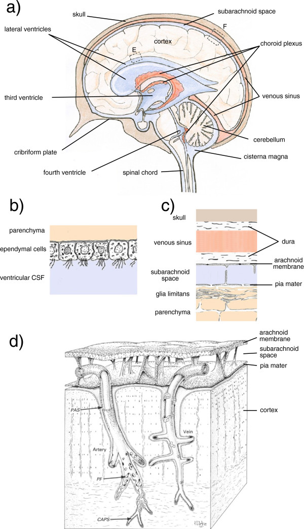
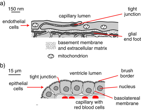
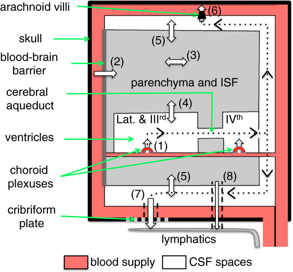
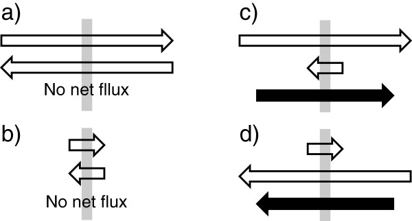
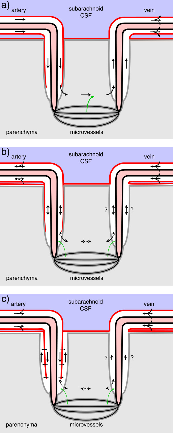
Interstitial fluid (ISF) surrounds the parenchymal cells of the brain and spinal cord while cerebrospinal fluid (CSF) fills the larger spaces within and around the CNS. Regulation of the composition and volume of these fluids is important for effective functioning of brain cells and is achieved by barriers that prevent free exchange between CNS and blood and by mechanisms that secrete fluid of controlled composition into the brain and distribute and reabsorb it. Structures associated with this regular fluid turnover include the choroid plexuses, brain capillaries comprising the blood-brain barrier, arachnoid villi and perineural spaces penetrating the cribriform plate. ISF flow, estimated from rates of removal of markers from the brain, has been thought to reflect rates of fluid secretion across the blood-brain barrier, although this has been questioned because measurements were made under barbiturate anaesthesia possibly affecting secretion and flow and because CSF influx to the parenchyma via perivascular routes may deliver fluid independently of blood-brain barrier secretion. Fluid secretion at the blood-brain barrier is provided by specific transporters that generate solute fluxes so creating osmotic gradients that force water to follow. Any flow due to hydrostatic pressures driving water across the barrier soon ceases unless accompanied by solute transport because water movements modify solute concentrations. CSF is thought to be derived primarily from secretion by the choroid plexuses. Flow rates measured using phase contrast magnetic resonance imaging reveal CSF movements to be more rapid and variable than previously supposed, even implying that under some circumstances net flow through the cerebral aqueduct may be reversed with net flow into the third and lateral ventricles. Such reversed flow requires there to be alternative sites for both generation and removal of CSF. Fluorescent tracer analysis has shown that fluid flow can occur from CSF into parenchyma along periarterial spaces. Whether this represents net fluid flow and whether there is subsequent flow through the interstitium and net flow out of the cortex via perivenous routes, described as glymphatic circulation, remains to be established. Modern techniques have revealed complex fluid movements within the brain. This review provides a critical evaluation of the data.
Overview
The claims and statistics of this paper haven’t been checked yet.
Run the check — free, no account needed. It reads the open-access text and checks the sources it cites.
Integrity signals
Registry
Cites
Statistics
Figures
Verification
Claims the paper makes
The claims and statistics of this paper haven’t been checked yet.
Run the check — free, no account needed. It reads the open-access text and checks the sources it cites.
Claims other papers make about this one
Each is checked against this paper’s own text: does it say what the citing paper says it does?
The cited source says what the claim attributes to it.
In the citing paper
“Other recent studies (252, 255, 547, 555), however, did not support the proposed glymphatic mechanism, nor the convective, pressure-driven fluid flow of CSF from para-arterial to paravenous spaces throughout the parenchymal ECS (31, 30, 255, 296, 547, 255).”
From Blood-Brain Barrier: From Physiology to Disease and Back (2019) · cites this paper as [252]
Self-check
Percentages, sums and reported differences re-computed from the paper’s own numbers.
The self-check needs the paper’s full text.
Citations
Cites
Intravenous Fluids and Acute Kidney Injury
Ding, X., Cheng, Z., & Qian, Q. (2017) · Blood Purification · doi:10.1159/000452702 · Cited by 6,868
Iliff, J. J., Wang, M., Liao, Y., Plogg, B. A., Peng, W., Gundersen, G. A., Benveniste, H., Vates, G. E., Deane, R., Goldman, S. A., et al. (2012) · Science Translational Medicine · doi:10.1126/scitranslmed.3003748 · Cited by 5,824
Sleep Drives Metabolite Clearance from the Adult Brain
Xie, L., Kang, H., Xu, Q., Chen, M. J., Liao, Y., Thiyagarajan, M., O’Donnell, J., Christensen, D. J., Nicholson, C., Iliff, J. J., et al. (2013) · Science · doi:10.1126/science.1241224 · Cited by 5,411
FINE STRUCTURAL LOCALIZATION OF A BLOOD-BRAIN BARRIER TO EXOGENOUS PEROXIDASE
Reese, T. S., & Karnovsky, M. J. (1967) · The Journal of Cell Biology · doi:10.1083/jcb.34.1.207 · Cited by 2,629
JUNCTIONS BETWEEN INTIMATELY APPOSED CELL MEMBRANES IN THE VERTEBRATE BRAIN
Brightman, M., & Reese, T. S. (1969) · The Journal of Cell Biology · doi:10.1083/jcb.40.3.648 · Cited by 2,457
Thermodynamic analysis of the permeability of biological membranes to non-electrolytes
Kedem, O., & Katchalsky, A. (1958) · Biochimica et Biophysica Acta · doi:10.1016/0006-3002(58)90330-5 · Cited by 2,214
On the Absorption of Fluids from the Connective Tissue Spaces
Starling, E. H. (1896) · The Journal of Physiology · doi:10.1113/jphysiol.1896.sp000596 · Cited by 2,126
Diffusion in Brain Extracellular Space
Syková, E., & Nicholson, C. (2008) · Physiological Reviews · doi:10.1152/physrev.00027.2007 · Cited by 1,481
Convection-enhanced delivery of macromolecules in the brain.
Bobo, R. H., Laske, D. W., Akbaşak, A., Morrison, P. F., Dedrick, R. L., & Oldfield, E. H. (1994) · Proceedings of the National Academy of Sciences · doi:10.1073/pnas.91.6.2076 · Cited by 1,442
Effect of anoxia on ion distribution in the brain
Hansen, A. J. (1985) · Physiological Reviews · doi:10.1152/physrev.1985.65.1.101 · Cited by 1,342
Cerebral Arterial Pulsation Drives Paravascular CSF–Interstitial Fluid Exchange in the Murine Brain
Iliff, J. J., Wang, M., Zeppenfeld, D. M., Venkataraman, A., Plog, B. A., Liao, Y., Deane, R., & Nedergaard, M. (2013) · The Journal of Neuroscience · doi:10.1523/jneurosci.1592-13.2013 · Cited by 1,319 · cited 3×
“The route by which the markers gained access to the periarterial space from CSF in the subarachnoid cisternae is not known [202,203].”
BackgroundMethodMicrovascular fluid exchange and the revised Starling principle
Levick, J. R., & Michel, C. C. (2010) · Cardiovascular Research · doi:10.1093/cvr/cvq062 · Cited by 1,183
Brain-wide pathway for waste clearance captured by contrast-enhanced MRI
Iliff, J. J., Lee, H., Yu, M., Feng, T., Logan, J., Nedergaard, M., & Benveniste, H. (2013) · Journal of Clinical Investigation · doi:10.1172/jci67677 · Cited by 1,148
Mokri, B. (2001) · Neurology · doi:10.1212/wnl.56.12.1746 · Cited by 1,026
Blood–brain barrier structure and function and the challenges for CNS drug delivery
Abbott, N. J. (2013) · Journal of Inherited Metabolic Disease · doi:10.1007/s10545-013-9608-0 · Cited by 951
Passage of Molecules Through Capillary Walls
Pappenheimer, J. R. (1953) · Physiological Reviews · doi:10.1152/physrev.1953.33.3.387 · Cited by 940
Extracellular space structure revealed by diffusion analysis
Nicholson, C., & Syková, E. (1998) · Trends in Neurosciences · doi:10.1016/s0166-2236(98)01261-2 · Cited by 914
Merritt, H. H., FREMONT-SMITH, F., & Ayer, J. B. (1938) · The American Journal of the Medical Sciences · doi:10.1097/00000441-193804000-00024 · Cited by 880
Multiplicity of cerebrospinal fluid functions: new challenges in health and disease
Johanson, C. E., Duncan, J. A., Klinge, P. M., Brinker, T., Stopa, E. G., & Silverberg, G. D. (2008) · Cerebrospinal Fluid Research · doi:10.1186/1743-8454-5-10 · Cited by 861
A new look at cerebrospinal fluid circulation
Brinker, T., Stopa, E., Morrison, J., & Klinge, P. (2014) · Fluids and Barriers of the CNS · doi:10.1186/2045-8118-11-10 · Cited by 819
The Concept of a Blood-Brain Barrier
(1980) · General Pharmacology The Vascular System · doi:10.1016/0306-3623(80)90073-7 · Cited by 805
The blood–brain and the blood–cerebrospinal fluid barriers: function and dysfunction
Engelhardt, B., & Sorokin, L. M. (2009) · Seminars in Immunopathology · doi:10.1007/s00281-009-0177-0 · Cited by 792
Evidence for bulk flow of brain interstitial fluid: significance for physiology and pathology
Abbott, N. J. (2004) · Neurochemistry International · doi:10.1016/j.neuint.2003.11.006 · Cited by 786
Nicholson, C., & Phillips, J. M. (1981) · The Journal of Physiology · doi:10.1113/jphysiol.1981.sp013981 · Cited by 785
Electrical resistance across the blood-brain barrier in anaesthetized rats: a developmental study.
Butt, A. M., Jones, H. C., & Abbott, N. J. (1990) · The Journal of Physiology · doi:10.1113/jphysiol.1990.sp018243 · Cited by 775
Rennels, M. L., Gregory, T. F., Blaumanis, O. R., Fujimoto, K., & Grady, P. A. (1985) · Brain Research · doi:10.1016/0006-8993(85)91383-6 · Cited by 752
Aquaporin water channels in the nervous system
Papadopoulos, M. C., & Verkman, A. S. (2013) · Nature reviews. Neuroscience · doi:10.1038/nrn3468 · Cited by 748
Advances in Comparative and Environmental Physiology
Houlihan, D. F., Livingstone, D. R., & Lee, R. F. (1991) · Advances in comparative and environmental physiology · doi:10.1007/978-3-642-75897-3 · Cited by 739
Nedergaard, M. (2013) · Science · doi:10.1126/science.1240514 · Cited by 718
Weller, R. O., Subash, M., Preston, S., Mazanti, I., & Carare, R. O. (2008) · Brain Pathology · doi:10.1111/j.1750-3639.2008.00133.x · Cited by 697
Weller, R. O., Massey, A., Newman, T. A., Hutchings, M., Kuo, Y.-M., & Roher, A. E. (1998) · The American Journal of Pathology · doi:10.1016/s0002-9440(10)65616-7 · Cited by 679
Carare, R. O., Bernardes‐Silva, M., Newman, T. A., Page, A. M., Nicoll, J. A. R., Perry, V. H., & Weller, R. O. (2008) · Neuropathology and Applied Neurobiology · doi:10.1111/j.1365-2990.2007.00926.x · Cited by 665
Luissint, A.-C., Artus, C., Glacial, F., Ganeshamoorthy, K., & Couraud, P.-O. (2012) · Fluids and Barriers of the CNS · doi:10.1186/2045-8118-9-23 · Cited by 654
Oldendorf, W. H., Cornford, M. E., & Brown, W. J. (1977) · Annals of Neurology · doi:10.1002/ana.410010502 · Cited by 651
Cerebrospinal fluid secretion by the choroid plexus
Damkier, H. H., Brown, P. D., & Praetorius, J. (2013) · Physiological Reviews · doi:10.1152/physrev.00004.2013 · Cited by 606 · cited 3×
“[15] the choroid plexus has all the hallmarks of a high capacity secretory epithelium whose product is isosmotic with plasma.”
BackgroundPhysiology of the CSF and Blood-Brain Barriers
Davson, H., & Segal, M. B. (1996) · Medical Entomology and Zoology · Cited by 582
Johnston, M., Zakharov, A., Papaiconomou, C., Salmasi, G., & Armstrong, D. (2004) · Cerebrospinal Fluid Research · doi:10.1186/1743-8454-1-2 · Cited by 572
Guyton, A. C., Granger, H. J., & Taylor, A. E. (1971) · Physiological Reviews · doi:10.1152/physrev.1971.51.3.527 · Cited by 567
The theory of measurement of osmotic pressure
Staverman, A. J. (1951) · Recueil des Travaux Chimiques des Pays-Bas · doi:10.1002/recl.19510700409 · Cited by 548
AN EXPERIMENTAL, CLINICAL AND PATHOLOGICAL STUDY
DANDY, W. E. (1914) · American journal of diseases of children · doi:10.1001/archpedi.1914.02180010416002 · Cited by 522
Kida, S., Pantazis, A., & Weller, R. O. (1993) · Neuropathology and Applied Neurobiology · doi:10.1111/j.1365-2990.1993.tb00476.x · Cited by 520
Physiology of the choroid plexus.
Cserr, H. F. (1971) · Physiological Reviews · doi:10.1152/physrev.1971.51.2.273 · Cited by 514
Pappenheimer, J. R., & Soto-Rivera, A. (1948) · American Journal of Physiology-Legacy Content · doi:10.1152/ajplegacy.1948.152.3.471 · Cited by 504
Electrical resistance of brain microvascular endothelium
Crone, C., & Olesen, S. P. (1982) · Brain Research · doi:10.1016/0006-8993(82)91227-6 · Cited by 491
Lymphatic drainage of the brain and the pathophysiology of neurological disease
Weller, R. O., Djuanda, E., Yow, H.-Y., & Carare, R. O. (2009) · Acta Neuropathologica · doi:10.1007/s00401-008-0457-0 · Cited by 488
FLOW THROUGH INTERSTITIUM AND OTHER FIBROUS MATRICES
Levick, J. R. (1987) · Quarterly Journal of Experimental Physiology · doi:10.1113/expphysiol.1987.sp003085 · Cited by 485
Internal hydrocephalus. An experimental, clinical and pathological study
DANDY, W. E. (1914) · Archives of Pediatrics and Adolescent Medicine · Cited by 481
(1990) · Europe PMC (PubMed Central) · Cited by 463
Junctions in the meninges and marginal glia.
Nabeshima, S., Reese, T. S., Landis, D. M. D., & Brightman, M. W. (1975) · Journal of Comparative Neurology · doi:10.1002/cne.901640202 · Cited by 444
Pulsatile brain movement and associated hydrodynamics studied by magnetic resonance phase imaging
Greitz, D., Wirestam, R., Franck, A., Nordell, B., Thomsen, C., & St�hlberg, F. (1992) · Neuroradiology · doi:10.1007/bf00596493 · Cited by 410
Radiological assessment of hydrocephalus: new theories and implications for therapy
Greitz, D. (2004) · Neurosurgical Review · doi:10.1007/s10143-004-0326-9 · Cited by 397
Bulk flow and diffusion in the cerebrospinal fluid system of the goat
Heisey, S. R., Held, D. R., & Pappenheimer, J. R. (1962) · American Journal of Physiology-Legacy Content · doi:10.1152/ajplegacy.1962.203.5.775 · Cited by 394
Non-equilibrium thermodynamics of membrane processes
Staverman, A. J. (1952) · Transactions of the Faraday Society · doi:10.1039/tf9524800176 · Cited by 384
Human brain motion and cerebrospinal fluid circulation demonstrated with MR velocity imaging.
Feinberg, D. A., & Mark, A. S. (1987) · Radiology · doi:10.1148/radiology.163.3.3575734 · Cited by 378
Warf, B. C. (2005) · Journal of neurosurgery · doi:10.3171/ped.2005.103.6.0475 · Cited by 373
The function and structure of the cerebrospinal fluid outflow system
Pollay, M. (2010) · Cerebrospinal Fluid Research · doi:10.1186/1743-8454-7-9 · Cited by 371
Enzmann, D. R., & Pelc, N. J. (1991) · Radiology · doi:10.1148/radiology.178.2.1987610 · Cited by 362
Drainage of interstitial fluid from different regions of rat brain
Szentistvanyi, I., Patlak, C. S., Ellis, R. A., & Cserr, H. F. (1984) · American Journal of Physiology-Renal Physiology · doi:10.1152/ajprenal.1984.246.6.f835 · Cited by 355
Orešković, D., & Klarica, M. (2010) · Brain Research Reviews · doi:10.1016/j.brainresrev.2010.04.006 · Cited by 349
Recent research into the nature of cerebrospinal fluid formation and absorption
McComb, J. G. (1983) · Journal of neurosurgery · doi:10.3171/jns.1983.59.3.0369 · Cited by 337
Drainage of cerebral interstitial fluid into deep cervical lymph of the rabbit
Bradbury, M. W., Cserr, H. F., & Westrop, R. J. (1981) · American Journal of Physiology-Renal Physiology · doi:10.1152/ajprenal.1981.240.4.f329 · Cited by 335
High-flow microinfusion: tissue penetration and pharmacodynamics
Morrison, P. F., Laske, D. W., Bobo, H., Oldfield, E. H., & Dedrick, R. L. (1994) · American Journal of Physiology-Regulatory, Integrative and Comparative Physiology · doi:10.1152/ajpregu.1994.266.1.r292 · Cited by 327
Role of pressure gradients and bulk flow in dynamics of vasogenic brain edema
Reulen, H. J., Graham, R., Spatz, M., & KLATZO, I. (1977) · Journal of neurosurgery · doi:10.3171/jns.1977.46.1.0024 · Cited by 325
Efflux of radiolabeled polyethylene glycols and albumin from rat brain
Cserr, H. F., Cooper, D. N., Suri, P. K., & Patlak, C. S. (1981) · American Journal of Physiology-Renal Physiology · doi:10.1152/ajprenal.1981.240.4.f319 · Cited by 323
Yang, L., Kress, B. T., Weber, H. J., Thiyagarajan, M., Wang, B., Deane, R., Benveniste, H., Iliff, J. J., & Nedergaard, M. (2013) · Journal of Translational Medicine · doi:10.1186/1479-5876-11-107 · Cited by 317
Hadaczek, P., Yamashita, Y., Mirek, H., Tamas, L. B., Bohn, M. C., Noble, C. O., Park, J. W., & Bankiewicz, K. S. (2006) · Molecular Therapy · doi:10.1016/j.ymthe.2006.02.018 · Cited by 312
Water permeability of lipid membranes.
Fettiplace, R., & Haydon, D. A. (1980) · Physiological Reviews · doi:10.1152/physrev.1980.60.2.510 · Cited by 306
Bering, E. A., & Sato, O. (1963) · Journal of neurosurgery · doi:10.3171/jns.1963.20.12.1050 · Cited by 306
Diffusion of Macromolecules in the Brain: Implications for Drug Delivery
Wolak, D. J., & Thorne, R. G. (2013) · Molecular Pharmaceutics · doi:10.1021/mp300495e · Cited by 299
Water homeostasis in the brain: Basic concepts
Kimelberg, H. K. (2004) · Neuroscience · doi:10.1016/j.neuroscience.2004.07.033 · Cited by 294
Mechanisms to explain the reverse perivascular transport of solutes out of the brain
Schley, D., Carare-Nnadi, R., Please, C. P., Perry, V. H., & Weller, R. O. (2006) · Journal of Theoretical Biology · doi:10.1016/j.jtbi.2005.07.005 · Cited by 293
Schlageter, K. E., Molnár, P., Lapin, G. D., & Groothuis, D. R. (1999) · Microvascular Research · doi:10.1006/mvre.1999.2188 · Cited by 292
Strazielle, N., & Ghersi‐Egea, J. (2013) · Molecular Pharmaceutics · doi:10.1021/mp300518e · Cited by 277
Nitz, W. R., Bradley, W. G., Watanabe, A. S., Lee, R. R., Burgoyne, B., O’Sullivan, R., & Herbst, M. D. (1992) · Radiology · doi:10.1148/radiology.183.2.1561340 · Cited by 272
Nicholson, C., & Tao, L. (1993) · Biophysical Journal · doi:10.1016/s0006-3495(93)81324-9 · Cited by 266
Interstitial fluid drainage is impaired in ischemic stroke and Alzheimer’s disease mouse models
Arbel‐Ornath, M., Hudry, E., Eikermann‐Haerter, K., Hou, S. S., Gregory, J. L., Zhao, L., Betensky, R. A., Frosch, M. P., Greenberg, S. M., & Bacskai, B. J. (2013) · Acta Neuropathologica · doi:10.1007/s00401-013-1145-2 · Cited by 265
Recent insights into a new hydrodynamics of the cerebrospinal fluid
Bulat, M., & Klarica, M. (2010) · Brain Research Reviews · doi:10.1016/j.brainresrev.2010.08.002 · Cited by 255
CSF hydrodynamic studies in man. 2 . Normal hydrodynamic variables related to CSF pressure and flow.
Ekstedt, J. (1978) · Journal of Neurology Neurosurgery & Psychiatry · doi:10.1136/jnnp.41.4.345 · Cited by 246
Preston, S., Steart, P. V., Wilkinson, A., Nicoll, J. A. R., & Weller, R. O. (2003) · Neuropathology and Applied Neurobiology · doi:10.1046/j.1365-2990.2003.00424.x · Cited by 245
Cserr, H. F., Cooper, D. N., & Milhorat, T. H. (1977) · Experimental Eye Research · doi:10.1016/s0014-4835(77)80041-9 · Cited by 239
Secretion of cerebrospinal fluid by the ventricular ependyma of the rabbit
Pollay, M., & Curl, F. D. (1967) · American Journal of Physiology-Legacy Content · doi:10.1152/ajplegacy.1967.213.4.1031 · Cited by 236
Anatomical relationships of the pia mater to cerebral blood vessels in man
Hutchings, M., & Weller, R. O. (1986) · Journal of Neurosurgery · doi:10.3171/jns.1986.65.3.0316 · Cited by 233
Barriers in the brain: a renaissance?
Saunders, N. R., Ek, C. J., Habgood, M. D., & Dzięgielewska, K. M. (2008) · Trends in Neurosciences · doi:10.1016/j.tins.2008.03.003 · Cited by 231
The third circulation revisited
Milhorat, T. H. (1975) · Journal of neurosurgery · doi:10.3171/jns.1975.42.6.0628 · Cited by 222
Extracellular space of brain as determined by diffusion of inulin from the ventricular system
Rall, D. P., OPPELT, W. W., & Patlak, C. S. (1962) · Life Sciences · doi:10.1016/0024-3205(62)90104-2 · Cited by 220
Handbook of Physiology, Section 2, Circulation
Briggs, D. I. (1964) · Physical Therapy · doi:10.1093/ptj/44.5.401 · Cited by 220
Paravascular microcirculation facilitates rapid lipid transport and astrocyte signaling in the brain
Thrane, V. R., Thrane, A. S., Plog, B. A., Thiyagarajan, M., Iliff, J. J., Deane, R., Nagelhus, E. A., & Nedergaard, M. (2013) · Scientific Reports · doi:10.1038/srep02582 · Cited by 215
Distribution of extracellular tracers in perivascular spaces of the rat brain
Ichimura, T., Fraser, P. A., & Cserr, H. F. (1991) · Brain Research · doi:10.1016/0006-8993(91)91275-6 · Cited by 215
The Choroid Plexus and Cerebrospinal Fluid: Emerging Roles in Development, Disease, and Therapy
Lehtinen, M. K., Bjornsson, C. S., Dymecki, S. M., Gilbertson, R. J., Holtzman, D. M., & Monuki, E. S. (2013) · Journal of Neuroscience · doi:10.1523/jneurosci.3258-13.2013 · Cited by 214
Interdependency of local capillary density, blood flow, and metabolism in rat brains
Klein, B. S., Kuschinsky, W., Schröck, H., & Vetterlein, F. (1986) · American Journal of Physiology-Heart and Circulatory Physiology · doi:10.1152/ajpheart.1986.251.6.h1333 · Cited by 212
Bradbury, M. W. B., & Westrop, R. J. (1983) · The Journal of Physiology · doi:10.1113/jphysiol.1983.sp014731 · Cited by 211
Bulk flow of brain interstitial fluid under normal and hyperosmolar conditions
Rosenberg, G. A., Kyner, W. T., & Estrada, E. (1980) · American Journal of Physiology-Renal Physiology · doi:10.1152/ajprenal.1980.238.1.f42 · Cited by 210
Carare, R. O., Hawkes, C. A., Jeffrey, M., Kalaria, R. N., & Weller, R. O. (2013) · Neuropathology and Applied Neurobiology · doi:10.1111/nan.12042 · Cited by 207
Measurements of dog blood-brain transfer constants by ventriculocisternal perfusion
Patlak, C. S., & Fenstermacher, J. D. (1975) · American Journal of Physiology-Legacy Content · doi:10.1152/ajplegacy.1975.229.4.877 · Cited by 206
Zhang, E. T., Richards, H. K., Kida, S., & Weller, R. O. (1992) · Acta Neuropathologica · doi:10.1007/bf00296784 · Cited by 205
Physiology and Pharmacology of the Blood-Brain Barrier
Bradbury, M. W. B. (1992) · Handbook of experimental pharmacology · doi:10.1007/978-3-642-76894-1 · Cited by 205
Rapid solute transport throughout the brain via paravascular fluid pathways.
Rennels, M. L., Blaumanis, O. R., & Grady, P. A. (1990) · PubMed · Cited by 204
Sucrose and inulin space measurements of cerebral cortex in four mammalian species
Levin, V. Y., Fenstermacher, J. D., & Patlak, C. S. (1970) · American Journal of Physiology-Legacy Content · doi:10.1152/ajplegacy.1970.219.5.1528 · Cited by 201
Filtration and reflection coefficients of the rabbit blood-brain barrier
Fenstermacher, J. D., & Johnson, J. (1966) · American Journal of Physiology-Legacy Content · doi:10.1152/ajplegacy.1966.211.2.341 · Cited by 199
Balédent, O., Gondry‐Jouet, C., Meyer, M.-É., de Marco, G., Gars, D. L., Henry–Feugeas, M.-C., & Idy-Peretti, I. (2003) · Investigative Radiology · doi:10.1097/01.rli.0000100892.87214.49 · Cited by 194
Cerebrospinal fluid flow measured by phase-contrast cine MR.
Enzmann, D. R., & Pelc, N. J. (1993) · PubMed Central · Cited by 190
Wagner, H. J., Pilgrim, C., & Brandl, J. (1974) · Acta Neuropathologica · doi:10.1007/bf00690695 · Cited by 189
Hawkes, C. A., Sullivan, P. M., Hands, S. L., Weller, R. O., Nicoll, J. A. R., & Carare, R. O. (2012) · PLoS ONE · doi:10.1371/journal.pone.0041636 · Cited by 182
Loo, D. D. F., Wright, E. M., & Zeuthen, T. (2002) · The Journal of Physiology · doi:10.1113/jphysiol.2002.018713 · Cited by 180
The role of the lymphatic system in drainage of cerebrospinal fluid and aqueous humour.
Bradbury, M. W. B., & Cole, D. F. (1980) · The Journal of Physiology · doi:10.1113/jphysiol.1980.sp013129 · Cited by 172
CSF hydrodynamic studies in man. 1. Method of constant pressure CSF infusion.
Ekstedt, J. (1977) · Journal of Neurology Neurosurgery & Psychiatry · doi:10.1136/jnnp.40.2.105 · Cited by 171
Hydrocephalus and the Cerebrospinal Fluid
Milhorat, T. H. (1972) · Cited by 170
Becker, N. H., Novikoff, A. B., & Zimmerman, H. M. (1967) · Journal of Histochemistry & Cytochemistry · doi:10.1177/15.3.160 · Cited by 162
Clearance of edema fluid into cerebrospinal fluid
Reulen, H. J., TSUYUMU, M., Tack, A., Fenske, A. R., & Prioleau, G. R. (1978) · Journal of neurosurgery · doi:10.3171/jns.1978.48.5.0754 · Cited by 158
Water Exchange of Central Nervous System and Cerebrospinal Fluid
Bering, E. A. (1952) · Journal of neurosurgery · doi:10.3171/jns.1952.9.3.0275 · Cited by 158
Hawkes, C. A., Gatherer, M., Sharp, M. M., Dorr, A. E., Yuen, H. M., Kalaria, R. N., Weller, R. O., & Carare, R. O. (2013) · Aging Cell · doi:10.1111/acel.12045 · Cited by 157
Barriers in the Immature Brain
Saunders, N. R., Knott, G. W., & Dzięgielewska, K. M. (2000) · Cellular and Molecular Neurobiology · doi:10.1023/a:1006991809927 · Cited by 156
Experimental biology of the lymphatic circulation
Johnston, M. G. (1985) · Elsevier eBooks · Cited by 154
Ghersi‐Egea, J., Finnegan, W., Chen, J.-L., & Fenstermacher, J. D. (1996) · Neuroscience · doi:10.1016/0306-4522(96)00281-3 · Cited by 151
Orešković, D., & Klarica, M. (2011) · Progress in Neurobiology · doi:10.1016/j.pneurobio.2011.05.005 · Cited by 151
Choroid Plexus and Cerebrospinal Fluid Production
Milhorat, T. H. (1969) · Science · doi:10.1126/science.166.3912.1514 · Cited by 148
Kedem, O. (1989) · Biochimica et Biophysica Acta (BBA) - General Subjects · doi:10.1016/s0006-3002(89)80036-8 · Cited by 147
A mathematical model of blood, cerebrospinal fluid and brain dynamics
Linninger, A. A., Xenos, M. A., Sweetman, B., Ponkshe, S., Guo, X., & Penn, R. D. (2009) · Journal of Mathematical Biology · doi:10.1007/s00285-009-0250-2 · Cited by 144
Greitz, D. (2007) · Child s Nervous System · doi:10.1007/s00381-007-0303-z · Cited by 142
Oldendorf, W. H. (1977) · Experimental Eye Research · doi:10.1016/s0014-4835(77)80016-x · Cited by 141
Drug “Diffusion” within the Braina
Fenstermacher, J. D., & Kaye, T. G. (1988) · Annals of the New York Academy of Sciences · doi:10.1111/j.1749-6632.1988.tb31809.x · Cited by 136
Mollanji, R., Bozanovic-Sosic, R., Zakharov, A., Makarian, L., & Johnston, M. G. (2002) · American Journal of Physiology-Regulatory, Integrative and Comparative Physiology · doi:10.1152/ajpregu.00695.2001 · Cited by 133
Filtration and diffusion of water across the blood-brain barrier in man
Paulson, O. B., Hertz, M. M., Bolwig, T. G., & Lassen, N. A. (1977) · Microvascular Research · doi:10.1016/0026-2862(77)90120-0 · Cited by 132
Water transport in the brain: Role of cotransporters
MacAulay, N., Hamann, S., & Zeuthen, T. (2004) · Neuroscience · doi:10.1016/j.neuroscience.2004.06.045 · Cited by 127
Murtha, L. A., Yang, Q., Parsons, M. W., Levi, C. R., Beard, D. J., Spratt, N. J., & McLeod, D. D. (2014) · Fluids and Barriers of the CNS · doi:10.1186/2045-8118-11-12 · Cited by 125
Gideon, P., St�hlberg, F., Thomsen, C., Gjerris, F., S�rensen, P. S., & Henriksen, O. M. (1994) · Neuroradiology · doi:10.1007/bf00588133 · Cited by 125
Kim, D.-S., Choi, J.-U., Huh, R., Yun, P.-H., & Kim, D. I. (1999) · Child s Nervous System · doi:10.1007/s003810050440 · Cited by 120
Fluids and barriers of the CNS: a historical viewpoint
Liddelow, S. A. (2011) · Fluids and Barriers of the CNS · doi:10.1186/2045-8118-8-2 · Cited by 113
Cerebrospinal Fluid Transport: a Lymphatic Perspective
Johnston, M., & Papaiconomou, C. (2002) · Physiology · doi:10.1152/nips.01400.2002 · Cited by 110
Bito, L. Z., & Davson, H. (1966) · Experimental Neurology · doi:10.1016/0014-4886(66)90114-2 · Cited by 108
Quantification of cerebrospinal fluid transport across the cribriform plate into lymphatics in rats
Nagra, G., Koh, L., Zakharov, A., Armstrong, D., & Johnston, M. G. (2006) · American Journal of Physiology-Regulatory, Integrative and Comparative Physiology · doi:10.1152/ajpregu.00235.2006 · Cited by 107
Johnston, M., Zakharov, A., Koh, L., & Armstrong, D. W. (2005) · Neuropathology and Applied Neurobiology · doi:10.1111/j.1365-2990.2005.00679.x · Cited by 106
Wåhlin, A., Ambarki, K., Hauksson, J. B., Birgander, R., Malm, J., & Eklund, A. (2011) · Journal of Magnetic Resonance Imaging · doi:10.1002/jmri.23527 · Cited by 106
Ventricular perfusion in cats with kaolin-induced hydrocephalus
Eisenberg, H. M., McLennan, J. E., & Welch, K. (1974) · Journal of neurosurgery · doi:10.3171/jns.1974.41.1.0020 · Cited by 106
Water influx into cerebrospinal fluid is primarily controlled by aquaporin-4, not by aquaporin-1
Igarashi, H., Tsujita, M., Kwee, I. L., & Nakada, T. (2013) · Neuroreport · doi:10.1097/wnr.0000000000000042 · Cited by 104
Papisov, M. I., Belov, V. V., & Gannon, K. S. (2013) · Molecular Pharmaceutics · doi:10.1021/mp300474m · Cited by 97
Groothuis, D. R., Vavra, M. W., Schlageter, K. E., Kang, E. W.-Y., Itskovich, A. C., Hertzler, S., Allen, C. V., & Lipton, H. L. (2006) · Journal of Cerebral Blood Flow & Metabolism · doi:10.1038/sj.jcbfm.9600315 · Cited by 95
The transport of potassium between blood, cerebrospinal fluid and brain.
Bradbury, M. W. B., & Davson, H. (1965) · The Journal of Physiology · doi:10.1113/jphysiol.1965.sp007752 · Cited by 94
Jones, H. C., & Keep, R. F. (1987) · The Journal of Physiology · doi:10.1113/jphysiol.1987.sp016419 · Cited by 93
GJEDDE, A. H., & Diemer, N. H. (1985) · Journal of Cerebral Blood Flow & Metabolism · doi:10.1038/jcbfm.1985.36 · Cited by 93
Mollanji, R., Bozanovic-Sosic, R., Silver, I., Li, B., Kim, C., Midha, R., & Johnston, M. (2001) · American Journal of Physiology-Regulatory, Integrative and Comparative Physiology · doi:10.1152/ajpregu.2001.280.5.r1573 · Cited by 90
Scarff, J. E. (1970) · Journal of neurosurgery · doi:10.3171/jns.1970.33.1.0001 · Cited by 89
Alvarez-Leefmans, F. J., & Delpire, E. J. (2009) · Journal of Bioresource Management · Cited by 89
Klarica, M., Orešković, D., Božić, B., Vukić, M., Butković, V., & Bulat, M. (2008) · Neuroscience · doi:10.1016/j.neuroscience.2008.11.041 · Cited by 86
Welch, K. A. (1975) · PubMed · Cited by 82
Absorption from the ventricles in experimentally produced internal hydrocephalus
Wislocki, G. B., & PUTNAM, T. J. (1921) · American Journal of Anatomy · doi:10.1002/aja.1000290302 · Cited by 81
(1955) · Europe PMC (PubMed Central) · Cited by 78
Transventricular and transpial absorption of cerebrospinal fluid into cerebral microvessels.
Bulat, M., Lupret, V., Orehković, D., & Klarica, M. (2008) · PubMed · Cited by 77
Sweet, W. H., Brownell, G. L., SCHOLL, J. A., Bowsher, D. R., Benda, P., & STICKLEY, E. E. (1955) · PubMed · Cited by 77
PERIVASCULAR SPACES OF THE MAMMALIAN CENTRAL NERVOUS SYSTEM
Woollam, D. H. M., & Millen, J. W. (1954) · Biological reviews/Biological reviews of the Cambridge Philosophical Society · doi:10.1111/j.1469-185x.1954.tb00597.x · Cited by 76
The Movements of Water and Solutes in the Brains of Mammals
Fenstermacher, J. D., & Patlak, C. S. (1976) · Springer eBooks · doi:10.1007/978-3-642-66524-0_16 · Cited by 76
Transport through the Ependymal Linings
Rall, D. P. (1968) · Progress in brain research · doi:10.1016/s0079-6123(08)64154-0 · Cited by 75
Ventricle wall movements and cerebrospinal fluid flow in hydrocephalus
Penn, R. D., Basati, S., Sweetman, B., Guo, X., & Linninger, A. A. (2011) · Journal of neurosurgery · doi:10.3171/2010.12.jns10926 · Cited by 75
Reassessment of the pathways responsible for cerebrospinal fluid absorption in the neonate
Papaiconomou, C., Zakharov, A., Azizi, N., Djenic, J., & Johnston, M. (2004) · Child s Nervous System · doi:10.1007/s00381-003-0840-z · Cited by 75
Nagaraja, T. N., Patel, P., Gorski, M., Gorevic, P. D., Patlak, C. S., & Fenstermacher, J. D. (2005) · Cerebrospinal Fluid Research · doi:10.1186/1743-8454-2-5 · Cited by 74
Bateman, G. A., & Brown, K. M. (2011) · Child s Nervous System · doi:10.1007/s00381-011-1617-4 · Cited by 70
The Anatomy of the Cerebrospinal Fluid
Aitken, J. T. (1962) · The Medical Journal of Australia · doi:10.5694/j.1326-5377.1962.tb24408.x · Cited by 70
Clearance of Edema Fluid into Cerebrospinal Fluid
Reulen, H. J., Prioleau, G. R., TSUYUMU, M., & Fenske, A. R. (1983) · Springer eBooks · doi:10.1007/978-1-4615-9269-3_50 · Cited by 67
Milhorat, T. H., Clark, R. G., & Hammock, M. K. (1970) · Journal of neurosurgery · doi:10.3171/jns.1970.32.4.0390 · Cited by 65
Biphasic Finite Element Model of Solute Transport for Direct Infusion into Nervous Tissue
Chen, X., & Sarntinoranont, M. (2007) · Annals of Biomedical Engineering · doi:10.1007/s10439-007-9371-1 · Cited by 64
Permeability of the choroid plexus of the rabbit to several solutes
Welch, K. A., & Sadler, K. M. (1966) · American Journal of Physiology-Legacy Content · doi:10.1152/ajplegacy.1966.210.3.652 · Cited by 63
Transport of potassium at the blood—brain barrier
Bradbury, M. W. B., Segal, M. B., & Wilson, J. P. (1972) · The Journal of Physiology · doi:10.1113/jphysiol.1972.sp009771 · Cited by 61
Chikly, B., & Quaghebeur, J. (2013) · Journal of Bodywork and Movement Therapies · doi:10.1016/j.jbmt.2013.02.002 · Cited by 59
Evidence for Choroid-Plexus Arsorption in Hydrocephalus
Milhorat, T. H., Mosher, M. B., Hammock, M. K., & Murphy, C. F. (1970) · New England Journal of Medicine · doi:10.1056/nejm197008062830604 · Cited by 58
Secretion and Bulk Flow of Interstitial Fluid
Cserr, H. F., & Patlak, C. S. (1992) · Handbook of experimental pharmacology · doi:10.1007/978-3-642-76894-1_9 · Cited by 58
Choroid plexus coagulation for hydrocephalus not due to CSF overproduction: a review
Zhu, X., & Rocco, C. D. (2012) · Child s Nervous System · doi:10.1007/s00381-012-1960-0 · Cited by 56
“Compensated hyperosmolarity” of cerebrospinal fluid and the development of hydrocephalus
Klarica, M., Miše, B., Vladić, A., Radoš, M., & Orešković, D. (2013) · Neuroscience · doi:10.1016/j.neuroscience.2013.06.022 · Cited by 55
Gross, P. M., Sposito, N. M., Pettersen, S. E., Panton, D. G., & Fenstermacher, J. D. (1987) · Journal of Cerebral Blood Flow & Metabolism · doi:10.1038/jcbfm.1987.38 · Cited by 55
Water, sodium, and thiourea transcapillary diffusion in the dog heart
Yudilevich, D. L., & Alvarez, O. (1967) · American Journal of Physiology-Legacy Content · doi:10.1152/ajplegacy.1967.213.2.308 · Cited by 55
Transport of water and electrolytes between brain and ventricular fluid in the rabbit
Curl, F. D., & Pollay, M. (1968) · Experimental Neurology · doi:10.1016/0014-4886(68)90109-x · Cited by 52
STUDIES ON CAPILLARY PERMEABILITY WITH TRACER SUBSTANCESPreprint
Flexner, L. B., Cowie, D. B., & Vosburgh, G. J. (1948) · Cold Spring Harbor Symposia on Quantitative Biology · doi:10.1101/sqb.1948.013.01.015 · Cited by 52
An Alternative Pathway of Cerebrospinal Fluid Absorption in Communicating Hydrocephalus
James, A. E., Strecker, E.-P., Sperber, E. E., Flor, W. J., MERZ, T., & Burns, B. (1974) · Radiology · doi:10.1148/111.1.143 · Cited by 48
Warf, B. C. (2011) · World Neurosurgery · doi:10.1016/j.wneu.2011.02.012 · Cited by 48
Failure of choroid plexectomy as treatment for hydrocephalus.
Milhorat, T. H. (1974) · PubMed · Cited by 48
Diffusion in the extracellular space in brain and tumors
Verkman, A. S. (2013) · Physical Biology · doi:10.1088/1478-3975/10/4/045003 · Cited by 46
Cerebrospinal fluid production rates determined by simultaneous albumin and inulin perfusion
Curran, R. E., Mosher, M. B., Owens, E. S., & Fenstermacher, J. D. (1970) · Experimental Neurology · doi:10.1016/0014-4886(70)90079-8 · Cited by 46
Ultrastructure of Brain Endothelium
Brightman, M. (1992) · Handbook of experimental pharmacology · doi:10.1007/978-3-642-76894-1_1 · Cited by 45
Brain stem penetration by horseradish peroxidase from the cerebrospinal fluid spaces in the cat
Borison, H. L., Borison, R., & McCarthy, L. E. (1980) · Experimental Neurology · doi:10.1016/0014-4886(80)90211-3 · Cited by 43
Orešković, D., Klarica, M., & Vukić, M. (2002) · Neuroscience Letters · doi:10.1016/s0304-3940(02)00395-6 · Cited by 43
Regulation of Brain Volume Under Isosmotic and Anisosmotic Conditions
Cserr, H. F., & Patlak, C. S. (1991) · Advances in comparative and environmental physiology · doi:10.1007/978-3-642-76226-0_3 · Cited by 43
Cerebrospinal fluid hydrodynamic studies in children.
Blomquist, H. K. S., Sundin, S., & Ekstedt, J. (1986) · Journal of Neurology Neurosurgery & Psychiatry · doi:10.1136/jnnp.49.5.536 · Cited by 38
Dynamic Aspects of Cerebrospinal Fluid*
Davson, H. (1972) · Developmental Medicine & Child Neurology · doi:10.1111/j.1469-8749.1972.tb09767.x · Cited by 38
Albumin outflow into deep cervical lymph from different regions of rabbit brain
Yamada, S., DePasquale, M., Patlak, C. S., & Cserr, H. F. (1991) · American Journal of Physiology-Heart and Circulatory Physiology · doi:10.1152/ajpheart.1991.261.4.h1197 · Cited by 37
Milhorat, T. H., & Clark, R. G. (1970) · Journal of neurosurgery · doi:10.3171/jns.1970.32.5.0522 · Cited by 37
Latent collagenase in the ulcerating rabbit cornea
Berman, M., Leary, R., & Gage, J. (1977) · Experimental Eye Research · doi:10.1016/0014-4835(77)90172-5 · Cited by 36
Aarabi, B., & Long, D. M. (1979) · Journal of neurosurgery · doi:10.3171/jns.1979.51.6.0779 · Cited by 36
Measurement of osmotic reflection coefficient for small molecules in cat hindlimbs
Wolf, M. B., & WATSON, P. D. (1989) · American Journal of Physiology-Heart and Circulatory Physiology · doi:10.1152/ajpheart.1989.256.1.h282 · Cited by 33
External hydrocephalus in infants: six cases with MR venogram and flow quantification correlation
Bateman, G. A., & Napier, B. D. (2011) · Child s Nervous System · doi:10.1007/s00381-011-1549-z · Cited by 33
Wh, S., Selverstone, B., Soloway, S., & Stetten, D. (1950) · PubMed · Cited by 31
Electrical impedance in the subicular area of rats during paradoxical sleep
Ranck, J. B. (1966) · Experimental Neurology · doi:10.1016/0014-4886(66)90107-5 · Cited by 30
K42 distribution in brain during simultaneous ventriculocisternal and subarachnoid perfusion
Pape, L. G., & Robert, K. (1972) · Brain Research · doi:10.1016/0006-8993(72)90589-6 · Cited by 27
Water filtration and diffusion across the blood brain barrier in man.
Paulson, O. B., Hertz, M. M., Tg, B., & Lassen, N. A. (1977) · PubMed · Cited by 26
Imaging of the cerebrospinal fluid circulation
Balédent, O. (2014) · Cambridge University Press eBooks · doi:10.1017/cbo9781139382816.013 · Cited by 23
The penetration of radiolabeled substances into rabbit brain from subarachnoid space
Levin, Е., & Wb, S. (1972) · Brain Research · doi:10.1016/0006-8993(72)90622-1 · Cited by 22
James, A. E., Flor, W. J., NOVAK, G. R., Strecker, E.-P., Burns, B., & Epstein, M. H. (1977) · Experimental Eye Research · doi:10.1016/s0014-4835(77)80040-7 · Cited by 21
Hill, A. E. (1979) · Quarterly Reviews of Biophysics · doi:10.1017/s0033583500002602 · Cited by 20
Overview of the CSF Dual Outflow System
Pollay, M. (2011) · Acta neurochirurgica. Supplementum · doi:10.1007/978-3-7091-0923-6_10 · Cited by 20
Clinical Brain Imaging: Normal Structure and Functional Anatomy
Hayman, L. A., & Hinck, V. C. (1992) · Medical Entomology and Zoology · Cited by 20
Piechnik, S. K., Summers, P. E., Jezzard, P., & Byrne, J. V. (2008) · Acta neurochirurgica. Supplementum · doi:10.1007/978-3-211-85578-2_50 · Cited by 19
Ion and Water Transport across the Blood–Brain Barrier
O’Donnell, M. E. (2010) · Elsevier eBooks · doi:10.1016/b978-0-12-374373-2.00030-3 · Cited by 17
Cited by
Sorted by citations
Blood–brain barrier breakdown in Alzheimer disease and other neurodegenerative disorders
Sweeney, M. D., Sagare, A. P., & Zlokovic, B. V. (2018) · Nature Reviews Neurology · doi:10.1038/nrneurol.2017.188 · Cited by 3,076
The Neurovascular Unit Coming of Age: A Journey through Neurovascular Coupling in Health and Disease
Iadecola, C. (2017) · Neuron · doi:10.1016/j.neuron.2017.07.030 · Cited by 2,480
Blood-Brain Barrier: From Physiology to Disease and Back
Sweeney, M. D., Zhao, Z., Montagne, A., Nelson, A. R., & Zlokovic, B. V. (2019) · Physiological Reviews · doi:10.1152/physrev.00050.2017 · Cited by 2,312
Clearance systems in the brain—implications for Alzheimer disease
Tarasoff-Conway, J. M., Carare, R. O., Osorio, R. S., Glodzik, L., Butler, T., Fieremans, E., Axel, L., Rusinek, H., Nicholson, C., Zlokovic, B. V., et al. (2015) · Nature Reviews Neurology · doi:10.1038/nrneurol.2015.119 · Cited by 1,678
The movers and shapers in immune privilege of the CNS
Engelhardt, B., Vajkoczy, P., & Weller, R. O. (2017) · Nature Immunology · doi:10.1038/ni.3666 · Cited by 893
Metadata
- Authors
- Hladky, S. B., & Barrand, M. A.
- Affiliations
- University of Cambridge (GB)
- Corresponding author
- Hladky, Stephen B
- ORCID
- Hladky, Stephen B
- Venue
- Fluids and Barriers of the CNS
- Published
- 2014
- Volume
- 11
- Issue
- 1
- Pages
- 26
- ISSN
- 2045-8118 (ISSN-L)
- PMC
- PMC4326185
- OpenAlex
- W2096525076
- Cited by
- 633
- References
- 215
- Citations by year
- 2026: 37 · 2025: 49 · 2024: 57 · 2023: 65 · 2022: 77 · 2021: 75 · 2020: 45 · 2019: 65 · 2018: 61 · 2017: 58 · 2016: 26 · 2015: 17
- FWCI
- 3.85
- Type
- journal article
- Language
- en
- Open access
- gold
- doi.org
- Licence
- cc-by
- Locations
- Fluids and Barriers of the CNS (publishedVersion, cc-by)
- Europe PMC (PubMed Central) (publishedVersion, cc-by)
- PubMed Central (publishedVersion, cc-by)
- PubMed (publishedVersion)
- Topics
- Health Sciences › Medicine › Clinical Neurology › Cerebrospinal fluid and hydrocephalus
- Life Sciences › Biochemistry, Genetics and Molecular Biology › Molecular Biology › Ion Transport and Channel Regulation
- Life Sciences › Neuroscience › Neurology › Barrier Structure and Function Studies
- Keywords
- interstitial fluid, cerebrospinal fluid, cerebrospinal fluid flow, blood-brain barrier, choroid plexus, arachnoid villi, perineural space, cribriform plate, cerebrospinal fluid production, CSF reabsorption, phase-contrast MRI, osmotic gradient, solute transport, perivascular spaces, barbiturate anesthesia
About these checks
Registry, retraction, Cites and statistics results are computed, not judged, with one exception: a printed reference that no index lists is read once by a model before it counts as a citation error. Claims are extracted by a model and kept only when their quote appears verbatim in the text; each cited source is then checked, with a model’s help, for whether it says what the claim relies on. Those verdicts can be wrong — read the claim’s quote and the cited source before relying on one.
Statistics and arithmetic are recomputed, not judged.
Confidence bands are shown only for fields calibrated on our annotated test set. None is calibrated yet, so no band is shown.
What counts as a claim ›How we measure confidence ›How we check this ›
Are you an author, or do you see a problem with this page? Request a review
