

The DOI matches its Crossref record; 215 of the 215 references it cites were found in a registry or an index.
Mechanisms of fluid movement into, through and out of the brain: evaluation of the evidence
Open accessAbstract
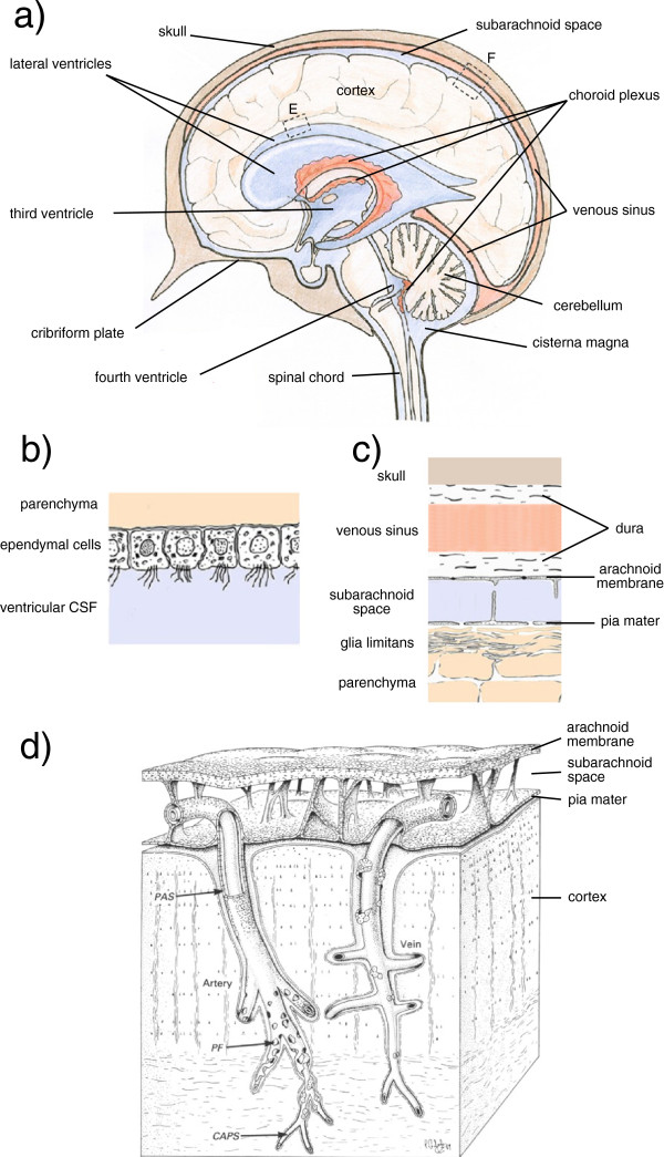
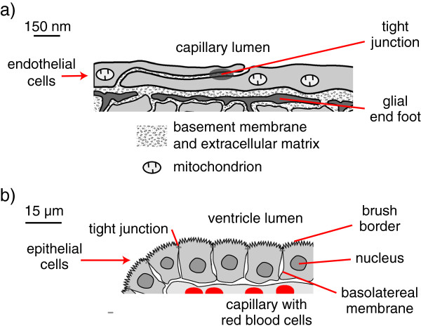
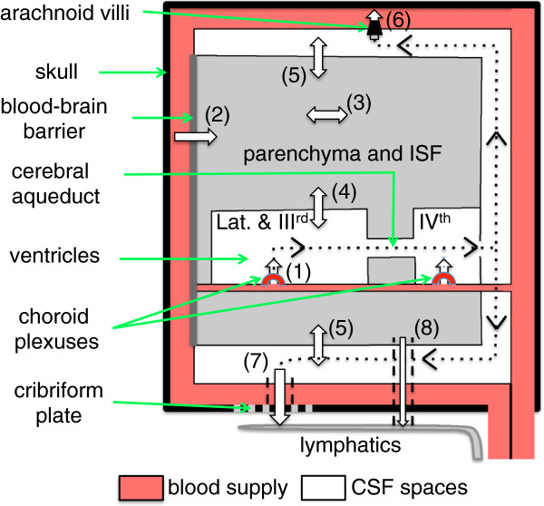
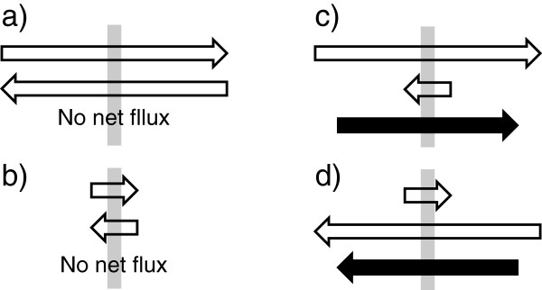
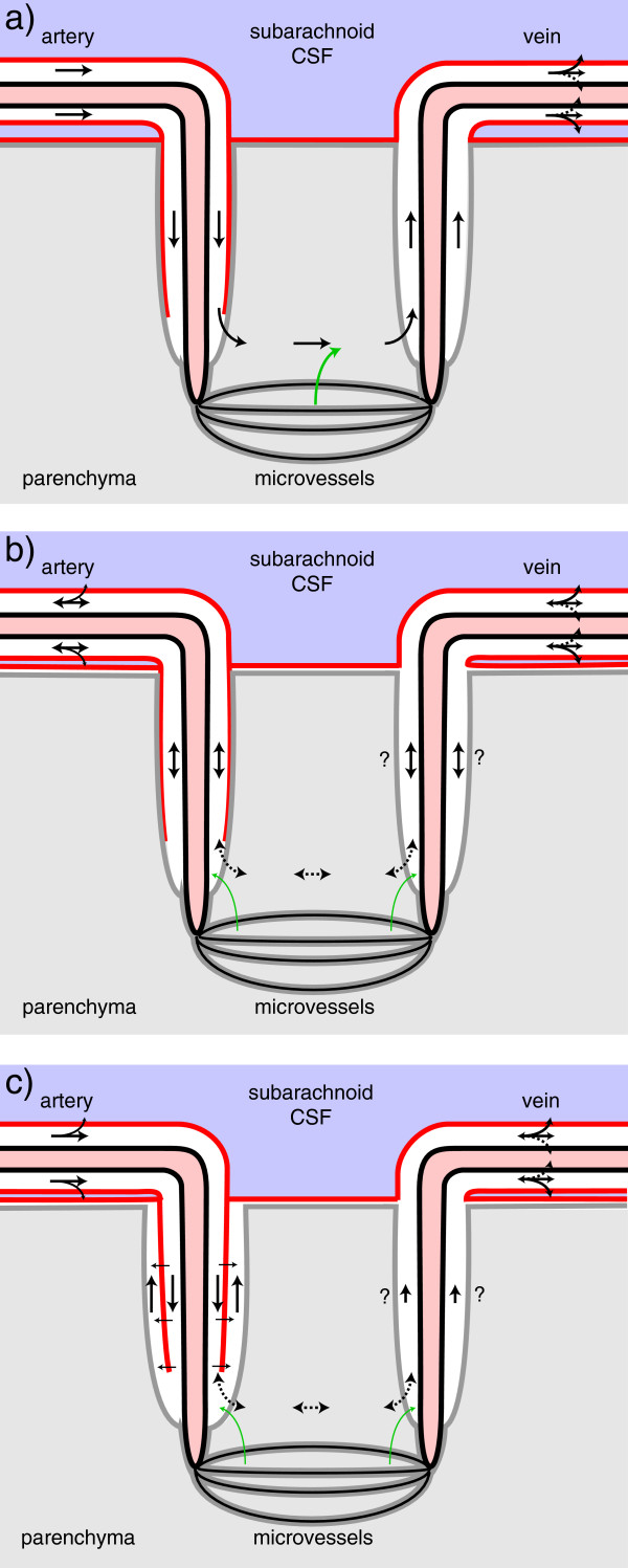
Interstitial fluid (ISF) surrounds the parenchymal cells of the brain and spinal cord while cerebrospinal fluid (CSF) fills the larger spaces within and around the CNS. Regulation of the composition and volume of these fluids is important for effective functioning of brain cells and is achieved by barriers that prevent free exchange between CNS and blood and by mechanisms that secrete fluid of controlled composition into the brain and distribute and reabsorb it. Structures associated with this regular fluid turnover include the choroid plexuses, brain capillaries comprising the blood-brain barrier, arachnoid villi and perineural spaces penetrating the cribriform plate. ISF flow, estimated from rates of removal of markers from the brain, has been thought to reflect rates of fluid secretion across the blood-brain barrier, although this has been questioned because measurements were made under barbiturate anaesthesia possibly affecting secretion and flow and because CSF influx to the parenchyma via perivascular routes may deliver fluid independently of blood-brain barrier secretion. Fluid secretion at the blood-brain barrier is provided by specific transporters that generate solute fluxes so creating osmotic gradients that force water to follow. Any flow due to hydrostatic pressures driving water across the barrier soon ceases unless accompanied by solute transport because water movements modify solute concentrations. CSF is thought to be derived primarily from secretion by the choroid plexuses. Flow rates measured using phase contrast magnetic resonance imaging reveal CSF movements to be more rapid and variable than previously supposed, even implying that under some circumstances net flow through the cerebral aqueduct may be reversed with net flow into the third and lateral ventricles. Such reversed flow requires there to be alternative sites for both generation and removal of CSF. Fluorescent tracer analysis has shown that fluid flow can occur from CSF into parenchyma along periarterial spaces. Whether this represents net fluid flow and whether there is subsequent flow through the interstitium and net flow out of the cortex via perivenous routes, described as glymphatic circulation, remains to be established. Modern techniques have revealed complex fluid movements within the brain. This review provides a critical evaluation of the data.
Overview
The claims and statistics of this paper haven’t been checked yet.
Run the check — free, no account needed. It reads the open-access text and checks the sources it cites.
Integrity signals
Registry
Cites
Statistics
Figures
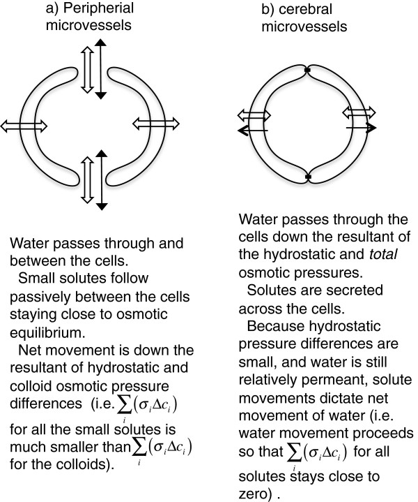
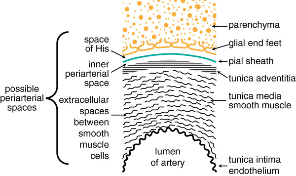
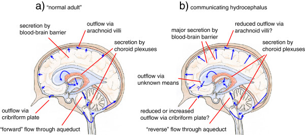
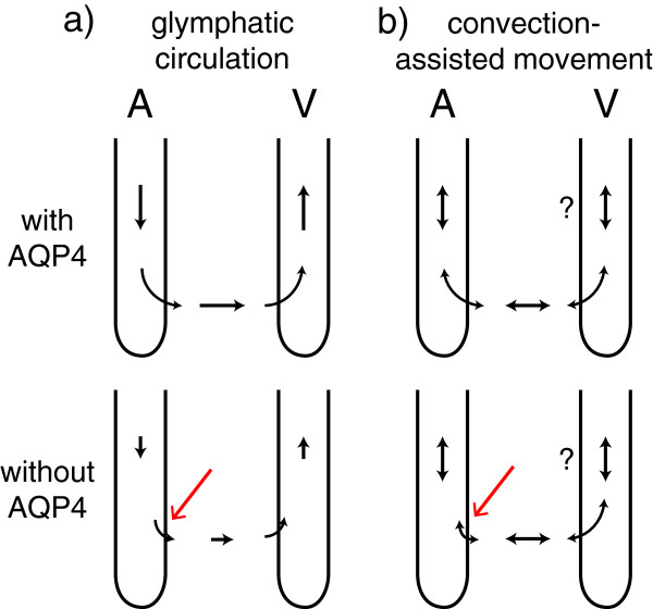
Verification
Claims the paper makes
The claims and statistics of this paper haven’t been checked yet.
Run the check — free, no account needed. It reads the open-access text and checks the sources it cites.
Claims other papers make about this one
Each is checked against this paper’s own text: does it say what the citing paper says it does?
The cited source says what the claim attributes to it.
In the citing paper
“Other recent studies (252, 255, 547, 555), however, did not support the proposed glymphatic mechanism, nor the convective, pressure-driven fluid flow of CSF from para-arterial to paravenous spaces throughout the parenchymal ECS (31, 30, 255, 296, 547, 255).”
From Blood-Brain Barrier: From Physiology to Disease and Back (2019) · cites this paper as [252]
Self-check
Percentages, sums and reported differences re-computed from the paper’s own numbers.
The self-check needs the paper’s full text.
Citations
Cites
Intravenous Fluids and Acute Kidney Injury
Ding, X., Cheng, Z., & Qian, Q. (2017) · Blood Purification · doi:10.1159/000452702 · Cited by 6,868
Iliff, J. J., Wang, M., Liao, Y., Plogg, B. A., Peng, W., Gundersen, G. A., Benveniste, H., Vates, G. E., Deane, R., Goldman, S. A., et al. (2012) · Science Translational Medicine · doi:10.1126/scitranslmed.3003748 · Cited by 5,824
Sleep Drives Metabolite Clearance from the Adult Brain
Xie, L., Kang, H., Xu, Q., Chen, M. J., Liao, Y., Thiyagarajan, M., O’Donnell, J., Christensen, D. J., Nicholson, C., Iliff, J. J., et al. (2013) · Science · doi:10.1126/science.1241224 · Cited by 5,411
FINE STRUCTURAL LOCALIZATION OF A BLOOD-BRAIN BARRIER TO EXOGENOUS PEROXIDASE
Reese, T. S., & Karnovsky, M. J. (1967) · The Journal of Cell Biology · doi:10.1083/jcb.34.1.207 · Cited by 2,629
JUNCTIONS BETWEEN INTIMATELY APPOSED CELL MEMBRANES IN THE VERTEBRATE BRAIN
Brightman, M., & Reese, T. S. (1969) · The Journal of Cell Biology · doi:10.1083/jcb.40.3.648 · Cited by 2,457
Thermodynamic analysis of the permeability of biological membranes to non-electrolytes
Kedem, O., & Katchalsky, A. (1958) · Biochimica et Biophysica Acta · doi:10.1016/0006-3002(58)90330-5 · Cited by 2,214
On the Absorption of Fluids from the Connective Tissue Spaces
Starling, E. H. (1896) · The Journal of Physiology · doi:10.1113/jphysiol.1896.sp000596 · Cited by 2,126
Diffusion in Brain Extracellular Space
Syková, E., & Nicholson, C. (2008) · Physiological Reviews · doi:10.1152/physrev.00027.2007 · Cited by 1,481
Convection-enhanced delivery of macromolecules in the brain.
Bobo, R. H., Laske, D. W., Akbaşak, A., Morrison, P. F., Dedrick, R. L., & Oldfield, E. H. (1994) · Proceedings of the National Academy of Sciences · doi:10.1073/pnas.91.6.2076 · Cited by 1,442
Effect of anoxia on ion distribution in the brain
Hansen, A. J. (1985) · Physiological Reviews · doi:10.1152/physrev.1985.65.1.101 · Cited by 1,342
Cited by
Sorted by citations
Items 501–600 of 636
Bellettieri, M. P. G., Anderloni, M., Rass, V., Kindl, P., Donadello, K., Taccone, F. S., Helbok, R., & Bogossian, E. G. (2023) · Clinical Neurology and Neurosurgery · doi:10.1016/j.clineuro.2023.108011 · Cited by 2
Advancements in Brain Lymphatic System and Its Involvement in Neurological Diseases
Bari, D., Das, U., Shevalkar, G. B., Kapadia, R., Singhai, V., & Pardeshi, C. V. (2024) · Springer eBooks · doi:10.1007/978-3-031-64828-1_2 · Cited by 2
Middlebrooks, E. H., Bennett, J., & Crow, A. O. (2015) · Fluids and Barriers of the CNS · doi:10.1186/s12987-015-0011-0 · Cited by 2
Blood-Brain Barrier Mechanisms of Edema Formation
O’Donnell, M. E., Wulff, H., & Chen, Y. (2017) · Elsevier eBooks · doi:10.1016/b978-0-12-803196-4.00007-2 · Cited by 2
CSF sodium at toxic levels precedes delirium in hip fracture patients
Hassel, B., Mariussen, E., Idland, A., Dahl, G. T., Raeder, J. C., Frihagen, F., Berg, J. P., Chaudhry, F. A., Wyller, T. B., & Watne, L. O. (2018) · NeuroToxicology · doi:10.1016/j.neuro.2018.08.010 · Cited by 2
Houdt, R. V., Vandecraen, J., Heylen, W., Leys, N., Monsieurs, P., Provoost, A., & Aertsen, A. (2021) · Frontiers in Microbiology · doi:10.3389/fmicb.2021.698330 · Cited by 2
Piña, Y., Gramatzki, D., Forsyth, P. A., Lee, E. Q., & Rhun, É. L. (2021) · Hematology/Oncology Clinics of North America · doi:10.1016/j.hoc.2021.08.009 · Cited by 3
Hydrocephalus: Generalities and Clinical Presentations
Rocco, C. D., & Frassanito, P. (2020) · Springer eBooks · doi:10.1007/978-3-319-72168-2_11 · Cited by 2
Fagiolino, P. L., & Vázquez, M. (2023) · Springer eBooks · doi:10.1007/978-3-031-36526-3_9 · Cited by 2
Reiber, H., & Uhr, M. (2020) · Springer Reference Medizin · doi:10.1007/978-3-662-60676-6_21 · Cited by 2
Choroid Plexus: Source of Cerebrospinal Fluid and Regulator of Brain Development and Function
Snodgrass, R., & Johanson, C. E. (2018) · Springer eBooks · doi:10.1007/978-3-319-31889-9_38-1 · Cited by 2
Paladini, D., Donarini, G., Conti, A. M. F., Angelis, L. C. D., Witte, M., Genesio, R., Bernas, M. J., Bellini, T., Boccardo, F. M., Ramenghi, L. A., et al. (2019) · Lymphology · doi:10.2458/lymph.4620 · Cited by 2
Choroid plexus and microcirculation of the brain in cerebral disorders in the highlands.
Шувалова, М. С. (2021) · KRSU Publishing House eBooks · doi:10.36979/978-9967-19-824-1-2021 · Cited by 2
The Lymphatic System in Neurological Disease and Alzheimer's Disease. A Brief Editorial
Pappolla, M. A., Carare, R. O., Poeggeler, B., Wısnıewskı, T., & Sambamurti, K. (2022) · Current Alzheimer Research · doi:10.2174/1567205020666221028111517 · Cited by 2
Brain barriers and brain fluid research in 2016: advances, challenges and controversies
Keep, R. F., Jones, H. C., & Drewes, L. R. (2017) · Fluids and Barriers of the CNS · doi:10.1186/s12987-017-0052-7 · Cited by 2
CSF turnover reshapes biomarker interpretation in neurodegeneration studiesPreprint
García‐González, P., Puerta, R., Dehairs, J., Yang, C., Wang, C., Timsina, J., de Rojas, I., Olivé, C., Valenzuela, A., Bayón-Buján, P., et al. (2026) · medRxiv · doi:10.64898/2026.02.02.26345363 · Cited by 3
García‐Cosamalón, J., Marchena, M., Villar‐Suárez, V., Ley, L., & de Castro, F. (2026) · Frontiers in Cell and Developmental Biology · doi:10.3389/fcell.2026.1785072 · Cited by 1
Ciampi, C., Govoni, S. M., Preda, S., Mannelli, L. D. C., Racchi, M., Martelli, A., & Lanni, C. (2026) · Frontiers in Cell and Developmental Biology · doi:10.3389/fcell.2026.1891553 · Cited by 1
Wang, Y., Ge, D., Jin, Z., Zhao, L., Wu, D., Lin, Z., & Initiative, A. D. N. (2026) · Journal of Magnetic Resonance Imaging · doi:10.1002/jmri.70584 · Cited by 1
Effects of External Ventricular Drainage on Serum Sodium Levels in Adult Neurosurgical Patients
Baran, O., Kemerdere, R., Yuksel, O., Sayyahmelli, S., AKKAYA, N., Tanrıverdi, T., & Kafadar, A. M. (2018) · Bezmialem Science · doi:10.14235/bs.2018.1252 · Cited by 1
Morphological changes in cerebrospinal fluid production
Paszkowycz, H., Olewnik, Ł. H., Gonera, B., Haładaj, R., & Zielińska, N. (2024) · Folia Morphologica · doi:10.5603/fm.99964 · Cited by 1
Lalou, A. D., Czosnyka, Z. H., Czosnyka, M., & Pickard, J. D. (2025) · Fluids and Barriers of the CNS · doi:10.1186/s12987-025-00720-2 · Cited by 1
Melanoma leptomeningeal disease: Advances in diagnosis and emerging therapeutic strategies
Middleton, M. L., & Lucke-Wold, B. P. (2026) · World Journal of Experimental Medicine · doi:10.5493/wjem.v16.i1.117938 · Cited by 1
Li, Y., Du, A., Zhang, Q., Xu, G., Xu, X. G., Ma, X., Chu, J. C. H., Thiyagarajan, V., Yu, Z., Chu, I. K., et al. (2026) · Analytical Chemistry · doi:10.1021/acs.analchem.6c01031 · Cited by 1
Ritter, P. S., Edelmann, K. A., Thelwall, P. E., Boumezbeur, F., Bauer, M., Werner, A., Hall, G., Kavanagh, O. N., & Cousins, D. A. (2026) · The Lancet Psychiatry · doi:10.1016/s2215-0366(26)00153-7 · Cited by 1
Osman, A. M., He, F. Y., Remšík, J., Snyder, J., Son, S., Toomey, S., Estrera, R., Wijetunga, N. A., Manoranjan, B., Nobre, A. R., et al. (2026) · Science Translational Medicine · doi:10.1126/scitranslmed.aeh7968 · Cited by 1
Fluorescence imaging of lymphatic outflow of cerebrospinal fluid in mice
Kwon, S., Janssen, C. F., Velasquez, F. C., & Sevick-Muraca, E. M. (2018) · SPIE Medical Imaging: Biomedical Applications in Molecular, Structural, and Functional Imaging · doi:10.1117/12.2293535 · Cited by 1
Electrokinetic Properties of a Hydrogel Based on PVA, Xanthan, and PEDOT:PSS
Bolshin, D. S., Gotovtsev, P. M., & Kashkarov, P. K. (2022) · Nanobiotechnology Reports · doi:10.1134/s2635167622060027 · Cited by 1
Bubeníková, A., Skalický, P., Whitley, H., & Bradáč, O. (2023) · Springer eBooks · doi:10.1007/978-3-031-36522-5_5 · Cited by 1
Superior Sagittal Sinus Thrombectomy in Pediatric Head Injury
Johansen, P. M., Ciavarra, B. M., McCormack, R., Kole, M. J., Spiegel, G. R., & Fletcher, S. A. (2024) · Pediatric Neurosurgery · doi:10.1159/000538184 · Cited by 1
Sharma, R., Monika, Tyagi, B., Gupta, D., Mazumder, R., & Mazumder, A. (2024) · Central Nervous System Agents in Medicinal Chemistry · doi:10.2174/0118715249329586240924105624 · Cited by 1
Zhou, L., Li, Y., & de Leon, M. J. (2025) · Neuroimaging Clinics of North America · doi:10.1016/j.nic.2024.12.001 · Cited by 1
General Introduction to Barrier Mechanisms in the Central Nervous System
Saunders, N. R., Dzięgielewska, K. M., Møllgård, K., & Habgood, M. D. (2017) · Springer eBooks · doi:10.1007/978-3-319-45514-3_1 · Cited by 1
Cerebrospinal Fluid Biomarkers of Hydrocephalus
Isaacs, A. M., & Limbrick, D. D. (2018) · Springer eBooks · doi:10.1007/978-3-319-97928-1_3 · Cited by 1
Fluid Management in Neurointensive Care
Dąbrowski, W., Wise, R., Rzecki, Z., & Malbrain, M. L. N. G. (2019) · Springer eBooks · doi:10.1007/978-981-13-3390-3_2 · Cited by 1
Raybaud, C. (2019) · Springer eBooks · doi:10.1007/978-3-319-27250-4_44 · Cited by 1
Reiber, H., & Uhr, M. (2017) · Springer eBooks · doi:10.1007/978-3-662-44768-0_21-1 · Cited by 1
Possible Role of the New Identified “Subarachnoid Lymphatic-Like Membrane” in Traumatic Brain Injury
Grasso, G., Sallì, M., Kim, H. S., & Torregrossa, F. (2023) · World Neurosurgery · doi:10.1016/j.wneu.2023.02.105 · Cited by 1
Fluid movement in the healthy and diseased brain
Shi, Y., & Keep, R. F. (2023) · Neurobiology of Disease · doi:10.1016/j.nbd.2023.106168 · Cited by 1
Reeves, B. C., Karimy, J. K., Duy, P. Q., & Kahle, K. T. (2024) · Cambridge University Press eBooks · doi:10.1017/9781108917339.025 · Cited by 1
Kuroda, S., Yamamoto, S., Hori, E., Kashiwazaki, D., & Noguchi, K. (2024) · Surgical Neurology International · doi:10.25259/sni_281_2024 · Cited by 1
Impact of in vivo cyclic reprogramming on the choroid plexusPreprint
Lopez, J. A., Abboud, C., Ibrahim, M., Ahumada, J. R., Avino, M., Plourde, M., Fernandes, K. J. L., Bentzinger, C. F., & Laurent, B. (2023) · bioRxiv (Cold Spring Harbor Laboratory) · doi:10.1101/2023.02.28.530533 · Cited by 1
Saha, D., & Tapuria, P. (2024) · Springer eBooks · doi:10.1007/978-981-99-8059-8_3 · Cited by 1
Owens, T. (2016) · Elsevier eBooks · doi:10.1016/b978-0-12-374279-7.03004-6
Wang, Q., Grater, R., Delva, L., Veizaj, E., Black, C., Rowbottom, C., Rusesabagina, R., Rohde, E., Schroeder, P. E., Bohnert, T., et al. (2018) · Journal of Neurology and Neuromedicine · doi:10.29245/2572.942x/2018/5.1217
Begcevic, I. (2018) · TSpace (University of Toronto)
The New Concepts of Cerebrospinal Fluid Physiology
Wang, J., & Wang, N. (2019) · Advances in visual science and eye diseases · doi:10.1007/978-981-13-2137-5_9
The postsynaptic protein neurogranin: a new item in the Alzheimer’s disease biomarker toolbox
Kvartsberg, H. (2019) · Gothenburg University Publications Electronic Archive (Gothenburg University)
Fluid Management in Neurosurgical Patients
Dąbrowski, W., Wise, R., & Malbrain, M. L. N. G. (2019) · Springer eBooks · doi:10.1007/978-981-13-3387-3_26
Transport of lipid vesicles via the cilia logistic network in the brain of mice
Günther, A.-K. (2019) · eDiss (Georg-August-Universität Göttingen) · doi:10.53846/goediss-7642
Diagnostica per immagini dell’idrocefalo del bambino
Brun, G., Reyre, A., Scavarda, D., Girard, N. J., & Brunel, H. (2020) · EMC - Neurologia · doi:10.1016/s1634-7072(20)43300-8
Imagerie de l'hydrocéphalie de l'enfant
Brun, G., Reyre, A., Scavarda, D., Girard, N. J., & Brunel, H. (2019)
Nanoparticle Drug Delivery to Brain Tumors: From Intravenous to Intrathecal
Householder, K. T. (2018)
Jhaveri, M. D., Salzman, K. L., Ross, J. S., Moore, K. R., Osborn, A. G., & Ho, C. Y. (2018) · Elsevier eBooks · doi:10.1016/b978-0-323-44308-1.50121-5
Metabolism and Cerebral Blood Flow
Dengl, M., & Schackert, G. (2021) · Springer eBooks · doi:10.1007/978-3-030-66572-2_2
Alshuhri, M. S. (2021) · Enlighten: Theses (The University of Glasgow) · doi:10.5525/gla.thesis.82303
Glymphatic Structure and Morphological Features of the Brain
Nur, İ. H., & Pérez, W. (2021) · Journal of Morphological Sciences · doi:10.51929/jms.38.43.2021
Scoz, A., Bertazzi, L., & Toro, E. F. (2021) · Applied Mathematics and Computation · doi:10.1016/j.amc.2021.126625
Hemodynamic Patterns of Spinal Cord Perfusion in Thoracoabdominal Aortic Aneurysm Repair
Rescigno, G. U., Banfı, C., Rossella, C., & Nazari, S. (2021) · Aorta · doi:10.1055/s-0041-1725121
Intraventrikuläre Hirnblutungen des Frühgeborenen
Thome, U. H., Knüpfer, M. M., & Krause, M. (2021) · Neuroradiologie Scan · doi:10.1055/a-1503-4222
Grigorescu, B.-L., Săplăcan, I., Gliga, F. I., & Fodor, R. Ș. (2021) · Internal Medicine · doi:10.2478/inmed-2021-0191
Neuroimaging in Pediatric Hydrocephalus
Raybaud, C. (2017) · Springer eBooks · doi:10.1007/978-3-319-31512-6_15-1 · Cited by 2
Spatio-temporal dynamics in the anchoring of cilia
Kapoor, S. (2019) · doi:10.53846/goediss-7668
da Silva, S. C. (2021) · Digital Library of Theses and Dissertations (Universidade de São Paulo) · doi:10.11606/t.17.2021.tde-28012022-112417
Rikitake, M., Hata, J., Iida, M., Seki, F., Ito, R., Komaki, Y., Yamada, C., Yoshimaru, D., Okano, H. J., & Shirakawa, T. (2022) · The Open Neuroimaging Journal · doi:10.2174/18744400-v15-e2202040
Imaging blood-brain barrier function in aging
Verheggen, I. C. M. (2021) · Research Publications (Maastricht University) · doi:10.26481/dis.20210601iv
(2016) · Elsevier eBooks · doi:10.1016/b978-0-323-44781-2.50129-7
(2016) · Elsevier eBooks · doi:10.1016/b978-0-323-37754-6.50186-x
Effect of Parenchymal Arachnoid on Brain Fluid Transport
Hansen, E. A., Janson, C., Romanova, L. G., & Lam, C. H. (2022) · Basic and Clinical Neuroscience Journal · doi:10.32598/bcn.2022.3089.1
Horie, T., Konta, N., Sakakibara, Y., Atsumi, H., Niwa, T., & Matsumae, M. (2023) · Japanese Journal of Radiological Technology · doi:10.6009/jjrt.2023-1324
Amara, A., & Ghersi‐Egea, J. (2023) · Masterclass in neuroendocrinology · doi:10.1007/978-3-031-21358-8_7
Zielinski, M. R., & Gibbons, A. J. (2023) · Elsevier eBooks · doi:10.1016/b978-0-323-85841-0.00003-1
Taylor, E., Cramberg, M. J., Parker, S., Scott, A., Sopko, S., Swords, A., & Young, B. A. (2023) · Journal of Anatomy · doi:10.1111/joa.13972
Targeted Drug Delivery to the CNS: Beyond the Intrathecal Space
Abrams, D. J., & Stewart, G. R. (2023) · Springer eBooks · doi:10.1007/978-3-031-39558-1_24
Jansson, D., & Iliff, J. J. (2023) · Springer eBooks · doi:10.1007/978-3-031-39558-1_8
A Novel Makeshift Method to Collect Leaking Epidural Fluid: A Case Report
Bais, K. S., Raghove, V., Kamath, S., & Yarmush, J. (2024) · Cureus · doi:10.7759/cureus.60553
Fluid Management in Neurocritical Care
van der Jagt, M. (2025) · Elsevier eBooks · doi:10.1016/b978-0-323-95702-1.00310-9
Physiologie der Liquorproduktion
Roskos, M. (2025) · Trillium Diagnostik · doi:10.47184/td.2025.01.09
Idiopathic Normal Pressure Hydrocephalus : Focus on Imaging and Clinical Symptoms
Eleftheriou, A. (2025) · Linköping University medical dissertations · doi:10.3384/9789181180916
Cerebrospinal Fluid Examination
Michelson, D. J. (2025) · Elsevier eBooks · doi:10.1016/b978-0-443-10944-7.00020-2
Zhou, L., Li, Y., & de Leon, M. J. (2025) · PET Clinics · doi:10.1016/j.cpet.2025.04.006
Pociūtė, A., & Pivoriūnas, A. (2025) · BioEssays · doi:10.1002/bies.70040
Toro, E. F. (2025) · Springer eBooks · doi:10.1007/978-3-031-92598-6_4
Fluid management in neurocritical care patients
Dąbrowski, W., Dangayach, N. S., Robba, C., & Malbrain, M. L. N. G. (2025) · Elsevier eBooks · doi:10.1016/b978-0-443-22273-3.00029-8
Fluid therapy for acute brain injury
Александрович, С. В., Tsentsiper, L. M., Sokolova, M. M., Lochekhina, E. B., Владимирович, К. В., & Кirov, М. Y. (2025) · Russian Journal of Anesthesiology and Reanimatology · doi:10.17116/anaesthesiology2025051108
Nartsissov, Y. R. (2025) · Biophysical Reviews · doi:10.1007/s12551-025-01366-y
The Biochemistry, Pathophysiology, And Clinical Importance of Cerebrospinal Fluid: A Review
Howaidy, S. H., & Abdulijabar, K. H. (2025) · International Journal of Medical Science and Dental Health · doi:10.55640/ijmsdh-11-11-23
Segmenting, meshing, and modeling CSF spaces
Hornkjøl, M., Valnes, L. M., & Dokken, J. S. (2025) · Simula SpringerBriefs on computing · doi:10.1007/978-3-032-00679-0_3
Nizari, S., Wells, J. A., Carare, R. O., Romero, I. A., & Hawkes, C. A. (2021) · Figshare · doi:10.6084/m9.figshare.13544422
Additional file 2 of Quantitative analysis of macroscopic solute transport in the murine brain
Ray, L., Pike, M. M., Simon, M. J., Iliff, J., & Heys, J. J. (2021) · Figshare · doi:10.6084/m9.figshare.17140756
Additional file 2 of Role of aquaporin-4 polarization in extracellular solute clearance
Bojarskaite, L., Nafari, S., Ravnanger, A. K., Frey, M. M., Skauli, N., Åbjørsbråten, K. S., Roth, L. C., Amiry-Moghaddam, M. R., Nagelhus, E. A., Ottersen, O. P., et al. (2024) · Figshare · doi:10.6084/m9.figshare.25485913
Additional file 1 of Altered brain fluid management in a rat model of arterial hypertension
Naessens, D. M. P., Coolen, B. F., de Vos, J., VanBavel, E., Strijkers, G. J., & Bakker, E. N. T. P. (2020) · Figshare · doi:10.6084/m9.figshare.12577445
Additional file 1 of Large-scale morphometry of the subarachnoid space of the optic nerve
Rossinelli, D., Killer, H. E., Meyer, P., Knott, G. W., Fourestey, G., Kurtcuoglu, V., Kohler, C., Gruber, P., Remonda, L., Neutzner, A., et al. (2024) · Figshare · doi:10.6084/m9.figshare.26576357
Additional file 2 of Intracranial pressure elevation alters CSF clearance pathways
Vinje, V., Eklund, A., Mardal, K.-A., Rognes, M. E., & Støverud, K.-H. (2020) · Figshare · doi:10.6084/m9.figshare.12144285
Additional file 2 of Altered brain fluid management in a rat model of arterial hypertension
Naessens, D. M. P., Coolen, B. F., de Vos, J., VanBavel, E., Strijkers, G. J., & Bakker, E. N. T. P. (2020) · Figshare · doi:10.6084/m9.figshare.12577451
Duclos, B. A., Roegge, C. S., Dobson, H., Haller, S., Bodner, J., Pannem, S., Gesaman, J., & Nourmohammadi, A. (2024) · Figshare · doi:10.6084/m9.figshare.26567408
Metadata
- Authors
- Hladky, S. B., & Barrand, M. A.
- Affiliations
- University of Cambridge (GB)
- Corresponding author
- Hladky, Stephen B
- ORCID
- Hladky, Stephen B
- Venue
- Fluids and Barriers of the CNS
- Published
- 2014
- Volume
- 11
- Issue
- 1
- Pages
- 26
- ISSN
- 2045-8118 (ISSN-L)
- PMC
- PMC4326185
- OpenAlex
- W2096525076
- Cited by
- 633
- References
- 215
- Citations by year
- 2026: 37 · 2025: 49 · 2024: 57 · 2023: 65 · 2022: 77 · 2021: 75 · 2020: 45 · 2019: 65 · 2018: 61 · 2017: 58 · 2016: 26 · 2015: 17
- FWCI
- 3.85
- Type
- journal article
- Language
- en
- Open access
- gold
- doi.org
- Licence
- cc-by
- Locations
- Fluids and Barriers of the CNS (publishedVersion, cc-by)
- Europe PMC (PubMed Central) (publishedVersion, cc-by)
- PubMed Central (publishedVersion, cc-by)
- PubMed (publishedVersion)
- Topics
- Health Sciences › Medicine › Clinical Neurology › Cerebrospinal fluid and hydrocephalus
- Life Sciences › Biochemistry, Genetics and Molecular Biology › Molecular Biology › Ion Transport and Channel Regulation
- Life Sciences › Neuroscience › Neurology › Barrier Structure and Function Studies
- Keywords
- interstitial fluid, cerebrospinal fluid, cerebrospinal fluid flow, blood-brain barrier, choroid plexus, arachnoid villi, perineural space, cribriform plate, cerebrospinal fluid production, CSF reabsorption, phase-contrast MRI, osmotic gradient, solute transport, perivascular spaces, barbiturate anesthesia
About these checks
Registry, retraction, Cites and statistics results are computed, not judged, with one exception: a printed reference that no index lists is read once by a model before it counts as a citation error. Claims are extracted by a model and kept only when their quote appears verbatim in the text; each cited source is then checked, with a model’s help, for whether it says what the claim relies on. Those verdicts can be wrong — read the claim’s quote and the cited source before relying on one.
Statistics and arithmetic are recomputed, not judged.
Confidence bands are shown only for fields calibrated on our annotated test set. None is calibrated yet, so no band is shown.
What counts as a claim ›How we measure confidence ›How we check this ›
Are you an author, or do you see a problem with this page? Request a review
