

The DOI matches its Crossref record; 215 of the 215 references it cites were found in a registry or an index.
Mechanisms of fluid movement into, through and out of the brain: evaluation of the evidence
Open accessAbstract
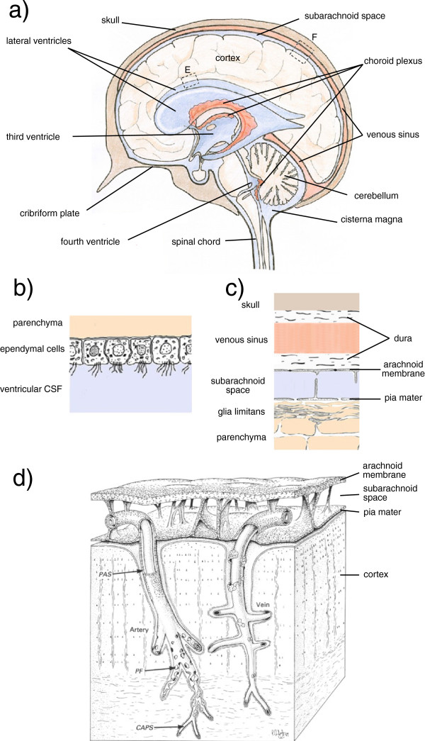
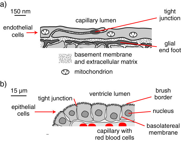
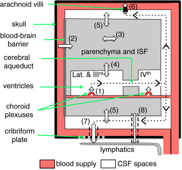
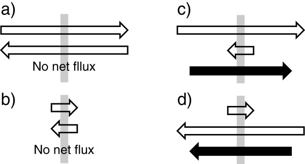
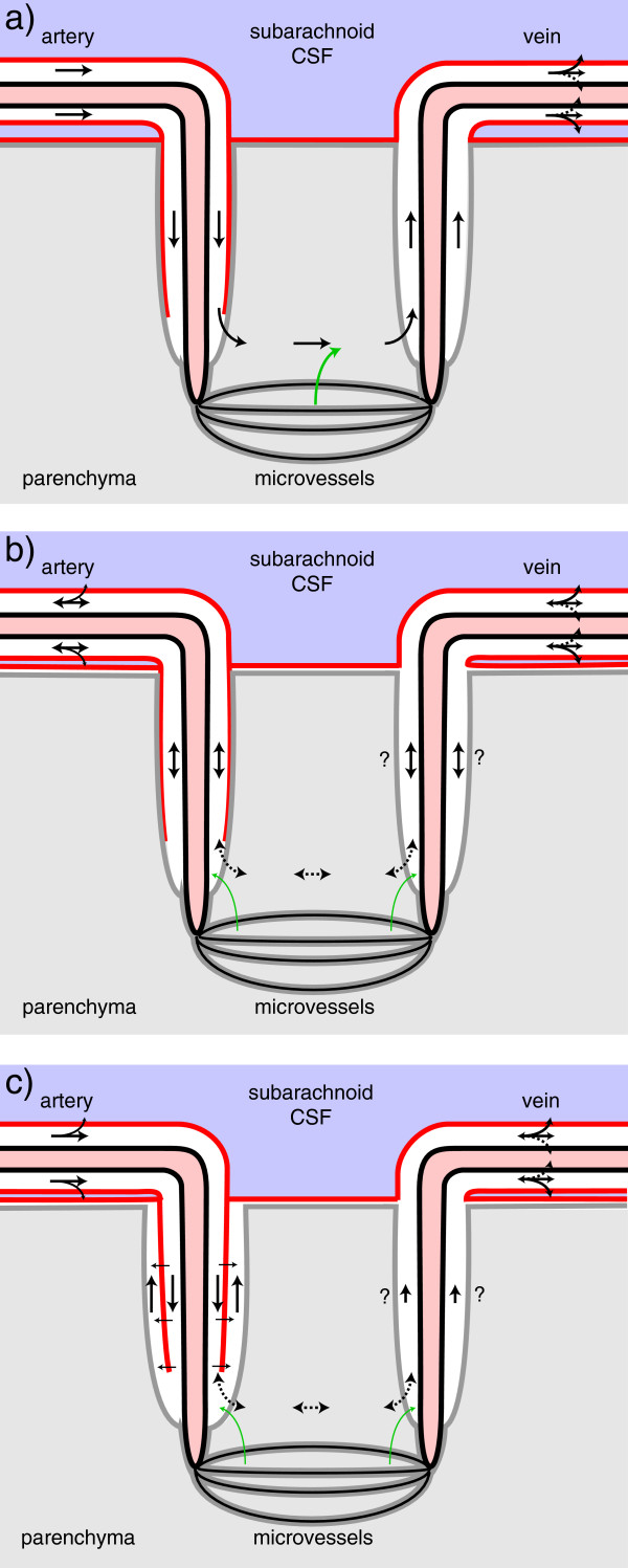
Interstitial fluid (ISF) surrounds the parenchymal cells of the brain and spinal cord while cerebrospinal fluid (CSF) fills the larger spaces within and around the CNS. Regulation of the composition and volume of these fluids is important for effective functioning of brain cells and is achieved by barriers that prevent free exchange between CNS and blood and by mechanisms that secrete fluid of controlled composition into the brain and distribute and reabsorb it. Structures associated with this regular fluid turnover include the choroid plexuses, brain capillaries comprising the blood-brain barrier, arachnoid villi and perineural spaces penetrating the cribriform plate. ISF flow, estimated from rates of removal of markers from the brain, has been thought to reflect rates of fluid secretion across the blood-brain barrier, although this has been questioned because measurements were made under barbiturate anaesthesia possibly affecting secretion and flow and because CSF influx to the parenchyma via perivascular routes may deliver fluid independently of blood-brain barrier secretion. Fluid secretion at the blood-brain barrier is provided by specific transporters that generate solute fluxes so creating osmotic gradients that force water to follow. Any flow due to hydrostatic pressures driving water across the barrier soon ceases unless accompanied by solute transport because water movements modify solute concentrations. CSF is thought to be derived primarily from secretion by the choroid plexuses. Flow rates measured using phase contrast magnetic resonance imaging reveal CSF movements to be more rapid and variable than previously supposed, even implying that under some circumstances net flow through the cerebral aqueduct may be reversed with net flow into the third and lateral ventricles. Such reversed flow requires there to be alternative sites for both generation and removal of CSF. Fluorescent tracer analysis has shown that fluid flow can occur from CSF into parenchyma along periarterial spaces. Whether this represents net fluid flow and whether there is subsequent flow through the interstitium and net flow out of the cortex via perivenous routes, described as glymphatic circulation, remains to be established. Modern techniques have revealed complex fluid movements within the brain. This review provides a critical evaluation of the data.
Overview
The claims and statistics of this paper haven’t been checked yet.
Run the check — free, no account needed. It reads the open-access text and checks the sources it cites.
Integrity signals
Registry
Cites
Statistics
Figures
Verification
Claims the paper makes
The claims and statistics of this paper haven’t been checked yet.
Run the check — free, no account needed. It reads the open-access text and checks the sources it cites.
Claims other papers make about this one
Each is checked against this paper’s own text: does it say what the citing paper says it does?
The cited source says what the claim attributes to it.
In the citing paper
“Other recent studies (252, 255, 547, 555), however, did not support the proposed glymphatic mechanism, nor the convective, pressure-driven fluid flow of CSF from para-arterial to paravenous spaces throughout the parenchymal ECS (31, 30, 255, 296, 547, 255).”
From Blood-Brain Barrier: From Physiology to Disease and Back (2019) · cites this paper as [252]
Self-check
Percentages, sums and reported differences re-computed from the paper’s own numbers.
The self-check needs the paper’s full text.
Citations
Cites
Intravenous Fluids and Acute Kidney Injury
Ding, X., Cheng, Z., & Qian, Q. (2017) · Blood Purification · doi:10.1159/000452702 · Cited by 6,868
Iliff, J. J., Wang, M., Liao, Y., Plogg, B. A., Peng, W., Gundersen, G. A., Benveniste, H., Vates, G. E., Deane, R., Goldman, S. A., et al. (2012) · Science Translational Medicine · doi:10.1126/scitranslmed.3003748 · Cited by 5,824
Sleep Drives Metabolite Clearance from the Adult Brain
Xie, L., Kang, H., Xu, Q., Chen, M. J., Liao, Y., Thiyagarajan, M., O’Donnell, J., Christensen, D. J., Nicholson, C., Iliff, J. J., et al. (2013) · Science · doi:10.1126/science.1241224 · Cited by 5,411
FINE STRUCTURAL LOCALIZATION OF A BLOOD-BRAIN BARRIER TO EXOGENOUS PEROXIDASE
Reese, T. S., & Karnovsky, M. J. (1967) · The Journal of Cell Biology · doi:10.1083/jcb.34.1.207 · Cited by 2,629
JUNCTIONS BETWEEN INTIMATELY APPOSED CELL MEMBRANES IN THE VERTEBRATE BRAIN
Brightman, M., & Reese, T. S. (1969) · The Journal of Cell Biology · doi:10.1083/jcb.40.3.648 · Cited by 2,457
Thermodynamic analysis of the permeability of biological membranes to non-electrolytes
Kedem, O., & Katchalsky, A. (1958) · Biochimica et Biophysica Acta · doi:10.1016/0006-3002(58)90330-5 · Cited by 2,214
On the Absorption of Fluids from the Connective Tissue Spaces
Starling, E. H. (1896) · The Journal of Physiology · doi:10.1113/jphysiol.1896.sp000596 · Cited by 2,126
Diffusion in Brain Extracellular Space
Syková, E., & Nicholson, C. (2008) · Physiological Reviews · doi:10.1152/physrev.00027.2007 · Cited by 1,481
Convection-enhanced delivery of macromolecules in the brain.
Bobo, R. H., Laske, D. W., Akbaşak, A., Morrison, P. F., Dedrick, R. L., & Oldfield, E. H. (1994) · Proceedings of the National Academy of Sciences · doi:10.1073/pnas.91.6.2076 · Cited by 1,442
Effect of anoxia on ion distribution in the brain
Hansen, A. J. (1985) · Physiological Reviews · doi:10.1152/physrev.1985.65.1.101 · Cited by 1,342
Cited by
Sorted by citations
Items 401–500 of 636
Metzger, F., Mischek, D., & Stoffers, F. (2017) · Frontiers in Neuroscience · doi:10.3389/fnins.2017.00241 · Cited by 8
Yatsushiro, S., Sunohara, S., Matsumae, M., Atsumi, H., Horie, T., Kajihara, N., & Kuroda, K. (2022) · Magnetic Resonance in Medical Sciences · doi:10.2463/mrms.mp.2021-0126 · Cited by 8
Mechanical filtration of the cerebrospinal fluid: procedures, systems, and applications
Menéndez‐González, M. (2023) · Expert Review of Medical Devices · doi:10.1080/17434440.2023.2181695 · Cited by 8
A Gateway to the brain: Shuttles for Brain Delivery of Macromolecules
Webster, C. I., & Stanimirovic, D. B. (2015) · Therapeutic Delivery · doi:10.4155/tde.15.78 · Cited by 8
CNS fluid and solute movement: physiology, modelling and imaging
Jones, H. C., Keep, R. F., & Drewes, L. R. (2020) · Fluids and Barriers of the CNS · doi:10.1186/s12987-020-0174-1 · Cited by 8
SERS-Based Microfluidic Bioscreening Platform for Selective Detection of β-Amyloid Peptides
Jaiswal, A., Mishra, S., Dwivedi, P. K., & Verma, S. K. (2024) · Langmuir · doi:10.1021/acs.langmuir.4c03042 · Cited by 7
Lin, X., Wei, L., Li, X., Zeng, L., Tang, Y., Wang, H., Lu, H., Li, C., Wang, H., Chen, J., et al. (2025) · Advanced Science · doi:10.1002/advs.202500362 · Cited by 7
Wu, K., Yun, Z., Xue, W., Yu, T., Dai, A., Han, I., Kotheeranurak, V., Limthongkul, W., Liu, Y., & Liu, Q. (2025) · Materials Today Bio · doi:10.1016/j.mtbio.2025.102514 · Cited by 7
Moore, M. E., Karakaya, E., Altınbaş, Ö., Bartlett, M. J., Adilbay, D., Ergul, A., Yağmurlu, K., Albayram, M., & Albayram, Ö. (2026) · Neurobiology of Aging · doi:10.1016/j.neurobiolaging.2026.02.002 · Cited by 7
Plasma Levels of Amyloid-β Peptides and Tau Protein in Mexican Patients with Alzheimer’s Disease
Castillo-Mendieta, T., Arana-Lechuga, Y., Campos‐Peña, V., Sosa, A. L., Orozco-Suárez, S. A., Pinto‐Almazán, R., Segura-Uribe, J. J., de Tagle, A. J. R.-S., Ruiz-Sánchez, E., & Guerra‐Araiza, C. (2021) · Journal of Alzheimer s Disease · doi:10.3233/jad-200912 · Cited by 7
Reynolds, W. F., Malle, E., & Maki, R. A. (2022) · Antioxidants · doi:10.3390/antiox11122342 · Cited by 7
The possible impact of cervical stenosis on cephalad neuronal dysfunction
Khachatryan, T., & Robinson, J. S. (2018) · Medical Hypotheses · doi:10.1016/j.mehy.2018.06.008 · Cited by 7
Guo, H., Kommidi, H., Maachani, U. B., Voronina, J. C., Zhang, W., Magge, R., Ivanidze, J., Wu, A. P., Souweidane, M. M., Aras, Ö., et al. (2019) · Molecular Pharmaceutics · doi:10.1021/acs.molpharmaceut.9b00485 · Cited by 7
Jaskiw, G. E., Xu, D., Obrenovich, M. E., & Donskey, C. J. (2022) · Metabolomics · doi:10.1007/s11306-021-01866-4 · Cited by 7
Wesselman, H. M., Arceri, L., Nguyen, T. K., Lara, C. M., & Wingert, R. A. (2023) · FEBS Journal · doi:10.1111/febs.17012 · Cited by 7
Bert, R. J., Hayek, S. M., & Yaksh, T. L. (2017) · Anesthesia & Analgesia · doi:10.1213/ane.0000000000002071 · Cited by 7
Raybaud, C. (2018) · Springer eBooks · doi:10.1007/978-3-319-31889-9_44-1 · Cited by 7
Advances in brain barriers and brain fluid research and news from Fluids and Barriers of the CNS
Drewes, L. R., Jones, H. C., & Keep, R. F. (2015) · Fluids and Barriers of the CNS · doi:10.1186/s12987-016-0026-1 · Cited by 7
Boulghobra, A., & Bonose, M. (2022) · Critical Reviews in Analytical Chemistry · doi:10.1080/10408347.2022.2151833 · Cited by 6
Graphene-based neural electrodes: from materials and device fabrication to properties
Doğan, M. Z., & Bayram, C. (2025) · Journal of Materials Chemistry B · doi:10.1039/d5tb01040c · Cited by 6
Advances in Computational Modelling of Head Injury Biomechanics – a Comprehensive Review
Sbriglio, C., Ptak, M., & Kwiatkowski, A. (2025) · Archives of Computational Methods in Engineering · doi:10.1007/s11831-025-10463-w · Cited by 7
Alcaide, D., Cacheux, J., Bancaud, A., Muramatsu, R., & Matsunaga, Y. T. (2023) · Biomaterials Science · doi:10.1039/d3bm00027c · Cited by 7
Morozova, A., Španiel, F., Škoch, A., Brabec, M., Zolotarov, G., Musil, V., & Zach, P. (2024) · NeuroImage · doi:10.1016/j.neuroimage.2024.120871 · Cited by 7
Bioelectronics hydrogels for implantable cardiac and brain disease medical treatment application
Meng, Y., Xu, L., & Cheng, G. (2025) · International Journal of Biological Macromolecules · doi:10.1016/j.ijbiomac.2025.139945 · Cited by 6
Unravelling disease complexity: integrative analysis of multi-omic data in clinical research
Cominetti, O., & Dayon, L. (2025) · Expert Review of Proteomics · doi:10.1080/14789450.2025.2491357 · Cited by 7
Visualization and Characterization of Cerebrospinal Fluid Motion Based on Magnetic Resonance Imaging
Yatsushiro, S., Sunohara, S., Atsumi, H., Matsumae, M., & Kuroda, K. (2018) · InTech eBooks · doi:10.5772/intechopen.73302 · Cited by 6
Brain lymphatic drainage system in fetus and newborn: Birth of a new era of exploration.
Angelis, L. C. D., Witte, M., Bellini, T., Bernas, M. J., Boccardo, F. M., Ramenghi, L. A., & Bellini, C. (2018) · PubMed · Cited by 6
Dudchenko, N. G., Chimagomedova, A. S., Васенина, Е. Е., & Levin, O. S. (2022) · S S Korsakov Journal of Neurology and Psychiatry · doi:10.17116/jnevro202212207120 · Cited by 6
The Emerging Field of Perivascular Flow Dynamics: Biological Relevance and Clinical Applications
Huffman, J. N., Phillips, S. M., Taylor, G. T., & Paul, R. (2016) · Technology & Innovation · doi:10.21300/18.1.2016.63 · Cited by 7
Definition and Pathomechanism of the Intracranial Compartment Syndrome
Tonetti, T., Biondini, S., Minardi, F., Rossi, S., & Picetti, E. (2020) · Hot topics in acute care surgery and trauma · doi:10.1007/978-3-030-55378-4_2 · Cited by 6
Solid-contact ion-selective electrodes for potentiometric determination of phenylethylamine in vitro
Ma, S., Wang, Y., Jiang, L., Hu, R., Luo, Z., & Li, G. (2021) · Measurement Science and Technology · doi:10.1088/1361-6501/ac1235 · Cited by 6
Fagiolino, P. L., & Vázquez, M. (2022) · Current Pharmaceutical Design · doi:10.2174/1381612828666220422091159 · Cited by 6
Cliver, R. N., Castro, N., Russomano, T., Lardieri, G., Quarrie, L., van der Merwe, H., & Vázquez, M. (2022) · Neuroglia · doi:10.3390/neuroglia3030006 · Cited by 6
Ji, S., Liu, X., Ha, J., Ai, L., & Li, Z. (2023) · Journal of Chromatography B · doi:10.1016/j.jchromb.2023.123680 · Cited by 6
Tang, K., Zhou, J., Yang, H., Tan, X., Xie, M., Tao, X., Ge, X., & Xu, J. (2010) · PubMed · Cited by 6
Mild chronic hypoxia and the brain: an ambiguous relationship
Wszędybył-Winklewska, M., Czuba, E., Malinowski, K. S., Waśkow, M., Michalak, K., & Winklewski, P. J. (2026) · Journal of Neuroinflammation · doi:10.1186/s12974-025-03652-8 · Cited by 5
Fujii, N., Nomura, S., Izuma, H., & Ishihara, H. (2024) · Neurologia medico-chirurgica · doi:10.2176/jns-nmc.2023-0277 · Cited by 5
Fluid flow and amyloid transport and aggregation in the brain interstitial space
Iorio, A., Melchionna, S., Derreumaux, P., & Sterpone, F. (2024) · PNAS Nexus · doi:10.1093/pnasnexus/pgae548 · Cited by 6
Hu, J., Xie, S., Chen, T., Liao, Y. L., Qian, Z., & Zhang, L. (2025) · Neuroscience · doi:10.1016/j.neuroscience.2025.02.039 · Cited by 5
Blood–brain mechanics: How brain tissue responds to pulsatile blood flow dynamics
Gao, X., Tian, Y., Liao, Z., Yang, Y., Wu, W., Zhan, W., & Dini, D. (2025) · International Journal of Mechanical Sciences · doi:10.1016/j.ijmecsci.2025.110278 · Cited by 5
An Osmosis-driven 3D-printed brain implant for drug delivery
Ullah, A., Bookwalter, J., Sant, H. J., Azapagic, A., Shea, J., Berlet, R., Jha, N., Bailes, J. E., & Gale, B. K. (2025) · Biomedical Microdevices · doi:10.1007/s10544-025-00759-w · Cited by 5
Liu, Y., Zhang, Y., Wu, C., Zhu, J., Wang, C. Y., Tomko, N., Linetsky, M. D., Salomon, R. G., Ramos-Estébanez, C., Wang, Y., et al. (2017) · Magnetic Resonance in Medicine · doi:10.1002/mrm.26669 · Cited by 5
Amyloid Beta Dynamics in Biological Fluids—Therapeutic Impact
Schreiner, T. G., & Popescu, B. O. (2021) · Journal of Clinical Medicine · doi:10.3390/jcm10245986 · Cited by 5
Warren, K. E., Coupland, K., Hood, R. J., Kang, L., Walker, F. R., & Spratt, N. J. (2023) · Fluids and Barriers of the CNS · doi:10.1186/s12987-023-00427-2 · Cited by 5
Brain Edema Formation in Traumatic Brain Injury
Lemarchant, S., & Badaut, J. (2017) · Elsevier eBooks · doi:10.1016/b978-0-12-803196-4.00013-8 · Cited by 5
Brain lymphatic drainage system – visualization opportunities and current state of the art
Yankova, G., & Bogomyakova, O. B. (2020) · Complex Issues of Cardiovascular Diseases · doi:10.17802/2306-1278-2020-9-3-81-89 · Cited by 5
Shephali, Singh, Y., Mishra, M., Sachan, S., Pathak, N. M., & Shankar, R. (2022) · Anaesthesia Pain & Intensive Care · doi:10.35975/apic.v26i3.1899 · Cited by 5
Biomechanical forces in the aged brain: Relationship to AD
Lee, G. Y., Kim, O., Kim, E. R., & Lee, H. J. (2022) · Life Sciences · doi:10.1016/j.lfs.2022.121237 · Cited by 5
Sharma, A., Gokulchandran, N., Sane, H. M., Kulkarni, P., Kannan, K., Shaikh, Z., Biju, H., Paranjape, A., D'sa, M., & Badhe, P. B. (2025) · Clinical Transplantation and Research · doi:10.4285/ctr.25.0009 · Cited by 4
Transport pathways across the blood-brain barrier for waste clearance and drug delivery
Gröbner, L. S., & Pietrzik, C. U. (2026) · Fluids and Barriers of the CNS · doi:10.1186/s12987-026-00812-7 · Cited by 4
Quantitative and Qualitative Analysis of CSF Flow Dynamics
Токарев, А. С., Talypova, D. A., Terekhin, I. A., & Grin, A. A. (2022) · Russian Sklifosovsky Journal Emergency Medical Care · doi:10.23934/2223-9022-2022-11-1-86-95 · Cited by 4
van der Voort, E. C., van der Plas, M. C. E., & Zwanenburg, J. J. M. (2025) · Interface Focus · doi:10.1098/rsfs.2024.0048 · Cited by 4
Trumbore, C. N. (2018) · Journal of Alzheimer s Disease · doi:10.3233/jad-171080 · Cited by 4
Corbo, C. P., & Fulop, Z. L. (2020) · Microscopy Research and Technique · doi:10.1002/jemt.23457 · Cited by 4
Eggers, C., Hoetelmans, R. M. W., & Läer, S. (2020) · AIDS · doi:10.1097/qad.0000000000002626 · Cited by 4
Neuroimaging in Pediatric Hydrocephalus
Raybaud, C. (2020) · Springer eBooks · doi:10.1007/978-3-319-72168-2_15 · Cited by 4
From neurons to the neuro-glio-vascular unit: Seizures and brain homeostasis in networks
Cresto, N., Janvier, A., & Marchi, N. (2023) · Revue Neurologique · doi:10.1016/j.neurol.2022.12.005 · Cited by 4
Cerebral furin deficiency causes hydrocephalus in mice
Xie, S., Xie, X., Tang, J., Luo, B., Chen, J., Wen, Q., Zhou, J., & Chen, G. (2023) · Genes & Diseases · doi:10.1016/j.gendis.2023.04.037 · Cited by 4
Semyachkina-Glushkovskaya, O. V., Sergeev, K., Игоревна, С. Н., Slepnev, A., Karavaev, A. S., Hramkov, A. N., Prokhorov, M. D., Borovkova, E. I., Blokhina, I., Fedosov, I. V., et al. (2023) · Biomolecules · doi:10.3390/biom13111605 · Cited by 4
Fourth Ventricular Lesions in Metastatic Gliomas: A Rare Predilection?
Khan, M. N., Poulin, A., & Essig, M. (2017) · Brain Tumor Research and Treatment · doi:10.14791/btrt.2017.5.1.24 · Cited by 4
Андреевна, Т. М., & Papin, A. A. (2019) · Journal of Physics Conference Series · doi:10.1088/1742-6596/1268/1/012053 · Cited by 4
Kurochkin, M. A., Fedosov, I. V., & Postnov, D. E. (2021) · The European Physical Journal Plus · doi:10.1140/epjp/s13360-021-01700-9 · Cited by 4
Risk factors for hippocampal cavities in a marginally housed population
Cheng, A., Stubbs, J. L., Barr, A. M., Gicas, K. M., Su, W., Thornton, A. E., Lang, D. J., Hamzah, Y., Leonova, O., MacEwan, W. G., et al. (2022) · Hippocampus · doi:10.1002/hipo.23450 · Cited by 5
The stop and go of glymphatic flow
Kılıç, K., & Devor, A. (2023) · Nature Neuroscience · doi:10.1038/s41593-023-01344-1 · Cited by 4
Wickramasinghe, C. D., Kim, S., & Li, J. (2025) · CPT Pharmacometrics & Systems Pharmacology · doi:10.1002/psp4.70026 · Cited by 3
Sleep is a therapeutic window for photostimulation of drainage of aging brain
Terskov, A. V., Adushkina, V., Alexander, S., Наволокин, Н. А., Blokhina, I., Zlatogorskaya, D., Anastasiia, S.-G., Sonina, K., Evsukova, A., Elizarova, I. V., et al. (2025) · Frontiers of Optoelectronics · doi:10.1007/s12200-025-00168-0 · Cited by 5
Predictive N -Glycan Signatures of Severe Traumatic Brain Injury in Biofluids Using LC–MS/MS
Solomon, J., Onigbinde, S. B., Adeniyi, M., Daramola, O. E., Reyes, C. D. G., Fowowe, M., Bhuiyan, M. M. A. A., Nwaiwu, J. I., Kobeissy, F. H., Mondello, S., et al. (2025) · ACS Omega · doi:10.1021/acsomega.5c10061 · Cited by 3
Diurnal changes of cerebrospinal fluid and global signal coupling
Müller, L., Berberich, C., Heinrich, P., Nuttall, R., Pilge, S., Zott, B., Sorg, C., Wohlschlaeger, A. M., Schneider, G., & Zimmermann, J. (2026) · NeuroImage · doi:10.1016/j.neuroimage.2026.121833 · Cited by 3
Piotrowska, A., Winter, K., Carare, R. O., & Bechmann, I. (2020) · Frontiers in Aging Neuroscience · doi:10.3389/fnagi.2020.00015 · Cited by 3
Tachycardia and hypertension enhance tracer efflux from the spinal cord
Liu, S., Bilston, L. E., Stoodley, M. A., & Hemley, S. J. (2021) · Fluids and Barriers of the CNS · doi:10.1186/s12987-021-00279-8 · Cited by 3
Reet, J. V., Tunnell, K., Anderson, K., Kim, H., Kim, E., Kowsari, K., & Yoo, S. (2023) · ULTRASONOGRAPHY · doi:10.14366/usg.23037 · Cited by 3
Park, S., Park, B. S., Lee, H., Heo, C. M., Ko, J., Lee, D. A., & Park, K. M. (2024) · Frontiers in Neurology · doi:10.3389/fneur.2024.1459356 · Cited by 3
Biological Fluid Flows: Signaling Mediums for Circadian Timing
Duyvesteyn, E., Vizcarra, V. S., Waight, E., Balbuena, E., & Hablitz, L. M. (2025) · Journal of Biological Rhythms · doi:10.1177/07487304251323318 · Cited by 4
Visualization of the periventricular Virchow–Robin spaces with ependymal openings
Tsutsumi, S., Onο, H., Ishii, H., & Yasumoto, Y. (2018) · Child s Nervous System · doi:10.1007/s00381-018-3793-y · Cited by 3
Protoporphyrin IX Based All-Solid-State Ion-Selective Electrodes for Choline Determination In Vitro
Wang, Y., Ma, S., Zou, H.-Q., Wu, Z., Luo, Z., & Li, G. (2021) · Applied Sciences · doi:10.3390/app11125549 · Cited by 3
Mentor, S., & Fisher, D. (2022) · International Journal of Molecular Sciences · doi:10.3390/ijms23052457 · Cited by 4
Rosenstand, N. J., Nielsen, A. S., Skøt, L., Anhøj, S. J., Nielsen, D. G., Højlund, M., & Mellentin, A. I. (2022) · Current Neuropharmacology · doi:10.2174/1570159x21666221229160300 · Cited by 3
Velocity of cerebrospinal fluid in the aqueduct measured by phase‐contrast MRI in rat
Ding, G., Li, L., Chopp, M., Zhang, L., Li, Q., Luo, H., Wei, M., Zhang, J., Boyd, E., Zhang, Z., et al. (2024) · NMR in Biomedicine · doi:10.1002/nbm.5233 · Cited by 5
Kunz, P. B., Maurer, M. A., Vollmer, J., Macháček, M., Weinmann, O., Klisic, J., & Schwab, M. E. (2024) · Neurotherapeutics · doi:10.1016/j.neurot.2024.e00484 · Cited by 3
Estimation of fluid flow velocities in cortical brain tissue driven by the microvasculature
Koch, T., & Mardal, K. (2025) · Interface Focus · doi:10.1098/rsfs.2024.0042 · Cited by 3
Blending Established and New Perspectives on Choroid Plexus-CSF Dynamics
Johanson, C. E., & Keep, R. F. (2020) · Physiology in health and disease · doi:10.1007/978-1-0716-0536-3_2 · Cited by 3
Chai, Y., Zhang, H., Robles, C., Kim, A. S., Janhanshad, N., Thompson, P. M., van der Werf, Y. D., van Heese, E. M., Kim, J., Joo, E. Y., et al. (2025) · medRxiv · doi:10.1101/2025.03.19.25324269 · Cited by 3
Hypoxemia exerts detrimental effects on the choroid plexuses and cerebrospinal fluid system in rats
Barakat, R., Al‐Sarraf, H., & Redzic, Z. B. (2025) · Fluids and Barriers of the CNS · doi:10.1186/s12987-024-00613-w · Cited by 2
Xu, G., Zhang, Q., Li, Y., Du, A., Xu, X., Bai, R., Bi, C., Li, Y., Zhang, T., Zhang, Z., et al. (2025) · ACS Omega · doi:10.1021/acsomega.5c03650 · Cited by 2
Slotman, D. J., Frijlingh, M., Juffermans, L. J. M., Bartels, L. W., MOONEN, C. T. W., den Bosch, T. V., Boomsma, M. F., & Huirne, J. A. F. (2025) · Insights into Imaging · doi:10.1186/s13244-025-02033-2 · Cited by 2
Wang, G., Feng, C., Ma, Z., Zhao, L., Yosri, M., Wang, Y., Gao, X., Zhu, W., Li, H., Song, H., et al. (2025) · Cell Reports · doi:10.1016/j.celrep.2025.116658 · Cited by 2
van der Voort, E. C., van der Plas, M. C. E., Gosselink, M. W. J. M., & Zwanenburg, J. (2025) · Fluids and Barriers of the CNS · doi:10.1186/s12987-025-00735-9 · Cited by 3
Rheological Characterization of Cerebrospinal Fluid Under Different Temperature Conditions
Bauer, T.-C., Bradt, E., Hild, S., Gruber, A., Rossmann, T., Ruiz-Navarro, F., Oberndorfer, J., Stefanits, H., & Kráčalík, M. (2026) · Fluids · doi:10.3390/fluids11020038 · Cited by 2
Brain Barriers in Normal Pressure Hydrocephalus: Mechanisms and Therapeutic Perspectives
Lee, M. J., Kim, Y. J., Chang, H. J., An, J. H., Pyo, C. H., Jeong, J., Kim, T., Chang, J., Oh, E., Chung, W., et al. (2026) · Journal of Advanced Research · doi:10.1016/j.jare.2026.04.026 · Cited by 2
Tools for studying drug transport and metabolism in the brain
Pitcher, M. R., & Quevedo, J. (2015) · Expert Opinion on Drug Metabolism & Toxicology · doi:10.1517/17425255.2016.1132307 · Cited by 2
Cerebrospinal and Interstitial Fluids: Production, Outflow, and Circulation
Hladky, S. B., & Barrand, M. A. (2023) · Springer eBooks · doi:10.1007/978-3-031-39558-1_7 · Cited by 2
Self-citationDiffusion Mediates Molecular Transport through the Perivascular Space in the Brain
Tanaka, M., Hirayoshi, Y., Minatani, S., Hasegawa, I., & Itoh, Y. (2024) · International Journal of Molecular Sciences · doi:10.3390/ijms25052480 · Cited by 2
Fagiolino, P. L., Talevi, A., Vázquez, M., & Ibarra, M. (2024) · Springer eBooks · doi:10.1007/978-3-031-50419-8_20 · Cited by 2
Navigating the nanoscale: Principles of body navigation
Abbasi, S., Dirisala, A., Gonzalez‐Carter, D., Paraiso, W. K. D., Li, J., & Tockary, T. A. (2024) · Elsevier eBooks · doi:10.1016/b978-0-12-823994-0.00002-5 · Cited by 2
Bonow, R. H., Hanak, B. W., & Browd, S. R. (2018) · Elsevier eBooks · doi:10.1016/b978-0-323-43140-8.00008-1 · Cited by 2
Electrokinesis hypothesis of neuron selection for synapse formation and pruning
Esen, E. (2021) · Medical Hypotheses · doi:10.1016/j.mehy.2021.110701 · Cited by 2
The Cerebral Venous System: New Pathophysiological Theories and Diseases Related to Veins Occlusion
Mantovani, G., & Scerrati, A. (2022) · IntechOpen eBooks · doi:10.5772/intechopen.102351 · Cited by 2
Fu, Y., Yun, W., Zhang, Z., Ma, Y., Xiao, L., Zhang, M., & Zhu, W. (2022) · BMC Neurology · doi:10.1186/s12883-022-02744-9 · Cited by 2
Duclos, B. A., Roegge, C. S., Dobson, H., Haller, S., Bodner, J., Pannem, S., Gesaman, J., & Nourmohammadi, A. (2023) · Translational Medicine Communications · doi:10.1186/s41231-023-00136-w · Cited by 2
Physiological brain pulsations
Kiviniemi, V. J. (2023) · Elsevier eBooks · doi:10.1016/b978-0-323-91688-2.00012-6 · Cited by 2
Metadata
- Authors
- Hladky, S. B., & Barrand, M. A.
- Affiliations
- University of Cambridge (GB)
- Corresponding author
- Hladky, Stephen B
- ORCID
- Hladky, Stephen B
- Venue
- Fluids and Barriers of the CNS
- Published
- 2014
- Volume
- 11
- Issue
- 1
- Pages
- 26
- ISSN
- 2045-8118 (ISSN-L)
- PMC
- PMC4326185
- OpenAlex
- W2096525076
- Cited by
- 633
- References
- 215
- Citations by year
- 2026: 37 · 2025: 49 · 2024: 57 · 2023: 65 · 2022: 77 · 2021: 75 · 2020: 45 · 2019: 65 · 2018: 61 · 2017: 58 · 2016: 26 · 2015: 17
- FWCI
- 3.85
- Type
- journal article
- Language
- en
- Open access
- gold
- doi.org
- Licence
- cc-by
- Locations
- Fluids and Barriers of the CNS (publishedVersion, cc-by)
- Europe PMC (PubMed Central) (publishedVersion, cc-by)
- PubMed Central (publishedVersion, cc-by)
- PubMed (publishedVersion)
- Topics
- Health Sciences › Medicine › Clinical Neurology › Cerebrospinal fluid and hydrocephalus
- Life Sciences › Biochemistry, Genetics and Molecular Biology › Molecular Biology › Ion Transport and Channel Regulation
- Life Sciences › Neuroscience › Neurology › Barrier Structure and Function Studies
- Keywords
- interstitial fluid, cerebrospinal fluid, cerebrospinal fluid flow, blood-brain barrier, choroid plexus, arachnoid villi, perineural space, cribriform plate, cerebrospinal fluid production, CSF reabsorption, phase-contrast MRI, osmotic gradient, solute transport, perivascular spaces, barbiturate anesthesia
About these checks
Registry, retraction, Cites and statistics results are computed, not judged, with one exception: a printed reference that no index lists is read once by a model before it counts as a citation error. Claims are extracted by a model and kept only when their quote appears verbatim in the text; each cited source is then checked, with a model’s help, for whether it says what the claim relies on. Those verdicts can be wrong — read the claim’s quote and the cited source before relying on one.
Statistics and arithmetic are recomputed, not judged.
Confidence bands are shown only for fields calibrated on our annotated test set. None is calibrated yet, so no band is shown.
What counts as a claim ›How we measure confidence ›How we check this ›
Are you an author, or do you see a problem with this page? Request a review
