

The DOI matches its Crossref record; 215 of the 215 references it cites were found in a registry or an index.
Mechanisms of fluid movement into, through and out of the brain: evaluation of the evidence
Open accessAbstract
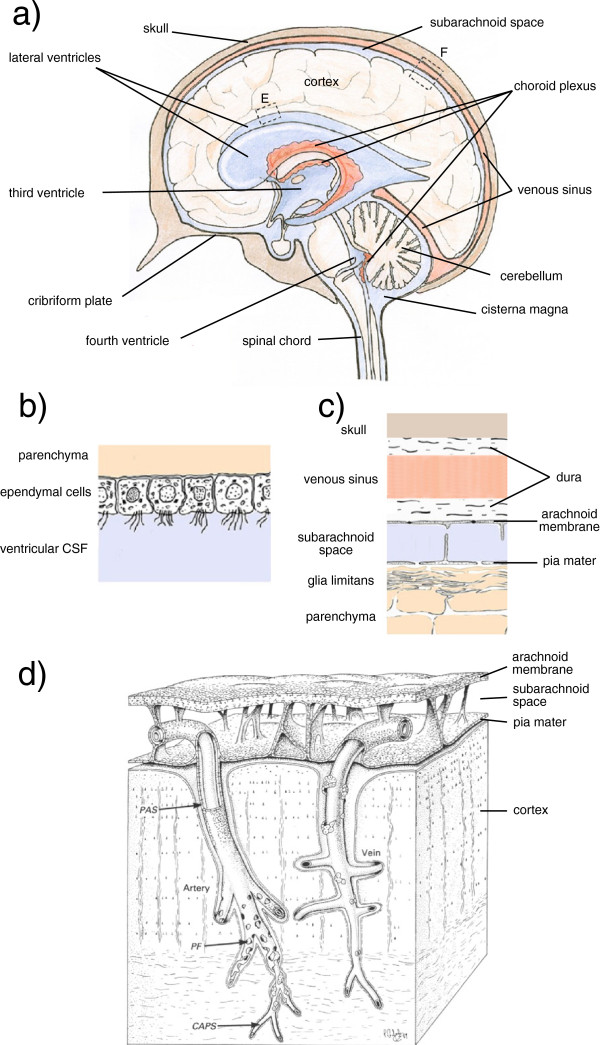
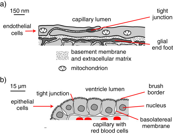
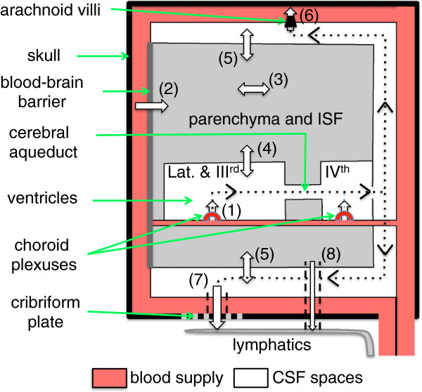
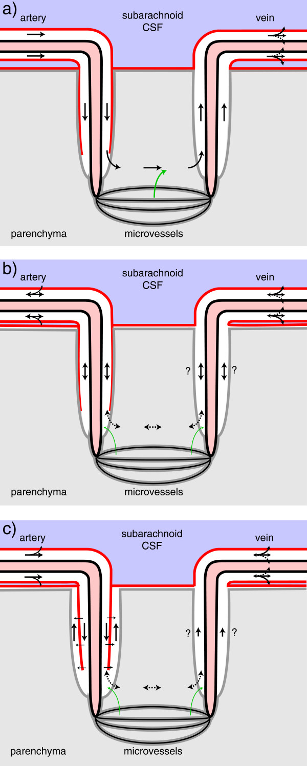
Interstitial fluid (ISF) surrounds the parenchymal cells of the brain and spinal cord while cerebrospinal fluid (CSF) fills the larger spaces within and around the CNS. Regulation of the composition and volume of these fluids is important for effective functioning of brain cells and is achieved by barriers that prevent free exchange between CNS and blood and by mechanisms that secrete fluid of controlled composition into the brain and distribute and reabsorb it. Structures associated with this regular fluid turnover include the choroid plexuses, brain capillaries comprising the blood-brain barrier, arachnoid villi and perineural spaces penetrating the cribriform plate. ISF flow, estimated from rates of removal of markers from the brain, has been thought to reflect rates of fluid secretion across the blood-brain barrier, although this has been questioned because measurements were made under barbiturate anaesthesia possibly affecting secretion and flow and because CSF influx to the parenchyma via perivascular routes may deliver fluid independently of blood-brain barrier secretion. Fluid secretion at the blood-brain barrier is provided by specific transporters that generate solute fluxes so creating osmotic gradients that force water to follow. Any flow due to hydrostatic pressures driving water across the barrier soon ceases unless accompanied by solute transport because water movements modify solute concentrations. CSF is thought to be derived primarily from secretion by the choroid plexuses. Flow rates measured using phase contrast magnetic resonance imaging reveal CSF movements to be more rapid and variable than previously supposed, even implying that under some circumstances net flow through the cerebral aqueduct may be reversed with net flow into the third and lateral ventricles. Such reversed flow requires there to be alternative sites for both generation and removal of CSF. Fluorescent tracer analysis has shown that fluid flow can occur from CSF into parenchyma along periarterial spaces. Whether this represents net fluid flow and whether there is subsequent flow through the interstitium and net flow out of the cortex via perivenous routes, described as glymphatic circulation, remains to be established. Modern techniques have revealed complex fluid movements within the brain. This review provides a critical evaluation of the data.
Overview
The claims and statistics of this paper haven’t been checked yet.
Run the check — free, no account needed. It reads the open-access text and checks the sources it cites.
Integrity signals
Registry
Cites
Statistics
Figures
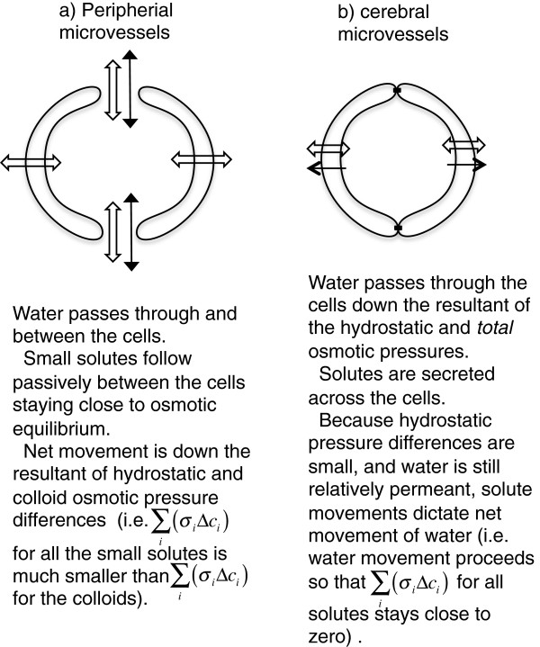
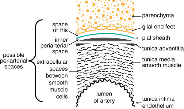
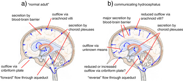
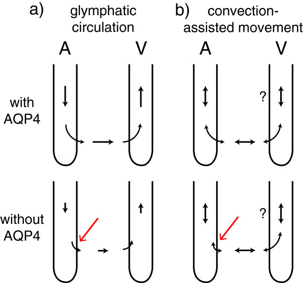
Verification
Claims the paper makes
The claims and statistics of this paper haven’t been checked yet.
Run the check — free, no account needed. It reads the open-access text and checks the sources it cites.
Claims other papers make about this one
Each is checked against this paper’s own text: does it say what the citing paper says it does?
The cited source says what the claim attributes to it.
In the citing paper
“Other recent studies (252, 255, 547, 555), however, did not support the proposed glymphatic mechanism, nor the convective, pressure-driven fluid flow of CSF from para-arterial to paravenous spaces throughout the parenchymal ECS (31, 30, 255, 296, 547, 255).”
From Blood-Brain Barrier: From Physiology to Disease and Back (2019) · cites this paper as [252]
Self-check
Percentages, sums and reported differences re-computed from the paper’s own numbers.
The self-check needs the paper’s full text.
Citations
Cites
Intravenous Fluids and Acute Kidney Injury
Ding, X., Cheng, Z., & Qian, Q. (2017) · Blood Purification · doi:10.1159/000452702 · Cited by 6,868
Iliff, J. J., Wang, M., Liao, Y., Plogg, B. A., Peng, W., Gundersen, G. A., Benveniste, H., Vates, G. E., Deane, R., Goldman, S. A., et al. (2012) · Science Translational Medicine · doi:10.1126/scitranslmed.3003748 · Cited by 5,824
Sleep Drives Metabolite Clearance from the Adult Brain
Xie, L., Kang, H., Xu, Q., Chen, M. J., Liao, Y., Thiyagarajan, M., O’Donnell, J., Christensen, D. J., Nicholson, C., Iliff, J. J., et al. (2013) · Science · doi:10.1126/science.1241224 · Cited by 5,411
FINE STRUCTURAL LOCALIZATION OF A BLOOD-BRAIN BARRIER TO EXOGENOUS PEROXIDASE
Reese, T. S., & Karnovsky, M. J. (1967) · The Journal of Cell Biology · doi:10.1083/jcb.34.1.207 · Cited by 2,629
JUNCTIONS BETWEEN INTIMATELY APPOSED CELL MEMBRANES IN THE VERTEBRATE BRAIN
Brightman, M., & Reese, T. S. (1969) · The Journal of Cell Biology · doi:10.1083/jcb.40.3.648 · Cited by 2,457
Thermodynamic analysis of the permeability of biological membranes to non-electrolytes
Kedem, O., & Katchalsky, A. (1958) · Biochimica et Biophysica Acta · doi:10.1016/0006-3002(58)90330-5 · Cited by 2,214
On the Absorption of Fluids from the Connective Tissue Spaces
Starling, E. H. (1896) · The Journal of Physiology · doi:10.1113/jphysiol.1896.sp000596 · Cited by 2,126
Diffusion in Brain Extracellular Space
Syková, E., & Nicholson, C. (2008) · Physiological Reviews · doi:10.1152/physrev.00027.2007 · Cited by 1,481
Convection-enhanced delivery of macromolecules in the brain.
Bobo, R. H., Laske, D. W., Akbaşak, A., Morrison, P. F., Dedrick, R. L., & Oldfield, E. H. (1994) · Proceedings of the National Academy of Sciences · doi:10.1073/pnas.91.6.2076 · Cited by 1,442
Effect of anoxia on ion distribution in the brain
Hansen, A. J. (1985) · Physiological Reviews · doi:10.1152/physrev.1985.65.1.101 · Cited by 1,342
Cited by
Sorted by citations
Items 101–200 of 636
Programming tissue-sensing T cells that deliver therapies to the brain
Simic, M., Watchmaker, P. B., Gupta, S., Wang, Y., Sagan, S. A., Duecker, J., Shepherd, C., Diebold, D., Pineo-Cavanaugh, P. L., Haegelin, J., et al. (2024) · Science · doi:10.1126/science.adl4237 · Cited by 98
Harnessing cerebrospinal fluid circulation for drug delivery to brain tissues
Naseri Kouzehgarani, G., Feldsien, T., Engelhard, H. H., Mirakhur, K. K., Phipps, C., Nimmrich, V., Clausznitzer, D., & Lefebvre, D. R. (2021) · Advanced Drug Delivery Reviews · doi:10.1016/j.addr.2021.03.002 · Cited by 95
Klebe, D. W., McBride, D. W., Krafft, P. R., Flores, J. J., Tang, J., & Zhang, J. H. (2019) · Journal of Neuroscience Research · doi:10.1002/jnr.24394 · Cited by 92
Crisp, S. J., Kullmann, D. M., & Vincent, A. C. (2016) · Nature reviews. Neuroscience · doi:10.1038/nrn.2015.27 · Cited by 92
AQP1 and AQP4 Contribution to Cerebrospinal Fluid Homeostasis
Trillo-Contreras, J. L., Toledo‐Aral, J. J., Echevarrı́a, M., & Villadiego, J. (2019) · Cells · doi:10.3390/cells8020197 · Cited by 95
Endo-lysosomal proteins and ubiquitin CSF concentrations in Alzheimer’s and Parkinson’s disease
Sjödin, S., Brinkmalm, G., Öhrfelt, A., Parnetti, L., Paciotti, S., Hansson, O., Hardy, J., Blennow, K., Zetterberg, H., & Brinkmalm, A. (2019) · Alzheimer s Research & Therapy · doi:10.1186/s13195-019-0533-9 · Cited by 94
Janigro, D., Bailey, D. M., Lehmann, S., Badaut, J., O’flynn, R., Hirtz, C., & Marchi, N. (2021) · Frontiers in Neurology · doi:10.3389/fneur.2020.577312 · Cited by 98
(2020) · International Review of Neurobiology · doi:10.1016/bs.irn.2020.02.006 · Cited by 87
A translational platform PBPK model for antibody disposition in the brain
Chang, H.-Y., Wu, S., Meno‐Tetang, G. M. L., & Shah, D. K. (2019) · Journal of Pharmacokinetics and Pharmacodynamics · doi:10.1007/s10928-019-09641-8 · Cited by 87
Sharp, M. K., Carare, R. O., & Martin, B. A. (2019) · Fluids and Barriers of the CNS · doi:10.1186/s12987-019-0132-y · Cited by 87
Antibody pharmacokinetics in rat brain determined using microdialysis
Chang, H.-Y., Morrow, K., Bonacquisti, E. E., Zhang, W., & Shah, D. K. (2018) · mAbs · doi:10.1080/19420862.2018.1473910 · Cited by 87
The need for mathematical modelling of spatial drug distribution within the brain
Vendel, E., Rottschäfer, V., & de Lange, E. C. M. (2019) · Fluids and Barriers of the CNS · doi:10.1186/s12987-019-0133-x · Cited by 87
Matsumae, M., Sato, O., Hirayama, A., Hayashi, N., TAKIZAWA, K., Atsumi, H., & Sorimachi, T. (2016) · Neurologia medico-chirurgica · doi:10.2176/nmc.ra.2016-0020 · Cited by 84
Melatonin Treatment Enhances Aβ Lymphatic Clearance in a Transgenic Mouse Model of Amyloidosis
Pappolla, M. A., Matsubara, E., Vidal, R., Pacheco‐Quinto, J., Pöeggeler, B., Zagorski, M. G., & Sambamurti, K. (2018) · Current Alzheimer Research · doi:10.2174/1567205015666180411092551 · Cited by 83
Eide, P. K., Valnes, L. M., Pripp, A. H., Mardal, K., & Ringstad, G. A. (2019) · Journal of Cerebral Blood Flow & Metabolism · doi:10.1177/0271678x19874790 · Cited by 83
Proulx, S. T., & Engelhardt, B. (2022) · Journal of Internal Medicine · doi:10.1111/joim.13469 · Cited by 86
Spijkerman, J. M., Geurts, L. J., Siero, J. C. W., Hendrikse, J., Luijten, P. R., & Zwanenburg, J. J. M. (2018) · Journal of Magnetic Resonance Imaging · doi:10.1002/jmri.26181 · Cited by 87
Fate of nanoparticles in the central nervous system after intrathecal injection in healthy mice
Householder, K. T., Dharmaraj, S., Sandberg, D. I., Wechsler-Reya, R. J., & Sirianni, R. W. (2019) · Scientific Reports · doi:10.1038/s41598-019-49028-w · Cited by 83
Webster, C. I., Caram‐Salas, N. L., Haqqani, A. S., Thom, G., Brown, L., Rennie, K., Yogi, Á., Costain, W. J., Brunette, E., & Stanimirovic, D. B. (2016) · The FASEB Journal · doi:10.1096/fj.201500078 · Cited by 83
Li, D., Liu, S., Yu, T., Liu, Z., Sun, S., Bragin, D. E., Shirokov, A. A., Наволокин, Н. А., Bragina, O. A., Hu, Z., et al. (2023) · Nature Communications · doi:10.1038/s41467-023-41710-y · Cited by 81
The Dynamic Relationship between the Glymphatic System, Aging, Memory, and Sleep
Voumvourakis, K. I., Sideri, E., Papadimitropoulos, G. N., Tsantzali, I., Hewlett, P., Kitsos, D. K., Stefanou, M., Bonakis, A., Giannopoulos, S., Tsivgoulis, G., et al. (2023) · Biomedicines · doi:10.3390/biomedicines11082092 · Cited by 84
Chowdhury, E. A., Meno‐Tetang, G. M. L., Chang, H. Y., Wu, S., Huang, H. W., Jamier, T., Chandran, J. S., & Shah, D. K. (2021) · Advanced Drug Delivery Reviews · doi:10.1016/j.addr.2021.01.017 · Cited by 80
Basal lamina changes in neurodegenerative disorders
Nguyen, B., Bix, G., & Yao, Y. (2021) · Molecular Neurodegeneration · doi:10.1186/s13024-021-00502-y · Cited by 75
Intracranial pressure elevation alters CSF clearance pathways
Vinje, V., Eklund, A., Mardal, K., Rognes, M. E., & Støverud, K.-H. (2020) · Fluids and Barriers of the CNS · doi:10.1186/s12987-020-00189-1 · Cited by 78
Effect of Systemic Inflammation in the CNS: A Silent History of Neuronal Damage
Solano, M. V. M., Lara, C. S., Sánchez-Garibay, C., Soto-Rojas, L. O., Escobedo-Ávila, I., Tena‐Suck, M. L., Ortiz-Butrón, R., Choreño-Parra, J. A., Romero‐López, J. P., & Meléndez-Camargo, M. E. (2023) · International Journal of Molecular Sciences · doi:10.3390/ijms241511902 · Cited by 78
Fluid Flow and Mass Transport in Brain Tissue
Ray, L., & Heys, J. J. (2019) · Fluids · doi:10.3390/fluids4040196 · Cited by 78
Pulsatile flow drivers in brain parenchyma and perivascular spaces: a resistance network model study
Rey, J. A., & Sarntinoranont, M. (2018) · Fluids and Barriers of the CNS · doi:10.1186/s12987-018-0105-6 · Cited by 74
A dynamic perfusion based blood-brain barrier model for cytotoxicity testing and drug permeation
Elbakary, B., & Badhan, R. K. S. (2020) · Scientific Reports · doi:10.1038/s41598-020-60689-w · Cited by 74
Dysregulation of Astrocyte Ion Homeostasis and Its Relevance for Stroke-Induced Brain Damage
van Putten, M. J. A. M., Fahlke, C. M., Kafitz, K. W., Hofmeijer, J., & Rose, C. R. (2021) · International Journal of Molecular Sciences · doi:10.3390/ijms22115679 · Cited by 79
Wang, Q., Delva, L., Weinreb, P. H., Pepinsky, R. B., Graham, D. L., Veizaj, E., Cheung, A. E., Chen, W., Nestorov, I. A., Rohde, E., et al. (2018) · Fluids and Barriers of the CNS · doi:10.1186/s12987-018-0093-6 · Cited by 71
Yamamoto, Y., Välitalo, P. A. J., van den Berg, D., Hartman, R., van den Brink, W., Wong, Y. C., Huntjens, D. R. H., Proost, J. H., Vermeulen, A., Krauwinkel, W. J. J., et al. (2016) · Pharmaceutical Research · doi:10.1007/s11095-016-2065-3 · Cited by 71
Chowdhury, E. A., Noorani, B., Alqahtani, F., Bhalerao, A., Raut, S., Sivandzade, F., & Cucullo, L. (2021) · Journal of Cerebral Blood Flow & Metabolism · doi:10.1177/0271678x20985946 · Cited by 77
Wostyn, P., Groot, V. D., Dam, D. V., Audenaert, K., Killer, H. E., & Deyn, P. P. D. (2017) · BioMed Research International · doi:10.1155/2017/5123148 · Cited by 70
Paravascular Pathways in the Eye: Is There an ‘Ocular Glymphatic System'?
Denniston, A. K., & Keane, P. A. (2015) · Investigative Ophthalmology & Visual Science · doi:10.1167/iovs.15-17243 · Cited by 73
Addressing Blood–Brain Barrier Impairment in Alzheimer’s Disease
Sharma, C., Woo, H., & Kim, S. R. (2022) · Biomedicines · doi:10.3390/biomedicines10040742 · Cited by 72
A real-time in vivo clearance assay for quantification of glymphatic efflux
Plá, V., Bork, P., Harnpramukkul, A., Olveda, G., Ladrón-de-Guevara, A., Giannetto, M. J., Hussain, R., Wang, W., Kelley, D. H., Hablitz, L. M., et al. (2022) · Cell Reports · doi:10.1016/j.celrep.2022.111320 · Cited by 64
Klarica, M., Radoš, M., & Orešković, D. (2019) · Neuroscience · doi:10.1016/j.neuroscience.2019.06.032 · Cited by 66
Role of aquaporin-4 polarization in extracellular solute clearance
Bojarskaite, L., Nafari, S., Ravnanger, A. K., Frey, M. M., Skauli, N., Åbjørsbråten, K. S., Roth, L. C., Amiry-Moghaddam, M. R., Nagelhus, E. A., Ottersen, O. P., et al. (2024) · Fluids and Barriers of the CNS · doi:10.1186/s12987-024-00527-7 · Cited by 65
Shape Memory Polyurethane and its Composites for Various Applications
Gupta, A., Maharjan, A., & Kim, B. S. (2019) · Applied Sciences · doi:10.3390/app9214694 · Cited by 65
Cerebrovascular activity is a major factor in the cerebrospinal fluid flow dynamics
Wang, Y., van Gelderen, P., de Zwart, J. A., Özbay, P. S., Mandelkow, H., Picchioni, D., & Duyn, J. H. (2022) · NeuroImage · doi:10.1016/j.neuroimage.2022.119362 · Cited by 63
Decker, Y., Krämer, J., Li, X., Müller, A., Scheller, A., Fassbender, K. C., & Proulx, S. T. (2021) · JCI Insight · doi:10.1172/jci.insight.150881 · Cited by 64
Chen, Z., Liu, C., Zhang, J., Relkin, N. R., Xing, Y., & Li, Y. (2017) · Fluids and Barriers of the CNS · doi:10.1186/s12987-017-0062-5 · Cited by 65
Tashima, T. (2020) · Molecules · doi:10.3390/molecules25215188 · Cited by 61
Fame, R. M., Cortés‐Campos, C., & Sive, H. L. (2020) · BioEssays · doi:10.1002/bies.201900186 · Cited by 64
Pham, W., Lynch, M., Spitz, G., O'Brien, T. J., Vivash, L. E., Sinclair, B., & Law, M. (2022) · Frontiers in Neuroscience · doi:10.3389/fnins.2022.1021311 · Cited by 63
A Kinetic Model for Blood Biomarker Levels After Mild Traumatic Brain Injury
Azizi, S., Hier, D. B., Allen, B., Obafemi-Ajayi, T., Olbricht, G. R., Thimgan, M. S., & Wunsch, D. C. (2021) · Frontiers in Neurology · doi:10.3389/fneur.2021.668606 · Cited by 58
Hladky, S. B., & Barrand, M. A. (2024) · Fluids and Barriers of the CNS · doi:10.1186/s12987-024-00532-w · Cited by 61
Self-citationBraun, C., Sakamoto, A., Fuchs, H., Ishiguro, N., Suzuki, S., Cui, Y., Klinder, K., Watanabe, M., Terasaki, T., & Sauer, A. (2017) · Molecular Pharmaceutics · doi:10.1021/acs.molpharmaceut.7b00449 · Cited by 58
Kumar, N. N., Pizzo, M. E., Nehra, G., Wilken-Resman, B., Boroumand, S., & Thorne, R. G. (2018) · Bioconjugate Chemistry · doi:10.1021/acs.bioconjchem.8b00548 · Cited by 60
Hersey, M., Samaranayake, S., Berger, S. N., Tavakoli, N. S., Mena, S., Nijhout, H. F., Reed, M. C., Best, J. A., Blakely, R., Reagan, L. P., et al. (2021) · Journal of Neuroscience · doi:10.1523/jneurosci.2618-20.2021 · Cited by 57
The glymphatic system’s role in traumatic brain injury-related neurodegeneration
Peters, M. E., & Lyketsos, C. G. (2023) · Molecular Psychiatry · doi:10.1038/s41380-023-02070-7 · Cited by 57
Assessment of the dynamics of human glymphatic system by near‐infrared spectroscopy
Myllylä, T., Harju, M., Korhonen, V. O., Bykov, A. V., Kiviniemi, V. J., & Meglinski, I. V. (2017) · Journal of Biophotonics · doi:10.1002/jbio.201700123 · Cited by 59
Cancel, L. M., Arias, K., Bikson, M., & Tarbell, J. M. (2018) · Scientific Reports · doi:10.1038/s41598-018-27524-9 · Cited by 61
Dlouhy, A. C., Bailey, D. K., Steimle, B. L., Parker, H. V., & Kosman, D. J. (2019) · Journal of Biological Chemistry · doi:10.1074/jbc.ra118.005142 · Cited by 57
van Osch, M. J. P., Wåhlin, A., Scheyhing, P., Mossige, I., Hirschler, L., Eklund, A., Mogensen, K., Gomolka, R. S., Radbruch, A., Qvarlander, S., et al. (2024) · NMR in Biomedicine · doi:10.1002/nbm.5159 · Cited by 58
Koschnitzky, J. E., Keep, R. F., Limbrick, D. D., McAllister, J. P., Morris, J. A., Strahle, J. M., & Yung, Y. C. (2018) · Fluids and Barriers of the CNS · doi:10.1186/s12987-018-0096-3 · Cited by 56
Neurovascular-glymphatic dysfunction and white matter lesions
Sabayan, B., & Westendorp, R. G. J. (2021) · GeroScience · doi:10.1007/s11357-021-00361-x · Cited by 58
Toro, E. F., Celant, M., Zhang, Q., Contarino, C., Agarwal, N., Linninger, A. A., & Müller, L. O. (2021) · International Journal for Numerical Methods in Biomedical Engineering · doi:10.1002/cnm.3532 · Cited by 52
Quantitative Water Permeability Mapping of Blood-Brain-Barrier Dysfunction in Aging
Ford, J. N., Zhang, Q., Sweeney, E. M., Merkler, A. E., de Leon, M. J., Gupta, A. K., Nguyen, T. D., & Ivanidze, J. (2022) · Frontiers in Aging Neuroscience · doi:10.3389/fnagi.2022.867452 · Cited by 53
Photodynamic opening of the blood‐brain barrier and pathways of brain clearing
Semyachkina-Glushkovskaya, O. V., Chekhonin, V. P., Borisova, E. G., Fedosov, I. V., Александрович, Н. А., Abdurashitov, A. S., Shirokov, A. A., Khlebtsov, B. N., Lyubun, Y. V., Наволокин, Н. А., et al. (2018) · Journal of Biophotonics · doi:10.1002/jbio.201700287 · Cited by 52
Mehta, N. H., Suss, R. A., Dyke, J. P., Theise, N. D., Chiang, G. C.-Y., Strauß, S., Saint-Louis, L. A., Li, Y., Pahlajani, S. S., Babaria, V., et al. (2022) · Neurobiology of Disease · doi:10.1016/j.nbd.2022.105776 · Cited by 51
Li, Z., Antila, S., Nurmi, H. J., Chilov, D., Korhonen, E. A., Fang, S., Karaman, S., Engelhardt, B., & Alitalo, K. K. (2023) · Science Immunology · doi:10.1126/sciimmunol.abq0375 · Cited by 51
Xiang, J., Routhe, L. G., Wilkinson, D. A., Hua, Y., Moos, T., Xi, G., & Keep, R. F. (2017) · Fluids and Barriers of the CNS · doi:10.1186/s12987-017-0056-3 · Cited by 51
Smith, A. J., & Verkman, A. S. (2019) · The Journal of Physiology · doi:10.1113/jp277636 · Cited by 52
Amyloid‐beta is present in human lymph nodes and greatly enriched in those of the cervical region
Nauen, D. W., & Troncoso, J. C. (2021) · Alzheimer s & Dementia · doi:10.1002/alz.12385 · Cited by 52
Fluid outflow in the rat spinal cord: the role of perivascular and paravascular pathways
Liu, S., Lam, M. A., Sial, A. W., Hemley, S. J., Bilston, L. E., & Stoodley, M. A. (2018) · Fluids and Barriers of the CNS · doi:10.1186/s12987-018-0098-1 · Cited by 52
Aquaporin‐4‐independent volume dynamics of astroglial endfeet during cortical spreading depression
Rosic, A. B., Dukefoss, D. B., Åbjørsbråten, K. S., Tang, W., Jensen, V., Ottersen, O. P., Enger, R., & Nagelhus, E. A. (2019) · Glia · doi:10.1002/glia.23604 · Cited by 51
Paravascular spaces: entry to or exit from the brain?
Bakker, E. N. T. P., Naessens, D. M. P., & VanBavel, E. (2018) · Experimental Physiology · doi:10.1113/ep087424 · Cited by 50
Uriarte, M., Francesco, P. N. D., Fernández, G., Castrogiovanni, D. C., D'Arcangelo, M., Imbernón, M., Cantel, S., Denoyelle, S., Fehrentz, J., Prætorius, J., et al. (2021) · Molecular and Cellular Endocrinology · doi:10.1016/j.mce.2021.111449 · Cited by 49
Quantitative analysis of macroscopic solute transport in the murine brain
Ray, L., Pike, M. M., Simon, M. J., Iliff, J. J., & Heys, J. J. (2021) · Fluids and Barriers of the CNS · doi:10.1186/s12987-021-00290-z · Cited by 50
Aquaporin Water Channels and Hydrocephalus
Verkman, A. S., Tradtrantip, L., Smith, A. J., & Yao, X. (2016) · Pediatric Neurosurgery · doi:10.1159/000452168 · Cited by 49
Croci, M., Vinje, V., & Rognes, M. E. (2019) · Fluids and Barriers of the CNS · doi:10.1186/s12987-019-0152-7 · Cited by 50
Computational modelling of fluid and solute transport in the brain
Martinac, A. D., & Bilston, L. E. (2019) · Biomechanics and Modeling in Mechanobiology · doi:10.1007/s10237-019-01253-y · Cited by 49
Cao, M. C., Cawston, E. E., Chen, G. L., Brooks, C., Douwes, J. E., McLean, D., Graham, E. S., Dragunow, M., & Scotter, E. L. (2022) · BMC Neurology · doi:10.1186/s12883-022-02730-1 · Cited by 49
Knowler, S. P., Galea, G. L., & Rusbridge, C. (2018) · Frontiers in Veterinary Science · doi:10.3389/fvets.2018.00171 · Cited by 47
Sullivan, J. M., Mazur, C., Wolf, D. A., Horky, L. L., Currier, N. V., Fitzsimmons, B., Hesterman, J. Y., Pauplis, R., Haller, S., Powers, B. E., et al. (2020) · Journal of Translational Medicine · doi:10.1186/s12967-020-02461-2 · Cited by 47
Muddying the water in brain edema?
Smith, A. J., Jin, B., & Verkman, A. S. (2015) · Trends in Neurosciences · doi:10.1016/j.tins.2015.04.006 · Cited by 47
Sun, Y.-R., Lv, Q.-K., Liu, J., Wang, F., & Liu, C. (2025) · Neurobiology of Disease · doi:10.1016/j.nbd.2025.106791 · Cited by 48
Removal of aquaporin-4 from glial and ependymal membranes causes brain water accumulation
Vindedal, G. F., Thoren, A. E., Jensen, V., Klungland, A., Zhang, Y., Holtzman, M. J., Ottersen, O. P., & Nagelhus, E. A. (2016) · Molecular and Cellular Neuroscience · doi:10.1016/j.mcn.2016.10.004 · Cited by 45
Neuroimaging in Pediatric Hydrocephalus
Krishnan, P., Raybaud, C., Palasamudram, S., & Shroff, M. M. (2019) · The Indian Journal of Pediatrics · doi:10.1007/s12098-019-02962-z · Cited by 47
Fournier, A. P., Gauberti, M., Quenault, A., Vivien, D., Macrez, R., & Docagne, F. (2018) · Journal of Cerebral Blood Flow & Metabolism · doi:10.1177/0271678x18754732 · Cited by 47
Monro-Kellie 4.0: moving from intracranial pressure to intracranial dynamics
Brasil, S., Patriota, G. C., Godoy, D. A., Paranhos, J. L. D. R., Rubiano, A. M., & Paiva, W. S. (2025) · Critical Care · doi:10.1186/s13054-025-05476-7 · Cited by 45
The role of circadian rhythm in choroid plexus functions
Quintela, T., Furtado, A., Duarte, A. C., Gonçalves, I., Myung, J., & Santos, C. R. A. (2021) · Progress in Neurobiology · doi:10.1016/j.pneurobio.2021.102129 · Cited by 45
Translational Approaches for Brain Delivery of Biologics via Cerebrospinal Fluid
Sadekar, S., Bowen, M., Cai, H., Jamalian, S., Rafidi, H., Shatz-Binder, W., Lafrance‐Vanasse, J., Chan, P., Meilandt, W. J., Oldendorp, A., et al. (2022) · Clinical Pharmacology & Therapeutics · doi:10.1002/cpt.2531 · Cited by 44
Intranasal Pathway for Nanoparticles to Enter the Central Nervous System
Kou, D., Gao, Y. L., Li, C., Zhou, D., Lu, K., Wang, N., Zhang, R., Yang, Z., Zhou, Y., Chen, L., et al. (2023) · Nano Letters · doi:10.1021/acs.nanolett.2c05056 · Cited by 42
Ozceylan, O., & Bayındır, Z. S. (2024) · ACS Omega · doi:10.1021/acsomega.4c01774 · Cited by 43
MR assessment of pediatric hydrocephalus: a road map
Raybaud, C. (2015) · Child s Nervous System · doi:10.1007/s00381-015-2888-y · Cited by 42
Blood–Brain Barrier, Lymphatic Clearance, and Recovery: Ariadne’s Thread in Labyrinths of Hypotheses
SEMYACHKINA-GLUSHKOVSKAYA, O. V., Postnov, D. E., & Kurths, J. (2018) · International Journal of Molecular Sciences · doi:10.3390/ijms19123818 · Cited by 42
Fluid metabolic pathways after subarachnoid hemorrhage
Zhou, J., Guo, P., Guo, Z., Sun, X., Chen, Y., & Feng, H. (2021) · Journal of Neurochemistry · doi:10.1111/jnc.15458 · Cited by 42
Impediment of Cerebrospinal Fluid Drainage Through Glymphatic System in Glioma
Xu, D., Zhou, J., Mei, H., Li, H., Sun, W., & Xu, H. (2022) · Frontiers in Oncology · doi:10.3389/fonc.2021.790821 · Cited by 42
Wolf, D. A., Hesterman, J. Y., Sullivan, J. M., Orcutt, K. D., Silva, M. D., Lobo, M. R., Wellman, T. J., Hoppin, J., & Verma, A. (2016) · JCI Insight · doi:10.1172/jci.insight.85311 · Cited by 42
Yoo, S., Kim, H., Kim, J., Kim, E., Kowsari, K., Reet, J. V., & Yoon, K. (2022) · Scientific Reports · doi:10.1038/s41598-022-17314-9 · Cited by 42
Emerging non-invasive MRI techniques for glymphatic system assessment in neurodegenerative disease
Botta, D., Hutuca, I., Ghoul, E. E., Sveikata, L., Assal, F., Lövblad, K., & Kurz, F. T. (2025) · Journal of Neuroradiology · doi:10.1016/j.neurad.2025.101322 · Cited by 42
Yan, Y., Song, L., Madinya, J. J., Ma, T., & Li, Y. (2017) · Tissue Engineering Part A · doi:10.1089/ten.tea.2016.0400 · Cited by 41
Fluid Management in Acute Brain Injury
Rossi, S., Picetti, E., Zoerle, T., Carbonara, M., Zanier, E. R., & Stocchetti, N. (2018) · Current Neurology and Neuroscience Reports · doi:10.1007/s11910-018-0885-8 · Cited by 43
Cilia-driven flows in the brain third ventricle
Gregor Eichele, C., Bodenschatz, E., Ditte, Z., Günther, A.-K., Kapoor, S., Wang, Y., & Westendorf, C. (2019) · Philosophical Transactions of the Royal Society B Biological Sciences · doi:10.1098/rstb.2019.0154 · Cited by 47
Martinez, J., Razo-Gutierrez, C., Le, C., Courville, R., Pimentel, C., Liu, C., Fung, S. E., Tuttobene, M. R., Phan, K., Vila, A. J., et al. (2021) · Scientific Reports · doi:10.1038/s41598-021-81714-6 · Cited by 41
Guidetti, M., Arlotti, M., Bocci, T., Bianchi, A. M., Parazzini, M., Ferrucci, R., & Priori, A. (2022) · Biomedicines · doi:10.3390/biomedicines10102333 · Cited by 41
CSF circulation and dispersion yield rapid clearance from intracranial compartments
Hornkjøl, M., Valnes, L. M., Ringstad, G. A., Rognes, M. E., Eide, P. K., Mardal, K., & Vinje, V. (2022) · Frontiers in Bioengineering and Biotechnology · doi:10.3389/fbioe.2022.932469 · Cited by 41
Diem, A. K., Carare, R. O., Weller, R. O., & Bressloff, N. W. (2018) · PLoS ONE · doi:10.1371/journal.pone.0205276 · Cited by 41
Metadata
- Authors
- Hladky, S. B., & Barrand, M. A.
- Affiliations
- University of Cambridge (GB)
- Corresponding author
- Hladky, Stephen B
- ORCID
- Hladky, Stephen B
- Venue
- Fluids and Barriers of the CNS
- Published
- 2014
- Volume
- 11
- Issue
- 1
- Pages
- 26
- ISSN
- 2045-8118 (ISSN-L)
- PMC
- PMC4326185
- OpenAlex
- W2096525076
- Cited by
- 633
- References
- 215
- Citations by year
- 2026: 37 · 2025: 49 · 2024: 57 · 2023: 65 · 2022: 77 · 2021: 75 · 2020: 45 · 2019: 65 · 2018: 61 · 2017: 58 · 2016: 26 · 2015: 17
- FWCI
- 3.85
- Type
- journal article
- Language
- en
- Open access
- gold
- doi.org
- Licence
- cc-by
- Locations
- Fluids and Barriers of the CNS (publishedVersion, cc-by)
- Europe PMC (PubMed Central) (publishedVersion, cc-by)
- PubMed Central (publishedVersion, cc-by)
- PubMed (publishedVersion)
- Topics
- Health Sciences › Medicine › Clinical Neurology › Cerebrospinal fluid and hydrocephalus
- Life Sciences › Biochemistry, Genetics and Molecular Biology › Molecular Biology › Ion Transport and Channel Regulation
- Life Sciences › Neuroscience › Neurology › Barrier Structure and Function Studies
- Keywords
- interstitial fluid, cerebrospinal fluid, cerebrospinal fluid flow, blood-brain barrier, choroid plexus, arachnoid villi, perineural space, cribriform plate, cerebrospinal fluid production, CSF reabsorption, phase-contrast MRI, osmotic gradient, solute transport, perivascular spaces, barbiturate anesthesia
About these checks
Registry, retraction, Cites and statistics results are computed, not judged, with one exception: a printed reference that no index lists is read once by a model before it counts as a citation error. Claims are extracted by a model and kept only when their quote appears verbatim in the text; each cited source is then checked, with a model’s help, for whether it says what the claim relies on. Those verdicts can be wrong — read the claim’s quote and the cited source before relying on one.
Statistics and arithmetic are recomputed, not judged.
Confidence bands are shown only for fields calibrated on our annotated test set. None is calibrated yet, so no band is shown.
What counts as a claim ›How we measure confidence ›How we check this ›
Are you an author, or do you see a problem with this page? Request a review
