

The DOI matches its Crossref record; 215 of the 215 references it cites were found in a registry or an index.
Mechanisms of fluid movement into, through and out of the brain: evaluation of the evidence
Open accessAbstract
Interstitial fluid (ISF) surrounds the parenchymal cells of the brain and spinal cord while cerebrospinal fluid (CSF) fills the larger spaces within and around the CNS. Regulation of the composition and volume of these fluids is important for effective functioning of brain cells and is achieved by barriers that prevent free exchange between CNS and blood and by mechanisms that secrete fluid of controlled composition into the brain and distribute and reabsorb it. Structures associated with this regular fluid turnover include the choroid plexuses, brain capillaries comprising the blood-brain barrier, arachnoid villi and perineural spaces penetrating the cribriform plate. ISF flow, estimated from rates of removal of markers from the brain, has been thought to reflect rates of fluid secretion across the blood-brain barrier, although this has been questioned because measurements were made under barbiturate anaesthesia possibly affecting secretion and flow and because CSF influx to the parenchyma via perivascular routes may deliver fluid independently of blood-brain barrier secretion. Fluid secretion at the blood-brain barrier is provided by specific transporters that generate solute fluxes so creating osmotic gradients that force water to follow. Any flow due to hydrostatic pressures driving water across the barrier soon ceases unless accompanied by solute transport because water movements modify solute concentrations. CSF is thought to be derived primarily from secretion by the choroid plexuses. Flow rates measured using phase contrast magnetic resonance imaging reveal CSF movements to be more rapid and variable than previously supposed, even implying that under some circumstances net flow through the cerebral aqueduct may be reversed with net flow into the third and lateral ventricles. Such reversed flow requires there to be alternative sites for both generation and removal of CSF. Fluorescent tracer analysis has shown that fluid flow can occur from CSF into parenchyma along periarterial spaces. Whether this represents net fluid flow and whether there is subsequent flow through the interstitium and net flow out of the cortex via perivenous routes, described as glymphatic circulation, remains to be established. Modern techniques have revealed complex fluid movements within the brain. This review provides a critical evaluation of the data.
Overview
The claims and statistics of this paper haven’t been checked yet.
Run the check — free, no account needed. It reads the open-access text and checks the sources it cites.
Integrity signals
Registry
Cites
Statistics
Figures
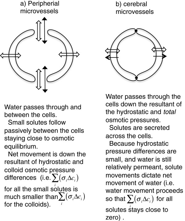
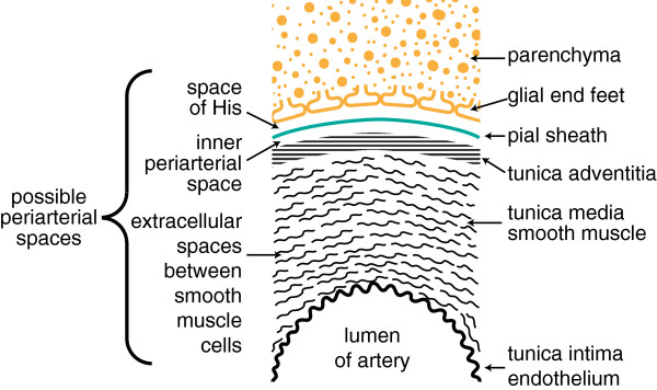
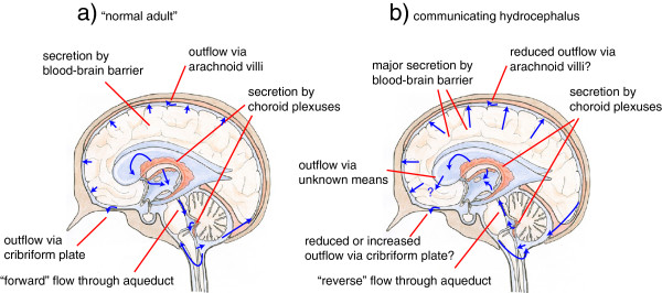
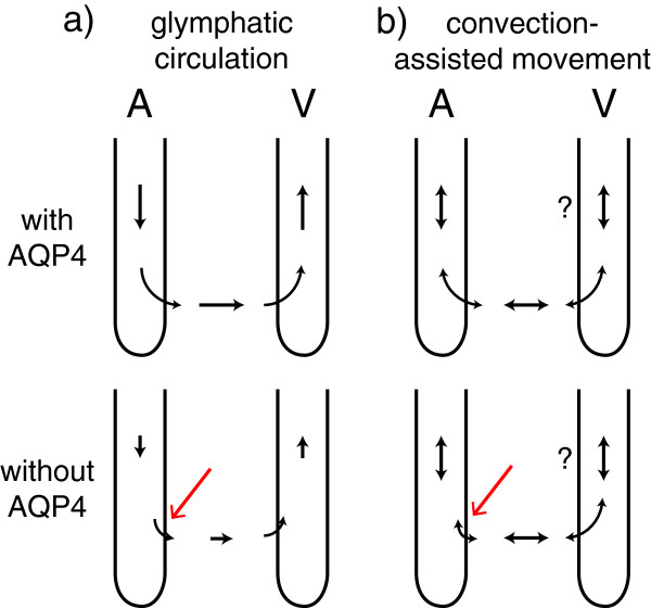
Verification
Claims the paper makes
The claims and statistics of this paper haven’t been checked yet.
Run the check — free, no account needed. It reads the open-access text and checks the sources it cites.
Claims other papers make about this one
Each is checked against this paper’s own text: does it say what the citing paper says it does?
The cited source says what the claim attributes to it.
In the citing paper
“Other recent studies (252, 255, 547, 555), however, did not support the proposed glymphatic mechanism, nor the convective, pressure-driven fluid flow of CSF from para-arterial to paravenous spaces throughout the parenchymal ECS (31, 30, 255, 296, 547, 255).”
From Blood-Brain Barrier: From Physiology to Disease and Back (2019) · cites this paper as [252]
Self-check
Percentages, sums and reported differences re-computed from the paper’s own numbers.
The self-check needs the paper’s full text.
Citations
Cites
Intravenous Fluids and Acute Kidney Injury
Ding, X., Cheng, Z., & Qian, Q. (2017) · Blood Purification · doi:10.1159/000452702 · Cited by 6,868
Iliff, J. J., Wang, M., Liao, Y., Plogg, B. A., Peng, W., Gundersen, G. A., Benveniste, H., Vates, G. E., Deane, R., Goldman, S. A., et al. (2012) · Science Translational Medicine · doi:10.1126/scitranslmed.3003748 · Cited by 5,824
Sleep Drives Metabolite Clearance from the Adult Brain
Xie, L., Kang, H., Xu, Q., Chen, M. J., Liao, Y., Thiyagarajan, M., O’Donnell, J., Christensen, D. J., Nicholson, C., Iliff, J. J., et al. (2013) · Science · doi:10.1126/science.1241224 · Cited by 5,411
FINE STRUCTURAL LOCALIZATION OF A BLOOD-BRAIN BARRIER TO EXOGENOUS PEROXIDASE
Reese, T. S., & Karnovsky, M. J. (1967) · The Journal of Cell Biology · doi:10.1083/jcb.34.1.207 · Cited by 2,629
JUNCTIONS BETWEEN INTIMATELY APPOSED CELL MEMBRANES IN THE VERTEBRATE BRAIN
Brightman, M., & Reese, T. S. (1969) · The Journal of Cell Biology · doi:10.1083/jcb.40.3.648 · Cited by 2,457
Thermodynamic analysis of the permeability of biological membranes to non-electrolytes
Kedem, O., & Katchalsky, A. (1958) · Biochimica et Biophysica Acta · doi:10.1016/0006-3002(58)90330-5 · Cited by 2,214
On the Absorption of Fluids from the Connective Tissue Spaces
Starling, E. H. (1896) · The Journal of Physiology · doi:10.1113/jphysiol.1896.sp000596 · Cited by 2,126
Diffusion in Brain Extracellular Space
Syková, E., & Nicholson, C. (2008) · Physiological Reviews · doi:10.1152/physrev.00027.2007 · Cited by 1,481
Convection-enhanced delivery of macromolecules in the brain.
Bobo, R. H., Laske, D. W., Akbaşak, A., Morrison, P. F., Dedrick, R. L., & Oldfield, E. H. (1994) · Proceedings of the National Academy of Sciences · doi:10.1073/pnas.91.6.2076 · Cited by 1,442
Effect of anoxia on ion distribution in the brain
Hansen, A. J. (1985) · Physiological Reviews · doi:10.1152/physrev.1985.65.1.101 · Cited by 1,342
Cited by
Sorted by citations
Blood–brain barrier breakdown in Alzheimer disease and other neurodegenerative disorders
Sweeney, M. D., Sagare, A. P., & Zlokovic, B. V. (2018) · Nature Reviews Neurology · doi:10.1038/nrneurol.2017.188 · Cited by 3,076
The Neurovascular Unit Coming of Age: A Journey through Neurovascular Coupling in Health and Disease
Iadecola, C. (2017) · Neuron · doi:10.1016/j.neuron.2017.07.030 · Cited by 2,480
Blood-Brain Barrier: From Physiology to Disease and Back
Sweeney, M. D., Zhao, Z., Montagne, A., Nelson, A. R., & Zlokovic, B. V. (2019) · Physiological Reviews · doi:10.1152/physrev.00050.2017 · Cited by 2,312
Clearance systems in the brain—implications for Alzheimer disease
Tarasoff-Conway, J. M., Carare, R. O., Osorio, R. S., Glodzik, L., Butler, T., Fieremans, E., Axel, L., Rusinek, H., Nicholson, C., Zlokovic, B. V., et al. (2015) · Nature Reviews Neurology · doi:10.1038/nrneurol.2015.119 · Cited by 1,678
The movers and shapers in immune privilege of the CNS
Engelhardt, B., Vajkoczy, P., & Weller, R. O. (2017) · Nature Immunology · doi:10.1038/ni.3666 · Cited by 893
Perivascular spaces in the brain: anatomy, physiology and pathology
Wardlaw, J. M., Benveniste, H., Nedergaard, M., Zlokovic, B. V., Mestre, H., Lee, H., Doubal, F. N., Brown, R., Ramirez, J., MacIntosh, B. J., et al. (2020) · Nature Reviews Neurology · doi:10.1038/s41582-020-0312-z · Cited by 870
The Glymphatic System in Central Nervous System Health and Disease: Past, Present, and Future
Plog, B. A., & Nedergaard, M. (2017) · Annual Review of Pathology Mechanisms of Disease · doi:10.1146/annurev-pathol-051217-111018 · Cited by 800
Aquaporin-4-dependent glymphatic solute transport in the rodent brain
Mestre, H., Hablitz, L. M., Xavier, A. L., Feng, W., Zou, W., Pu, T., Monai, H., Murlidharan, G., Castellanos Rivera, R. M., Simon, M. J., et al. (2018) · eLife · doi:10.7554/elife.40070 · Cited by 738
Glymphatic MRI in idiopathic normal pressure hydrocephalus
Ringstad, G., Vatnehol, S. A. S., & Eide, P. K. (2017) · Brain · doi:10.1093/brain/awx191 · Cited by 722
Development and functions of the choroid plexus–cerebrospinal fluid system
Lun, M. P., Monuki, E. S., & Lehtinen, M. K. (2015) · Nature reviews. Neuroscience · doi:10.1038/nrn3921 · Cited by 719
Alzheimer’s disease: A matter of blood–brain barrier dysfunction?
Montagne, A., Zhao, Z., & Zlokovic, B. V. (2017) · Journal of Experimental Medicine · doi:10.1084/jem.20171406 · Cited by 672
Understanding the functions and relationships of the glymphatic system and meningeal lymphatics
Louveau, A., Plog, B. A., Antila, S., Alitalo, K., Nedergaard, M., & Kipnis, J. (2017) · Journal of Clinical Investigation · doi:10.1172/jci90603 · Cited by 640
Rasmussen, M. K., Mestre, H., & Nedergaard, M. (2021) · Physiological Reviews · doi:10.1152/physrev.00031.2020 · Cited by 638
The role of brain barriers in fluid movement in the CNS: is there a ‘glymphatic’ system?
Abbott, N. J., Pizzo, M. E., Preston, J. E., Janigro, D., & Thorne, R. G. (2018) · Acta Neuropathologica · doi:10.1007/s00401-018-1812-4 · Cited by 593
The Glymphatic System and Waste Clearance with Brain Aging: A Review
Benveniste, H., Liu, X., Koundal, S., Sanggaard, S., Lee, H., & Wardlaw, J. (2019) · Gerontology · doi:10.1159/000490349 · Cited by 517
A balanced view of the cerebrospinal fluid composition and functions: Focus on adult humans
Spector, R., Snodgrass, S. R., & Johanson, C. E. (2015) · Experimental Neurology · doi:10.1016/j.expneurol.2015.07.027 · Cited by 467
Blood-Brain Barrier and Delivery of Protein and Gene Therapeutics to Brain
Pardridge, W. M. (2020) · Frontiers in Aging Neuroscience · doi:10.3389/fnagi.2019.00373 · Cited by 405
Proulx, S. T. (2021) · Cellular and Molecular Life Sciences · doi:10.1007/s00018-020-03706-5 · Cited by 412
Erickson, M. A., & Banks, W. A. (2018) · Pharmacological Reviews · doi:10.1124/pr.117.014647 · Cited by 398
The glymphatic system: Current understanding and modeling
Bohr, T., Hjorth, P. G., Holst, S. C., Hrabětová, S., Kiviniemi, V. J., Lilius, T. O., Lundgaard, I., Mardal, K., Martens, E. A., Mori, Y., et al. (2022) · iScience · doi:10.1016/j.isci.2022.104987 · Cited by 394
The development and functions of multiciliated epithelia
Spassky, N., & Meunier, A. (2017) · Nature Reviews Molecular Cell Biology · doi:10.1038/nrm.2017.21 · Cited by 390
Choroid plexus and the blood–cerebrospinal fluid barrier in disease
Solár, P., Zamani, A., Kubíčková, L., Dubový, P., & Joukal, M. (2020) · Fluids and Barriers of the CNS · doi:10.1186/s12987-020-00196-2 · Cited by 365
Smith, A. J., Yao, X., Dix, J. A., Jin, B.-J., & Verkman, A. S. (2017) · eLife · doi:10.7554/elife.27679 · Cited by 362
Bakker, E. N. T. P., Bacskai, B. J., Arbel-Ornath, M., Aldea, R., Bedussi, B., Morris, A. W. J., Weller, R. O., & Carare, R. O. (2016) · Cellular and Molecular Neurobiology · doi:10.1007/s10571-015-0273-8 · Cited by 353
Simon, M. J., & Iliff, J. J. (2015) · Biochimica et Biophysica Acta (BBA) - Molecular Basis of Disease · doi:10.1016/j.bbadis.2015.10.014 · Cited by 349
Cerebrovascular Smooth Muscle Cells as the Drivers of Intramural Periarterial Drainage of the Brain
Aldea, R., Weller, R. O., Wilcock, D. M., Carare, R. O., & Richardson, G. W. (2019) · Frontiers in Aging Neuroscience · doi:10.3389/fnagi.2019.00001 · Cited by 340
Cerebrospinal fluid dynamics and intracranial pressure elevation in neurological diseases
Bothwell, S. W., Janigro, D., & Patabendige, A. (2019) · Fluids and Barriers of the CNS · doi:10.1186/s12987-019-0129-6 · Cited by 348
Brain Extracellular Space: The Final Frontier of Neuroscience
Nicholson, C., & Hrabětová, S. (2017) · Biophysical Journal · doi:10.1016/j.bpj.2017.06.052 · Cited by 321
Boehler, C., Carli, S., Fadiga, L., Stieglitz, T., & Asplund, M. (2020) · Nature Protocols · doi:10.1038/s41596-020-0389-2 · Cited by 324
The blood–brain barrier in psychosis
Pollak, T. A., Drndarski, S., Stone, J. M., David, A. S., McGuire, P. K., & Abbott, N. J. (2017) · The Lancet Psychiatry · doi:10.1016/s2215-0366(17)30293-6 · Cited by 311
Eide, P. K., Vatnehol, S. A. S., Emblem, K. E., & Ringstad, G. (2018) · Scientific Reports · doi:10.1038/s41598-018-25666-4 · Cited by 299
Hladky, S. B., & Barrand, M. A. (2016) · Fluids and Barriers of the CNS · doi:10.1186/s12987-016-0040-3 · Cited by 304
Self-citationInterstitial solute transport in 3D reconstructed neuropil occurs by diffusion rather than bulk flow
Holter, K. E., Kehlet, B., Devor, A., Sejnowski, T. J., Dale, A. M., Omholt, S. W., Ottersen, O. P., Nagelhus, E. A., Mardal, K.-A., & Pettersen, K. H. (2017) · Proceedings of the National Academy of Sciences · doi:10.1073/pnas.1706942114 · Cited by 282
Glymphatic solute transport does not require bulk flow
Asgari, M., de Zélicourt, D., & Kurtcuoglu, V. (2016) · Scientific Reports · doi:10.1038/srep38635 · Cited by 286
Pizzo, M. E., Wolak, D. J., Kumar, N. N., Brunette, E., Brunnquell, C. L., Hannocks, M., Abbott, N. J., Meyerand, M. E., Sorokin, L., Stanimirovic, D. B., et al. (2018) · The Journal of Physiology · doi:10.1113/jp275105 · Cited by 272
The glymphatic hypothesis: the theory and the evidence
Hladky, S. B., & Barrand, M. A. (2022) · Fluids and Barriers of the CNS · doi:10.1186/s12987-021-00282-z · Cited by 278
Self-citationT cell responses in the central nervous system
Korn, T., & Kallies, A. (2017) · Nature Reviews Immunology · doi:10.1038/nri.2016.144 · Cited by 268
Fatigue, Sleep, and Autoimmune and Related Disorders
Zielinski, M. R., Systrom, D. M., & Rose, N. R. (2019) · Frontiers in Immunology · doi:10.3389/fimmu.2019.01827 · Cited by 279
Lymphatic drainage system of the brain: A novel target for intervention of neurological diseases
Sun, B., Wang, L.-H., Yang, T., Sun, J., Mao, L., Yang, M., Yuan, H., Colvin, R. A., & Yang, X. (2017) · Progress in Neurobiology · doi:10.1016/j.pneurobio.2017.08.007 · Cited by 263
Cerebrospinal Fluid Clearance in Alzheimer Disease Measured with Dynamic PET
de Leon, M. J., Li, Y., Okamura, N., Tsui, W. H., Saint-Louis, L. A., Glodzik, L., Osorio, R. S., Fortea, J., Butler, T., Pirraglia, E., et al. (2017) · Journal of Nuclear Medicine · doi:10.2967/jnumed.116.187211 · Cited by 246
The glymphatic system: implications for drugs for central nervous system diseases
Lohela, T. J., Lilius, T. O., & Nedergaard, M. (2022) · Nature Reviews Drug Discovery · doi:10.1038/s41573-022-00500-9 · Cited by 254
Brain Microvascular Pericytes in Vascular Cognitive Impairment and Dementia
Uemura, M. T., Maki, T., Ihara, M., Lee, V. M. ‐., & Trojanowski, J. Q. (2020) · Frontiers in Aging Neuroscience · doi:10.3389/fnagi.2020.00080 · Cited by 246
Non-invasive MR imaging of human brain lymphatic networks with connections to cervical lymph nodes
Albayram, S., Smith, G., Tufan, F., Tuna, İ. S., Bostancıklıoğlu, M., Zile, M. R., & Albayram, Ö. (2022) · Nature Communications · doi:10.1038/s41467-021-27887-0 · Cited by 243
Inflammation in acquired hydrocephalus: pathogenic mechanisms and therapeutic targets
Karimy, J. K., Reeves, B. C., Damisah, E. C., Duy, P. Q., Antwi, P., David, W. B., Wang, K. X.-L., Schiff, S. J., Limbrick, D. D., Alper, S. L., et al. (2020) · Nature Reviews Neurology · doi:10.1038/s41582-020-0321-y · Cited by 234
Zhang, L., & Hargens, A. R. (2017) · Physiological Reviews · doi:10.1152/physrev.00017.2016 · Cited by 234
Molecular mechanisms of brain water transport
MacAulay, N. (2021) · Nature reviews. Neuroscience · doi:10.1038/s41583-021-00454-8 · Cited by 230
Rapid lymphatic efflux limits cerebrospinal fluid flow to the brain
Ma, Q., Ries, M., Decker, Y., Müller, A., Riner, C., Bücker, A., Fassbender, K., Detmar, M., & Proulx, S. T. (2019) · Acta Neuropathologica · doi:10.1007/s00401-018-1916-x · Cited by 210
Hladky, S. B., & Barrand, M. A. (2018) · Fluids and Barriers of the CNS · doi:10.1186/s12987-018-0113-6 · Cited by 206
Self-citationJin, B.-J., Smith, A. J., & Verkman, A. S. (2016) · Journal of General Physiology · doi:10.1085/jgp.201611684 · Cited by 204
Biofluid biomarkers of traumatic brain injury
Agoston, D. V., Shutes‐David, A., & Peskind, E. R. (2017) · Brain Injury · doi:10.1080/02699052.2017.1357836 · Cited by 202
Hydrocephalus: historical analysis and considerations for treatment
Hochstetler, A., Raskin, J. S., & Blazer‐Yost, B. L. (2022) · European journal of medical research · doi:10.1186/s40001-022-00798-6 · Cited by 190
General Anesthesia Inhibits the Activity of the “Glymphatic System”
Gakuba, C., Gaberel, T., Goursaud, S., Bourges, J., Di Palma, C., Quenault, A., de Lizarrondo, S. M., Vivien, D., & Gauberti, M. (2018) · Theranostics · doi:10.7150/thno.19154 · Cited by 190
Paravascular spaces at the brain surface: Low resistance pathways for cerebrospinal fluid flow
Bedussi, B., Almasian, M., de Vos, J., VanBavel, E., & Bakker, E. N. (2018) · Journal of Cerebral Blood Flow & Metabolism · doi:10.1177/0271678x17737984 · Cited by 181
How Do Meningeal Lymphatic Vessels Drain the CNS?
Raper, D. M. S., Louveau, A., & Kipnis, J. (2016) · Trends in Neurosciences · doi:10.1016/j.tins.2016.07.001 · Cited by 184
Magnesium Is a Key Player in Neuronal Maturation and Neuropathology
Yamanaka, R., Shindo, Y., & Oka, K. (2019) · International Journal of Molecular Sciences · doi:10.3390/ijms20143439 · Cited by 184
Glymphatic System Function in Relation to Anesthesia and Sleep States
Benveniste, H., Heerdt, P. M., Fontes, M. L., Rothman, D. L., & Volkow, N. D. (2019) · Anesthesia & Analgesia · doi:10.1213/ane.0000000000004069 · Cited by 181
Brain clearance is reduced during sleep and anesthesia
Miao, A., Luo, T., Hsieh, B., Edge, C. J., Gridley, M., Wong, R. T. C., Constandinou, T. G., Wisden, W., & Franks, N. P. (2024) · Nature Neuroscience · doi:10.1038/s41593-024-01638-y · Cited by 172
Boespflug, E. L., & Iliff, J. J. (2017) · Biological Psychiatry · doi:10.1016/j.biopsych.2017.11.031 · Cited by 179
Interaction between blood-brain barrier and glymphatic system in solute clearance
Verheggen, I. C. M., van Boxtel, M. P. J., Verhey, F. R. J., Jansen, J. F. A., & Backes, W. H. (2018) · Neuroscience & Biobehavioral Reviews · doi:10.1016/j.neubiorev.2018.03.028 · Cited by 171
Harrison, I. F., Siow, B., Akilo, A. B., Evans, P. G., Ismail, O., Ohene, Y., Nahavandi, P., Thomas, D. L., Lythgoe, M. F., & Wells, J. A. (2018) · eLife · doi:10.7554/elife.34028 · Cited by 170
Bacyinski, A., Xu, M., Wang, W., & Hu, J. (2017) · Frontiers in Neuroanatomy · doi:10.3389/fnana.2017.00101 · Cited by 170
Precision drug delivery to the central nervous system using engineered nanoparticles
Gao, J., Xia, Z. J., Gunasekar, S., Jiang, C., Karp, J. M., & Joshi, N. (2024) · Nature Reviews Materials · doi:10.1038/s41578-024-00695-w · Cited by 163
Ramirez, J., Berezuk, C., McNeely, A. A., Gao, F., McLaurin, J., & Black, S. E. (2016) · Cellular and Molecular Neurobiology · doi:10.1007/s10571-016-0343-6 · Cited by 164
Smith, A. J., & Verkman, A. S. (2017) · The FASEB Journal · doi:10.1096/fj.201700999 · Cited by 162
Neurovascular coupling: motive unknown
Drew, P. J. (2022) · Trends in Neurosciences · doi:10.1016/j.tins.2022.08.004 · Cited by 176
Aquaporin-4 in glymphatic system, and its implication for central nervous system disorders
Peng, S., Liu, J., Liang, C.-T., Yang, L., & Wang, G. (2023) · Neurobiology of Disease · doi:10.1016/j.nbd.2023.106035 · Cited by 163
Sundaram, S., Hughes, R. L., Peterson, E., Müller-Oehring, E. M., Brontë-Stewart, H. M., Poston, K. L., Faerman, A., Bhowmick, C., & Schulte, T. (2019) · Neuroscience & Biobehavioral Reviews · doi:10.1016/j.neubiorev.2019.05.016 · Cited by 152
Cerebrospinal Fluid Mechanics and Its Coupling to Cerebrovascular Dynamics
Linninger, A. A., Tangen, K. M., Hsu, C., & Frim, D. M. (2015) · Annual Review of Fluid Mechanics · doi:10.1146/annurev-fluid-122414-034321 · Cited by 152
Bedussi, B., van der Wel, N. N., de Vos, J., van Veen, H. A., Siebes, M., VanBavel, E., & Bakker, E. N. T. P. (2016) · Journal of Cerebral Blood Flow & Metabolism · doi:10.1177/0271678x16655550 · Cited by 145
Nonsurgical therapy for hydrocephalus: a comprehensive and critical review
Bigio, M. R. D., & Curzio, D. L. D. (2015) · Fluids and Barriers of the CNS · doi:10.1186/s12987-016-0025-2 · Cited by 144
Pu, T., Zou, W., Feng, W., Zhang, Y., Wang, L., Wang, H., & Xiao, M. (2019) · Experimental Neurobiology · doi:10.5607/en.2019.28.1.104 · Cited by 143
Wichmann, T. O., Damkier, H. H., & Pedersen, M. (2022) · Frontiers in Human Neuroscience · doi:10.3389/fnhum.2021.737217 · Cited by 141
A new glaucoma hypothesis: a role of glymphatic system dysfunction
Wostyn, P., Dam, D. V., Audenaert, K., Killer, H. E., Deyn, P. P. D., & Groot, V. D. (2015) · Fluids and Barriers of the CNS · doi:10.1186/s12987-015-0012-z · Cited by 137
Role of choroid plexus in cerebrospinal fluid hydrodynamics
Orešković, D., Radoš, M., & Klarica, M. (2017) · Neuroscience · doi:10.1016/j.neuroscience.2017.04.025 · Cited by 133
Teixeira, M. I., Lopes, C. M., Amaral, M. H., & Costa, P. C. (2020) · European Journal of Pharmaceutics and Biopharmaceutics · doi:10.1016/j.ejpb.2020.01.005 · Cited by 133
Yang, Q. Z., Wei, X., Deng, B., Chang, Z., Jin, D., Huang, Y., Zhang, J. H., Yenari, M. A., Jin, K., & Wang, Q. (2022) · Neurobiology of Disease · doi:10.1016/j.nbd.2022.105750 · Cited by 136
Arterial pulsations drive oscillatory flow of CSF but not directional pumping
Kedarasetti, R. T., Drew, P. J., & Costanzo, F. (2020) · Scientific Reports · doi:10.1038/s41598-020-66887-w · Cited by 133
Blood biomarkers for mild traumatic brain injury: a selective review of unresolved issues
Hier, D. B., Obafemi-Ajayi, T., Thimgan, M. S., Olbricht, G. R., Azizi, S., Allen, B., Hadi, B., & Wunsch, D. C. (2021) · Biomarker Research · doi:10.1186/s40364-021-00325-5 · Cited by 130
Klostranec, J. M., Vucevic, D., Bhatia, K. D., Kortman, H. G. J., Krings, T., Murphy, K., terBrugge, K. G., & Mikulis, D. J. (2021) · Radiology · doi:10.1148/radiol.2021202043 · Cited by 130
Decreased pH in the aging brain and Alzheimer's disease
Decker, Y., Németh, E., Schomburg, R., Chemla, A., Fülöp, L., Menger, M. D., Liu, Y., & Fassbender, K. C. (2021) · Neurobiology of Aging · doi:10.1016/j.neurobiolaging.2020.12.007 · Cited by 129
Coles, J. A., Myburgh, E., Brewer, J. M., & McMenamin, P. G. (2017) · Progress in Neurobiology · doi:10.1016/j.pneurobio.2017.05.002 · Cited by 123
Non-Invasive MRI of Blood–Cerebrospinal Fluid Barrier Function
Evans, P., Sokolska, M., Alves, A., Harrison, I. F., Ohene, Y., Nahavandi, P., Ismail, O., Miranda, E., Lythgoe, M. F., Thomas, D. L., et al. (2020) · Nature Communications · doi:10.1038/s41467-020-16002-4 · Cited by 122
Schubert, J. J., Veronese, M., Marchitelli, L., Bodini, B., Tonietto, M., Stankoff, B., Brooks, D. J., Bertoldo, A., Edison, P., & Turkheimer, F. E. (2019) · Journal of Nuclear Medicine · doi:10.2967/jnumed.118.223834 · Cited by 121
Klostranec, J. M., Vucevic, D., Bhatia, K. D., Kortman, H. G. J., Krings, T., Murphy, K., terBrugge, K. G., & Mikulis, D. J. (2021) · Radiology · doi:10.1148/radiol.2021204088 · Cited by 121
Sikdar, P. P., Dip, T. M., Dhar, A. K., Bhattacharjee, M., Hoque, M. S., & Ali, S. B. (2022) · Journal of Applied Polymer Science · doi:10.1002/app.52832 · Cited by 118
Approaches to CNS Drug Delivery with a Focus on Transporter-Mediated Transcytosis
Razzak, R. A., Florence, G. J., & Gunn‐Moore, F. J. (2019) · International Journal of Molecular Sciences · doi:10.3390/ijms20123108 · Cited by 118
Quantitative Gd‐DOTA uptake from cerebrospinal fluid into rat brain using 3D VFA‐SPGR at 9.4T
Lee, H., Mortensen, K., Sanggaard, S., Koch, P., Brunner, H., Quistorff, B., Nedergaard, M., & Benveniste, H. (2018) · Magnetic Resonance in Medicine · doi:10.1002/mrm.26779 · Cited by 110
Lochhead, J. J., & Davis, T. P. (2019) · Pharmaceutics · doi:10.3390/pharmaceutics11110598 · Cited by 111
Meningeal Lymphatics: From Anatomy to Central Nervous System Immune Surveillance
Papadopoulos, Z., Herz, J., & Kipnis, J. (2020) · The Journal of Immunology · doi:10.4049/jimmunol.1900838 · Cited by 114
Kelley, D. H., & Thomas, J. H. (2022) · Annual Review of Fluid Mechanics · doi:10.1146/annurev-fluid-120720-011638 · Cited by 109
Goetzl, E. J., Elahi, F. M., Mustapić, M., Kapogiannis, D., Pryhoda, M. K., Gilmore, A., Gorgens, K. A., Davidson, B. S., Granholm, A., & Ledreux, A. (2019) · The FASEB Journal · doi:10.1096/fj.201802319r · Cited by 104
Glymphatic Cerebrospinal Fluid and Solute Transport Quantified by MRI and PET Imaging
Benveniste, H., Lee, H., Ozturk, B., Chen, X., Koundal, S., Vaska, P., Tannenbaum, A., & Volkow, N. D. (2020) · Neuroscience · doi:10.1016/j.neuroscience.2020.11.014 · Cited by 106
The glymphatic system and meningeal lymphatics of the brain: new understanding of brain clearance
Yankova, G., Bogomyakova, O. B., & Тулупов, А. А. (2021) · Reviews in the Neurosciences · doi:10.1515/revneuro-2020-0106 · Cited by 110
Kaur, J., Fahmy, L. M., Davoodi‐Bojd, E., Zhang, L., Ding, G., Hu, J., Zhang, Z., Chopp, M., & Jiang, Q. (2021) · Frontiers in Neuroanatomy · doi:10.3389/fnana.2021.665803 · Cited by 108
Autonomic arousals contribute to brain fluid pulsations during sleep
Picchioni, D., Özbay, P. S., Mandelkow, H., de Zwart, J. A., Wang, Y., van Gelderen, P., & Duyn, J. H. (2022) · NeuroImage · doi:10.1016/j.neuroimage.2022.118888 · Cited by 104
Clearance from the mouse brain by convection of interstitial fluid towards the ventricular system
Bedussi, B., van Lier, M. G. J. T. B., Bartstra, J. W., de Vos, J., Siebes, M., VanBavel, E., & Bakker, E. N. T. P. (2015) · Fluids and Barriers of the CNS · doi:10.1186/s12987-015-0019-5 · Cited by 102
Molecular isoforms of high-mobility group box 1 are mechanistic biomarkers for epilepsy
Walker, L. E., Frigerio, F., Ravizza, T., Ricci, E., Tse, K., Jenkins, R. E., Sills, G. J., Jorgensen, A., Porcu, L., Thippeswamy, T., et al. (2017) · Journal of Clinical Investigation · doi:10.1172/jci92001 · Cited by 102
RetractedIs bulk flow plausible in perivascular, paravascular and paravenous channels?
Faghih, M. M., & Sharp, M. K. (2018) · Fluids and Barriers of the CNS · doi:10.1186/s12987-018-0103-8 · Cited by 101
Human and mouse cortical astrocytes differ in aquaporin-4 polarization toward microvessels
Eidsvaag, V. A., Enger, R., Hansson, H., Eide, P. K., & Nagelhus, E. A. (2017) · Glia · doi:10.1002/glia.23138 · Cited by 100
Emami, A., Tepper, J., Short, B. G., Yaksh, T. L., Bendele, A. M., Ramani, T., Cisternas, A. F., Chang, J., & Mellon, R. D. (2017) · International Journal of Toxicology · doi:10.1177/1091581817741840 · Cited by 97
Metadata
- Authors
- Hladky, S. B., & Barrand, M. A.
- Affiliations
- University of Cambridge (GB)
- Corresponding author
- Hladky, Stephen B
- ORCID
- Hladky, Stephen B
- Venue
- Fluids and Barriers of the CNS
- Published
- 2014
- Volume
- 11
- Issue
- 1
- Pages
- 26
- ISSN
- 2045-8118 (ISSN-L)
- PMC
- PMC4326185
- OpenAlex
- W2096525076
- Cited by
- 633
- References
- 215
- Citations by year
- 2026: 37 · 2025: 49 · 2024: 57 · 2023: 65 · 2022: 77 · 2021: 75 · 2020: 45 · 2019: 65 · 2018: 61 · 2017: 58 · 2016: 26 · 2015: 17
- FWCI
- 3.85
- Type
- journal article
- Language
- en
- Open access
- gold
- doi.org
- Licence
- cc-by
- Locations
- Fluids and Barriers of the CNS (publishedVersion, cc-by)
- Europe PMC (PubMed Central) (publishedVersion, cc-by)
- PubMed Central (publishedVersion, cc-by)
- PubMed (publishedVersion)
- Topics
- Health Sciences › Medicine › Clinical Neurology › Cerebrospinal fluid and hydrocephalus
- Life Sciences › Biochemistry, Genetics and Molecular Biology › Molecular Biology › Ion Transport and Channel Regulation
- Life Sciences › Neuroscience › Neurology › Barrier Structure and Function Studies
- Keywords
- interstitial fluid, cerebrospinal fluid, cerebrospinal fluid flow, blood-brain barrier, choroid plexus, arachnoid villi, perineural space, cribriform plate, cerebrospinal fluid production, CSF reabsorption, phase-contrast MRI, osmotic gradient, solute transport, perivascular spaces, barbiturate anesthesia
About these checks
Registry, retraction, Cites and statistics results are computed, not judged, with one exception: a printed reference that no index lists is read once by a model before it counts as a citation error. Claims are extracted by a model and kept only when their quote appears verbatim in the text; each cited source is then checked, with a model’s help, for whether it says what the claim relies on. Those verdicts can be wrong — read the claim’s quote and the cited source before relying on one.
Statistics and arithmetic are recomputed, not judged.
Confidence bands are shown only for fields calibrated on our annotated test set. None is calibrated yet, so no band is shown.
What counts as a claim ›How we measure confidence ›How we check this ›
Are you an author, or do you see a problem with this page? Request a review
