

The DOI matches its Crossref record; 49 of the 49 references it cites were found in a registry or an index.
Shared expression of phenotypic markers in systemic sclerosis indicates a convergence of pericytes and fibroblasts to a myofibroblast lineage in fibrosis
Open accessAbstract
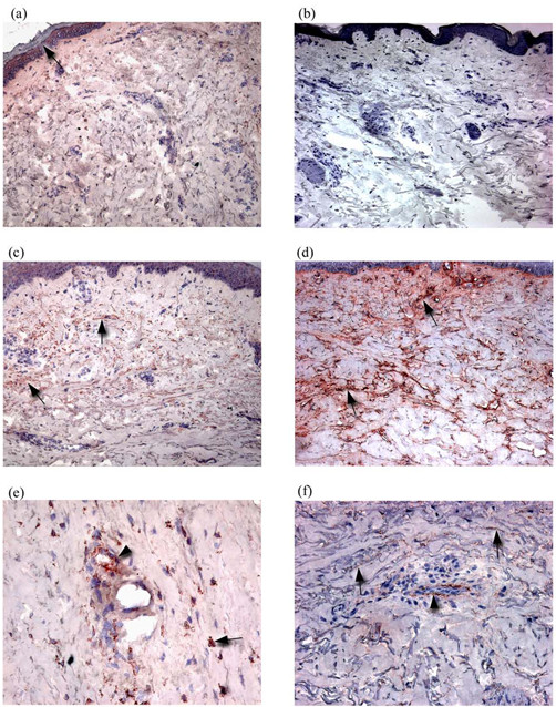
The mechanisms by which microvascular damage leads to dermal fibrosis in diffuse cutaneous systemic sclerosis (dcSSc) are unclear. We hypothesized that microvascular pericytes constitute a cellular link between microvascular damage and fibrosis by transdifferentiating into myofibroblasts. We used a combination of immunohistochemistry and double immunofluorescence labelling of frozen skin biopsies taken from normal and dcSSc patients to determine whether a phenotypic link between pericytes and myofibroblasts exists in dcSSc. Using α-smooth muscle actin, the ED-A splice variant of fibronectin (ED-A FN) and Thy-1 to identify myofibroblasts, we demonstrated the presence of myofibroblasts in fibrotic dcSSc skin. Myofibroblasts were totally absent from control skin, atrophic stage dcSSc skin and non-lesional skin. Using double immunofluorescence labelling, both myofibroblasts and pericytes were shown to express ED-A FN and Thy-1 in dcSSc skin but not in control skin. Proliferating cell nuclear antigen was also expressed by myofibroblasts and pericytes in dcSSc skin while being absent in control skin. These observations suggest that the presence of myofibroblasts may represent a transitional phase during the fibrotic stages of dcSSc and that Thy-1 +ve pericytes participate in the fibrogenic development of dcSSc by synthesizing ED-A FN, which may be associated with a proliferation and transition of pericytes and fibroblasts to myofibroblasts, thus linking microvascular damage and fibrosis.
Overview
The claims and statistics of this paper haven’t been checked yet.
Free: it reads the open-access text and checks the first 5 claims that cite a source we can check. Create a free account to check every claim.
Integrity signals
Registry
Cites
Statistics
Figures



Verification
Claims the paper makes
The claims and statistics of this paper haven’t been checked yet.
Free: it reads the open-access text and checks the first 5 claims that cite a source we can check. Create a free account to check every claim.
Claims other papers make about this one
Each is checked against this paper’s own text: does it say what the citing paper says it does?
The source backs part of the claim, not all of it — or backs it for a different population, condition or comparison.
In the citing paper
“Pericytes already express αSMA, and can become myofibroblasts if they leave their cellular niche and start to express proteins such as collagen type I and FN1-EDA.”
From Unraveling SSc Pathophysiology; The Myofibroblast (2018) · cites this paper as [27]
Experimental — accuracy not yet measuredIn the citing paper
“However, of note, in late stage fibrotic atrophic SSc skin these cells can no longer be detected (27).”
From Unraveling SSc Pathophysiology; The Myofibroblast (2018) · cites this paper as [27]
In the citing paper
“That this process occurs in SSc is suggested by a study that shows that pericytes in SSc skin, but not in healthy skin, express FN1-EDA and other myofibroblast markers (27).”
From Unraveling SSc Pathophysiology; The Myofibroblast (2018) · cites this paper as [27]
Self-check
Percentages, sums and reported differences re-computed from the paper’s own numbers.
The self-check needs the paper’s full text.
Citations
Cites
Myofibroblasts and mechano-regulation of connective tissue remodelling
Tomasek, J. J., Gabbiani, G., Hinz, B., Chaponnier, C., & Brown, R. A. (2002) · Nature Reviews Molecular Cell Biology · doi:10.1038/nrm809 · Cited by 4,290 · cited 1×
“Originally described in wound tissue, the primary role of myofibroblasts is contraction of early granulation tissue [6].”
BackgroundScleroderma (systemic sclerosis): classification, subsets and pathogenesis
(2001) · Classic Papers in Rheumatology · doi:10.3109/9780203214237-95 · Cited by 3,950
Self-citationThe myofibroblast in wound healing and fibrocontractive diseases
Gabbiani, G. (2003) · The Journal of Pathology · doi:10.1002/path.1427 · Cited by 1,630 · cited 1×
“Like pericytes, myofibroblasts express alpha smooth muscle actin (αSMA) and are strongly associated with fibrotic tissue [5].”
BackgroundDesmoulière, A., Redard, M., Darby, I. A., & Gabbiani, G. (1995) · Europe PMC (PubMed Central) · Cited by 1,448
PCNA: structure, functions and interactions
Kelman, Z. (1997) · Oncogene · doi:10.1038/sj.onc.1200886 · Cited by 929 · cited 1×
“Proliferating cells were labelled with a rabbit polyclonal antibody against proliferating cell nuclear antigen (PCNA) (Abcam, UK) [34].”
MethodSerini, G., Bochaton-Piallat, M.-L., Ropraz, P., Geinoz, A., Borsi, L., Zardi, L., & Gabbiani, G. (1998) · The Journal of Cell Biology · doi:10.1083/jcb.142.3.873 · Cited by 847 · cited 1×
“Thus, the de novo synthesis of ED-A FN appears to be a pre-requisite of α-SMA expression and myofibroblast differentiation [ 10 ].”
BackgroundBone marrow–derived progenitor cells in pulmonary fibrosis
Hashimoto, N., Jin, H., Liu, T., Chensue, S. W., & Phan, S. H. (2004) · Journal of Clinical Investigation · doi:10.1172/jci18847 · Cited by 720 · cited 1×
“This corroborates two recent studies of murine lung fibrosis in which collagen-synthesizing cells were found to be distinct from α-SMA+ve myofibroblasts [ 42 ,43] and a previous analysis of dcSSc skin, in which the presence of myofibroblasts did not correlate with α1(I) procollagen mRNA [14].”
ResultHinz, B., Mastrangelo, D., Iselin, C. E., Chaponnier, C., & Gabbiani, G. (2001) · The American Journal of Pathology · doi:10.1016/s0002-9440(10)61776-2 · Cited by 640 · cited 1×
“As ED-A FN is a pre-requisite for myofibroblast formation [9], the expression of ED-A FN by pericytes is likely to be of significance in the differentiation of perivascular fibroblasts and pericytes into myofibroblasts, and may represent a key step in linking microvascular damage and fibrosis.”
BackgroundBone marrow–derived progenitor cells in pulmonary fibrosis
Hashimoto, N., Jin, H., Liu, T., et al. (2004) · Journal of Clinical Investigation · doi:10.1172/jci200418847 · Cited by 621
Nailfold videocapillaroscopy assessment of microvascular damage in systemic sclerosis.
Houtman, N. M. (2000) · PubMed · Cited by 601
Research works printed but not linked: 1
- Jelaska A, Korn JH: Role of apoptosis and transforming growth factor beta1 in fibroblast selection and activation in systemic sclerosis. Arthritis Rheum. 2000, 43: 2230-2239. 10.1002/1529-0131(200010)43:10<2230::AID-ANR10>3.0.CO;2-8. · doi:10.1002/1529-0131(200010)43:10<2230::aid-anr10>3.0.co;2-8
Cited by
Hinz, B., Phan, S. H., Thannickal, V. J., Galli, A., Bochaton-Piallat, M.-L., & Gabbiani, G. (2007) · The American Journal of Pathology · doi:10.2353/ajpath.2007.070112 · Cited by 2,068
Formation and function of the myofibroblast during tissue repair
Hinz, B. (2007) · Journal of Investigative Dermatology · doi:10.1038/sj.jid.5700613 · Cited by 1,658
“Pericytes and SMC from the vasculature have been proposed as another local myofibroblast source in scleroderma (Rajkumar et al., 2005) as well as liver and glomerular fibrosis (Desmouliere et al., 2003).”
BackgroundFate Tracing Reveals the Pericyte and Not Epithelial Origin of Myofibroblasts in Kidney Fibrosis
Humphreys, B. D., Lin, S.-L., Kobayashi, A., Hudson, T. E., Nowlin, B. T., Bonventre, J. V., Valerius, M. T., McMahon, A. P., & Duffield, J. S. (2010) · The American Journal of Pathology · doi:10.2353/ajpath.2010.090517 · Cited by 1,442
Gabrielli, A., Avvedimento, E. V., & Krieg, T. (2009) · New England Journal of Medicine · doi:10.1056/nejmra0806188 · Cited by 1,384
Recent Developments in Myofibroblast Biology
Hinz, B., Phan, S. H., Thannickal, V. J., Prunotto, M., Desmoulière, A., Varga, J., De Wever, O., Mareel, M., & Gabbiani, G. (2012) · The American Journal of Pathology · doi:10.1016/j.ajpath.2012.02.004 · Cited by 1,193
“The extradomain A fibronectin splice variant, which is critical for myofibroblast differentiation, shows increased deposition.(93) Other structural ECM molecules that are aberrantly expressed in scleroderma include fibronectin, proteoglycans, fibrillin, and elastin fibrils.”
Background
Metadata
- Authors
- Rajkumar, V. S., Howell, K., Csiszar, K., Denton, C. P., Black, C. M., & Abraham, D. J.
- Affiliations
- The Royal Free Hospital (GB)
- Corresponding author
- Abraham, David J
- Venue
- Arthritis Research & Therapy
- Published
- 2005-07-21
- Volume
- 7
- Issue
- 5
- Pages
- R1113-23
- ISSN
- 1478-6354 (ISSN-L)
- PMC
- PMC1257439
- OpenAlex
- W2138211373
- Cited by
- 242
- References
- 48
- Citations by year
- 2026: 6 · 2025: 4 · 2024: 8 · 2023: 9 · 2022: 14 · 2021: 16 · 2020: 9 · 2019: 13 · 2018: 9 · 2017: 6 · 2016: 14 · 2015: 18 · 2014: 14 · 2013: 13 · 2012: 13
- FWCI
- 7.04
- Type
- journal article
- Language
- en
- Open access
- gold
- doi.org
- Licence
- cc-by
- creativecommons.org/licenses/by/2.0/ (tdm)
- creativecommons.org/licenses/by/2.0/ (vor)
- Locations
- Arthritis Research & Therapy (publishedVersion, cc-by)
- PubMed Central (publishedVersion)
- PubMed (publishedVersion)
- UCL Discovery (University College London) (submittedVersion)
- Funders
- Rosetrees Trust
- Jean Shanks Foundation
- National Institutes of Health
- National Institute of Arthritis and Musculoskeletal and Skin Diseases (S11AR047713, AR47713)
- Topics
- Health Sciences › Medicine › Rheumatology › Systemic Sclerosis and Related Diseases
- Health Sciences › Medicine › Rheumatology › Dupuytren's Contracture and Treatments
- Life Sciences › Biochemistry, Genetics and Molecular Biology › Genetics › Connective tissue disorders research
- Keywords
- diffuse cutaneous systemic sclerosis, myofibroblasts, pericytes, fibroblasts, skin fibrosis, microvascular damage, ED-A fibronectin, CD90, alpha-smooth muscle actin, proliferating cell nuclear antigen
- MeSH terms
- Actins, Adult, Aged, Collagen, Female, Fibroblasts, Fibronectins, Fibrosis, Humans, Male, Middle Aged, Phenotype, Scleroderma, Systemic, Skin, Biomarkers, Thy-1 Antigens, Proliferating Cell Nuclear Antigen, Cell Lineage, Pericytes, Myoblasts
About these checks
Registry, retraction, Cites and statistics results are computed, not judged, with one exception: a printed reference that no index lists is read once by a model before it counts as a citation error. Claims are extracted by a model and kept only when their quote appears verbatim in the text; each cited source is then checked, with a model’s help, for whether it says what the claim relies on. Those verdicts can be wrong — read the claim’s quote and the cited source before relying on one.
Statistics and arithmetic are recomputed, not judged.
Confidence bands are shown only for fields calibrated on our annotated test set. None is calibrated yet, so no band is shown.
What counts as a claim ›How we measure confidence ›How we check this ›
Are you an author, or do you see a problem with this page? Request a review
How to cite
Cite CoChat for the checks on this page, and the paper for its own findings.
- This page
- CoChat. (2026). Citation check: Shared expression of phenotypic markers in systemic sclerosis indicates a convergence of pericytes and fibroblasts to a myofibroblast lineage in fibrosis. https://app.cochat.ai/papers/10.1186/ar1790
- The paper
- Rajkumar, V. S., Howell, K., Csiszar, K., Denton, C. P., Black, C. M., & Abraham, D. J. (2005). Shared expression of phenotypic markers in systemic sclerosis indicates a convergence of pericytes and fibroblasts to a myofibroblast lineage in fibrosis. Arthritis Research & Therapy. https://doi.org/10.1186/ar1790
Data sources
Bibliographic data: Semantic Scholar, OpenAlex, Crossref.
Contains information from the Semantic Scholar Academic Graph Datasets, made available under the ODC Attribution License (ODC-BY 1.0).
Semantic Scholar’s data is described in: Kinney, R., et al. (2023). The Semantic Scholar Open Data Platform. arXiv:2301.10140.
