

The DOI matches its Crossref record; 195 of the 196 references it cites were found in a registry or an index; none of the 57 sources its claims cite is checked for support yet.
The overlap between vascular disease and Alzheimer’s disease - lessons from pathology
Open accessAbstract
Recent epidemiological and clinico-pathological data indicate considerable overlap between cerebrovascular disease (CVD) and Alzheimer's disease (AD) and suggest additive or synergistic effects of both pathologies on cognitive decline. The most frequent vascular pathologies in the aging brain and in AD are cerebral amyloid angiopathy and small vessel disease. Up to 84% of aged subjects show morphological substrates of CVD in addition to AD pathology. AD brains with minor CVD, similar to pure vascular dementia, show subcortical vascular lesions in about two-thirds, while in mixed type dementia (AD plus vascular dementia), multiple larger infarcts are more frequent. Small infarcts in patients with full-blown AD have no impact on cognitive decline but are overwhelmed by the severity of Alzheimer pathology, while in early stages of AD, cerebrovascular lesions may influence and promote cognitive impairment, lowering the threshold for clinically overt dementia. Further studies are warranted to elucidate the many hitherto unanswered questions regarding the overlap between CVD and AD as well as the impact of both CVD and AD pathologies on the development and progression of dementia.
Overview
AI snapshot
Written from the paper’s full text.
TL;DR
This review synthesizes pathological and clinicopathological evidence that cerebrovascular disease (CVD) and Alzheimer’s disease (AD) frequently overlap, with additive or synergistic effects on cognitive decline. It emphasizes that vascular lesions may lower the threshold for dementia in early AD, while severe AD pathology often overwhelms the cognitive impact of small vascular lesions.
Key findings
- Up to 84% of aged subjects show morphological substrates of CVD in addition to AD pathology; cerebral amyloid angiopathy (CAA) is present in 82% to 98% of AD patients, and small vessel disease (SVD) is common in both aging and AD.
- In pure vascular dementia (VaD), about 68% of cases show lacunar/strategic subcortical infarcts and 32.5% show multiple large cortico-subcortical infarcts; in mixed dementia (AD + severe CVD), 56.6% show multiple large/lobar or cortico-subcortical lesions and 43.4% show small subcortical lacunar, micro-infarcts, or strategic infarcts.
- Small infarcts in full-blown AD have no impact on cognitive decline and are overwhelmed by Alzheimer pathology, whereas in early AD, cerebrovascular lesions may promote cognitive impairment and lower the threshold for clinically overt dementia.
- Converging autopsy, amyloid PET, functional MRI, and CSF biomarker evidence indicates that AD and CVD exert additive rather than interactive adverse effects on cognitive health, though interactions between vascular factors and amyloidosis/tauopathy remain unresolved.
Methods
- Narrative review of epidemiological, clinico-pathological, and autopsy studies on the overlap between CVD and AD, including data from large cohorts such as the National Alzheimer’s Coordinating Center (NACC) database and the Rush Memory and Aging Project/Religious Orders Study.
- The paper summarizes prevalence and topographical distribution of cerebrovascular lesions across AD, VaD, mixed dementia, and aged controls, using tables from prior autopsy series (e.g., VaD total 188; mixed dementia n = 83; NACC 5,715 autopsy cases).
- Main analysis is qualitative synthesis of reported frequencies, odds ratios, and clinicopathological correlations; no new primary data or systematic meta-analysis are presented.
Limitations
- The paper states that further studies are warranted to elucidate unanswered questions about CVD–AD overlap and that there is still a lack of consensus on incorporating vascular disease into clinical and neuropathological classification schemes for dementias.
- It notes that sample selection may strongly bias results: community samples tend to show greater cerebrovascular pathology than hospital-based samples, and mixed AD/CVD prevalence was higher in the community-based RUSH Memory and Aging Project (44%) than in the RUSH Religious Order Study (28%).
- The text also raises doubts about PiB-PET as representative of Aβ loads in the living brain and states that differentiation of mixed AD/CVD with CSF biomarkers may be difficult; evident from the text, this is a narrative review without a systematic search or quantitative pooling, so findings depend on heterogeneous autopsy cohorts and diagnostic criteria.
Model-written summary. Check it against the paper before relying on it.
Integrity signals
Registry
Cites
Statistics
Figures
Verification
Claims the paper makes
51 Not checked yet · 12 Own interpretation · 6 No basis given · 1 Own result
The paper states this without a source or data behind it here, so there is nothing to check it against. The quote shows it is in the paper.
In the paper
“In AD brains with minor CVD the majority of CVLs are lacunar infarcts in basal ganglia and white matter, and multiple micro-infarcts.”
§ The overlap between vascular disease and Alzheimer’s disease - lessons from pathology > Review > Topographical distribution of cerebrovascular lesions
plain- Asserted: passes — The authors put it forward as true.
- Checkable: passes — It can be checked against evidence.
- Load-bearing: passes — The paper’s conclusions depend on it.
- Based: passes — The paper gives a reason why it holds.
The paper states this without a source or data behind it here, so there is nothing to check it against. The quote shows it is in the paper.
In the paper
“while in mixed type dementia (AD plus vascular dementia), multiple larger infarcts are more frequent.”
§ The overlap between vascular disease and Alzheimer’s disease - lessons from pathology > Abstract
plaincorrelational- Asserted: passes — The authors put it forward as true.
- Checkable: passes — It can be checked against evidence.
- Load-bearing: unclear — It is unclear whether the paper’s conclusions depend on it.
- Based: passes — The paper gives a reason why it holds.
A result the paper reports from its own data — it cites no other source for it, so there is none to check. The quote shows it is in the paper.
In the paper
“By contrast, mixed dementia (AD + severe CVD), according to our experience, is more frequently characterized by large or lobar infarcts, and multiple cortico-subcortical lesions (56.6%) than small subcortical lacunar infarcts, micro-infarcts, or strategic infarcts (43.4%, Table 2)”
§ The overlap between vascular disease and Alzheimer’s disease - lessons from pathology > Review > Topographical distribution of cerebrovascular lesions
hedgedcorrelational- Asserted: passes — The authors put it forward as true.
- Checkable: passes — It can be checked against evidence.
- Load-bearing: passes — The paper’s conclusions depend on it.
- Based: passes — The paper gives a reason why it holds.
The paper states this without a source or data behind it here, so there is nothing to check it against. The quote shows it is in the paper.
In the paper
“AD brains with minor CVD, similar to pure vascular dementia, show subcortical vascular lesions in about two-thirds”
§ The overlap between vascular disease and Alzheimer’s disease - lessons from pathology > Abstract
plaincorrelational- Asserted: passes — The authors put it forward as true.
- Checkable: passes — It can be checked against evidence.
- Load-bearing: unclear — It is unclear whether the paper’s conclusions depend on it.
- Based: passes — The paper gives a reason why it holds.
In the paper
“Up to 84% of aged subjects show morphological substrates of CVD in addition to AD pathology.”
§ The overlap between vascular disease and Alzheimer’s disease - lessons from pathology > Abstract
What the sources say
- Not checked yetcites [1] Petrovitch H, Ross GW, Steinhorn SC, Abbott RD, Markesberry W, Davis DG, Nelson J, Hardman J, Masaki KH, Vogt MR, Launer LJ, White LR: AD lesions and infarcts in demented and no-demented Japanese-American men. Ann Neurol. 2005, 57: 98-103. source
hedgedcorrelational- Asserted: passes — The authors put it forward as true.
- Checkable: passes — It can be checked against evidence.
- Load-bearing: passes — The paper’s conclusions depend on it.
- Based: passes — The paper gives a reason why it holds.
None of the sources cited for this claim have been checked yet.
In the paper
“These data add further evidence for AD pathology (mainly neurofibrillary tangles and neuritic plaques) being the main morphological substrate of clinical dementia [51],[79],[80].”
§ The overlap between vascular disease and Alzheimer’s disease - lessons from pathology > Review > Coincidence between cerebrovascular disease and Alzheimer’s disease
What the sources say
- Not checked yetcites [51] Bennett DA, Wilson RS, Arvanitakis Z, Boyle PA, de Toledo-Morrell L, Schneider JA: Selected findings from the religious orders study and rush memory and aging project. J Alzheimers Dis. 2013, 33: S397-S403. source
- Not checked yetcites [79] Nelson PT, Jicha GA, Schmitt FA, Liu H, Davis DG, Mendiondo MS, Abner EL, Markesbery WR: Clinicopathologic correlations in a large Alzheimer disease center autopsy cohort: neuritic plaques and neurofibrillary tangles “do count” when staging disease severity. J Neuropathol Exp Neurol. 2007, 66: 1136-1146. source
- Not checked yetcites [80] Nelson PT, Pious NM, Jicha GA, Wilcock DM, Fardo DW, Estus S, Rebeck GW: APOE-epsilon2 and APOE-epsilon4 correlate with increased amyloid accumulation in cerebral vasculature. J Neuropathol Exp Neurol. 2013, 72: 708-715. source
plaindirect- Asserted: passes — The authors put it forward as true.
- Checkable: passes — It can be checked against evidence.
- Load-bearing: passes — The paper’s conclusions depend on it.
- Based: passes — The paper gives a reason why it holds.
A conclusion the paper draws from its own results — it cites no other source for it, so there is none to check. The quote shows it is in the paper.
In the paper
“while small CVLs alone, seen in 10% to 50% of aged cognitively unimpaired controls, are not likely to account for a single cause of dementia.”
§ The overlap between vascular disease and Alzheimer’s disease - lessons from pathology > Review > Topographical distribution of cerebrovascular lesions
hedged- Asserted: passes — The authors put it forward as true.
- Checkable: passes — It can be checked against evidence.
- Load-bearing: passes — The paper’s conclusions depend on it.
- Based: passes — The paper gives a reason why it holds.
In the paper
“However, AD pathology alone more frequently accounts for dementia than both macroscopic and microscopic infarcts [15]”
§ The overlap between vascular disease and Alzheimer’s disease - lessons from pathology > Review > Coincidence between cerebrovascular disease and Alzheimer’s disease
What the sources say
- Not checked yetcites [15] Troncoso JC, Zonderman AB, Resnick SM, Crain B, Pletnikova O, O’Brien RJ: Effect of infarcts on dementia in the Baltimore longitudinal study of aging. Ann Neurol. 2008, 64: 168-176. source
plaindirect- Asserted: passes — The authors put it forward as true.
- Checkable: passes — It can be checked against evidence.
- Load-bearing: passes — The paper’s conclusions depend on it.
- Based: passes — The paper gives a reason why it holds.
A conclusion the paper draws from its own results — it cites no other source for it, so there is none to check. The quote shows it is in the paper.
In the paper
“while in early stages of AD, cerebrovascular lesions may influence and promote cognitive impairment, lowering the threshold for clinically overt dementia.”
§ The overlap between vascular disease and Alzheimer’s disease - lessons from pathology > Abstract
hedgedconditional- Asserted: passes — The authors put it forward as true.
- Checkable: passes — It can be checked against evidence.
- Load-bearing: passes — The paper’s conclusions depend on it.
- Based: passes — The paper gives a reason why it holds.
A conclusion the paper draws from its own results — it cites no other source for it, so there is none to check. The quote shows it is in the paper.
In the paper
“The combination of two or more pathological processes may influence the severity of cognitive deficits, unmasking preclinical dementia due to mild AD lesions”
§ The overlap between vascular disease and Alzheimer’s disease - lessons from pathology > Review > Topographical distribution of cerebrovascular lesions
hedgedconditional- Asserted: passes — The authors put it forward as true.
- Checkable: passes — It can be checked against evidence.
- Load-bearing: passes — The paper’s conclusions depend on it.
- Based: passes — The paper gives a reason why it holds.
- It may state two separate claims as one. 7
- Probably not a substantial claim. 1
A conclusion the paper draws from its own results — it cites no other source for it, so there is none to check. The quote shows it is in the paper.
In the paper
“Recent epidemiological and clinico-pathological data indicate considerable overlap between cerebrovascular disease (CVD) and Alzheimer’s disease (AD) and suggest additive or synergistic effects of both pathologies on cognitive decline.”
§ The overlap between vascular disease and Alzheimer’s disease - lessons from pathology > Abstract
hedgedcorrelational- Asserted: passes — The authors put it forward as true.
- Checkable: passes — It can be checked against evidence.
- Load-bearing: passes — The paper’s conclusions depend on it.
- Based: passes — The paper gives a reason why it holds.
It may state two separate claims as one.
In the paper
“There are considerable differences in the pathological burden in relation to age of onset of dementia, suggesting that late onset is associated with increased vascular pathology and lower AD burden [161],[162].”
§ The overlap between vascular disease and Alzheimer’s disease - lessons from pathology > Review > Cerebrovascular and Alzheimer’s disease pathology in demented and non-demented elderly
What the sources say
- Not checked yetcites [161],[162]
hedgedcorrelational- Asserted: passes — The authors put it forward as true.
- Checkable: passes — It can be checked against evidence.
- Load-bearing: passes — The paper’s conclusions depend on it.
- Based: passes — The paper gives a reason why it holds.
It may state two separate claims as one.
In the paper
“A lacunar state in basal ganglia and/or white matter was observed in 73%, hippocampal sclerosis in 3%, and mixed cerebral pathologies in 6%, whereas only 9% were free of CVLs [169].”
§ The overlap between vascular disease and Alzheimer’s disease - lessons from pathology > Review > Cerebrovascular and Alzheimer’s disease pathology in demented and non-demented elderly
What the sources say
- Not checked yetcites [169] Jellinger KA, Attems J: Neuropathology and general autopsy findings in nondemented aged subjects. Clin Neuropathol. 2012, 31: 87-98. source
plain- Asserted: passes — The authors put it forward as true.
- Checkable: passes — It can be checked against evidence.
- Load-bearing: fails — The paper’s conclusions may not depend on it.
- Based: passes — The paper gives a reason why it holds.
Probably not a substantial claim. The paper’s conclusions may not depend on it.
In the paper
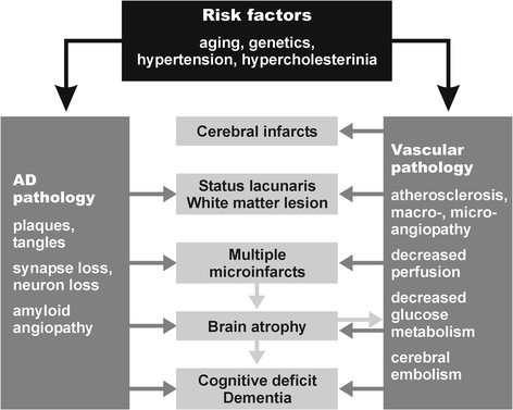
“The pathogenic chain of these and other deleterious effects, in a vicious circle, finally produces either structural cerebral disintegration (lacunes, infarcts, WMLs) with compromised neuronal metabolism, mitochondrial deficiency, oxidative stress, protein degradation, failure promoting cytoskeletal lesions with deposition of Aβ, and formation of neuritic lesions (e.g., neurofibrillary tangles).”
§ The overlap between vascular disease and Alzheimer’s disease - lessons from pathology > Review > Pathogenic factors
What the sources say
- Not checked yetcites [147] Jellinger KA, Attems J: Neuropathological evaluation of mixed dementia. J Neurol Sci. 2007, 257: 80-87. source
plaindirect- Asserted: passes — The authors put it forward as true.
- Checkable: passes — It can be checked against evidence.
- Load-bearing: passes — The paper’s conclusions depend on it.
- Based: passes — The paper gives a reason why it holds.
It may state two separate claims as one.
In the paper
“Microvascular changes in the aged brain and in AD induce impairment of cerebral perfusion, in particular decrease of regional blood flow, reduction of glucose transport and utilization, loss of vascular innervation with special impact on the cholinergic and transmitter deficits in AD [171], impairment of neurovascular regulation, ultrastructural changes in capillaries and basement membranes due to”
§ The overlap between vascular disease and Alzheimer’s disease - lessons from pathology > Review > Pathogenic factors
What the sources say
- Not checked yetcites [171] Roman GC, Kalaria RN: Vascular determinants of cholinergic deficits in Alzheimer disease and vascular dementia. Neurobiol Aging. 2006, 27: 1769-1785. source
plaindirect- Asserted: passes — The authors put it forward as true.
- Checkable: passes — It can be checked against evidence.
- Load-bearing: passes — The paper’s conclusions depend on it.
- Based: passes — The paper gives a reason why it holds.
It may state two separate claims as one.
In the paper
“These factors induce brain atrophy with cognitive and memory impairment (Figure 1) [147], although the complex cascade of these and other noxious factors needs further elucidation.”
§ The overlap between vascular disease and Alzheimer’s disease - lessons from pathology > Review > Pathogenic factors
What the sources say
- Not checked yetcites [147] Jellinger KA, Attems J: Neuropathological evaluation of mixed dementia. J Neurol Sci. 2007, 257: 80-87. source
plaindirect- Asserted: passes — The authors put it forward as true.
- Checkable: passes — It can be checked against evidence.
- Load-bearing: passes — The paper’s conclusions depend on it.
- Based: passes — The paper gives a reason why it holds.
It may state two separate claims as one.
In the paper
“Both hypertension and CAA are associated with an increased prevalence of CVLs [157]”
§ The overlap between vascular disease and Alzheimer’s disease - lessons from pathology > Review > Pathogenic factors
What the sources say
- Not checked yetcites [157] Jellinger KA, Attems J: Prevalence and pathogenic role of cerebrovascular lesions in Alzheimer’s disease. J Neurol Sci. 2005, 229-230: 37-41. source
plaincorrelational- Asserted: passes — The authors put it forward as true.
- Checkable: passes — It can be checked against evidence.
- Load-bearing: unclear — It is unclear whether the paper’s conclusions depend on it.
- Based: passes — The paper gives a reason why it holds.
It may state two separate claims as one.
In the paper
“CVD has been suggested to be an important cause of cognitive impairment in the elderly, both by itself or as a catalyst for the conversion of low-grade AD to overt dementia [186].”
§ The overlap between vascular disease and Alzheimer’s disease - lessons from pathology > Conclusions
What the sources say
- Not checked yetcites [186] Kalaria RN, Kenny RA, Ballard CG, Perry R, Ince P, Polvikoski T: Towards defining the neuropathological substrates of vascular dementia. J Neurol Sci. 2004, 226: 75-80. source
hedgedconditional- Asserted: passes — The authors put it forward as true.
- Checkable: passes — It can be checked against evidence.
- Load-bearing: passes — The paper’s conclusions depend on it.
- Based: passes — The paper gives a reason why it holds.
It may state two separate claims as one.
The authors state this as an assumption.
WMLs contribute to cognitive decline.
“and, thus, are assumed to contribute to cognitive decline [184].”
Claims other papers make about this one
Each is checked against this paper’s own text: does it say what the citing paper says it does?
The cited source says what the claim attributes to it.
In the citing paper
“Studies have consistently found a positive epidemiological correlation between Alzheimer’s disease (AD) and cardiometabolic traits, yet the biological mechanisms behind this correlation is not well understood [1–4].”
From Multi-trait association studies discover pleiotropic loci between Alzheimer’s disease and cardiometabolic traits (2021) · cites this paper as [1]
The cited source says what the claim attributes to it.
In the citing paper
“Studies have consistently found a positive epidemiological correlation between Alzheimer’s disease (AD) and cardiometabolic traits, yet the biological mechanisms behind this correlation is not well understood [1–4].”
From Multi-trait association studies discover pleiotropic loci between Alzheimer’s disease and cardiometabolic traits (2021) · cites this paper as [1]
In the citing paper
“Moreover, the growing recognition of overlapping pathologies (e.g., Lewy body inclusions, TDP-43 pathology, vascular lesions, etc.) among dementia patients indicates a wide range of biological heterogeneity within AD that necessitates a broader approach to scientific investigations of its pathophysiology [37–39].”
From Systems-based proteomics to resolve the biology of Alzheimer’s disease beyond amyloid and tau (2021) · cites this paper as [38]
Self-check
Percentages, sums and reported differences re-computed from the paper’s own numbers.
Each contradiction between two passages listed here was confirmed by an independent check.
Rated minor: it would not touch a result — a value no result rests on, a p-value that stays on the same side of the significance level, or a conflict that is clerical, resolved elsewhere in the paper, or outside its own findings.
In the paper
“Multiple infarcts (61 = 32.5%)”
§ The overlap between vascular disease and Alzheimer’s disease - lessons from pathology > Review > Topographical distribution of cerebrovascular lesions
Reported 61 · recomputed 58
Rated minor: it would not touch a result — a value no result rests on, a p-value that stays on the same side of the significance level, or a conflict that is clerical, resolved elsewhere in the paper, or outside its own findings.
In the paper
§ The overlap between vascular disease and Alzheimer’s disease - lessons from pathology > Review > Topographical distribution of cerebrovascular lesions
“Table: Table 3 Common lesions in AD, VaD, MIX, and aged controls (from[130])”
§ The overlap between vascular disease and Alzheimer’s disease - lessons from pathology > Review > Topographical distribution of cerebrovascular lesions
“The type and average prevalence of CVLs in AD, VaD, mixed dementia, and aged controls is shown in Table 3[147].”
Rated minor: it would not touch a result — a value no result rests on, a p-value that stays on the same side of the significance level, or a conflict that is clerical, resolved elsewhere in the paper, or outside its own findings.
In the paper
§ The overlap between vascular disease and Alzheimer’s disease - lessons from pathology > Review > Cerebrovascular and Alzheimer’s disease pathology in demented and non-demented elderly
“compared to 66.8% in age-matched controls, all showing low Braak stages (P <0.01).”
§ The overlap between vascular disease and Alzheimer’s disease - lessons from pathology > Review > Cerebrovascular and Alzheimer’s disease pathology in demented and non-demented elderly
“Minor and moderate vascular pathology in AD were about twice as frequent as in controls (26.2% vs. 12.2% and 20.9% vs. 11.3%; P <0.01). On the other hand, severe vascular pathology did not significantly differ between both groups (10.2% vs. 12.2%).”
Citations
Cites
Wardlaw, J. M., Smith, E. E., Biessels, G. J., Cordonnier, C., Fazekas, F., Frayne, R., Lindley, R. I., O'Brien, J. T., Barkhof, F., Benavente, O. R., et al. (2013) · The Lancet Neurology · doi:10.1016/s1474-4422(13)70124-8 · Cited by 5,544 · cited 1×
“SVD is a frequent cause of white matter lesions (WMLs; leukoaraiosis) that are increasingly detected by neuroimaging [118-121].”
BackgroundGorelick, P. B., Scuteri, A., Black, S. E., DeCarli, C., Greenberg, S. M., Iadecola, C., Launer, L. J., Laurent, S., Lopez, O. L., Nyenhuis, D., et al. (2011) · Stroke · doi:10.1161/str.0b013e3182299496 · Cited by 3,811 · cited 3×
“Epidemiological studies have shown that AD and CVD share common risk factors such as hypertension during midlife, diabetes mellitus, smoking, apolipoprotein E (ApoE) ε4 isoforms, hypercholesterolemia, homocysteinemia, and, in particular, age [16,30-34].”
BackgroundPantoni, L. (2010) · The Lancet Neurology · doi:10.1016/s1474-4422(10)70104-6 · Cited by 3,405 · cited 1×
“SVD affects small arteries and arterioles and refers to pathological changes similar to atherosclerosis that are termed small vessel arteriosclerosis/atherosclerosis, lipoor fibrohyalinosis, or hypertensive arteriopathy [115].”
BackgroundNeurovascular pathways to neurodegeneration in Alzheimer’s disease and other disorders
Zlokovic, B. V. (2011) · Nature Reviews Neuroscience · doi:10.1038/nrn3114 · Cited by 2,969 · cited 1×
“CVD has been suggested to contribute to AD neuropathological changes including selective brain atrophy and accumulation of abnormal proteins such as Aβ [24,35,84,85].”
BackgroundThe projected effect of risk factor reduction on Alzheimer's disease prevalence
Barnes, D. E., & Yaffe, K. S. (2011) · The Lancet Neurology · doi:10.1016/s1474-4422(11)70072-2 · Cited by 2,846
Mixed brain pathologies account for most dementia cases in community-dwelling older persons
Schneider, J. A., Arvanitakis, Z., Bang, W., & Bennett, D. A. (2007) · Neurology · doi:10.1212/01.wnl.0000271090.28148.24 · Cited by 1,992 · cited 2×
“The burden of vascular and AD-type pathologies are leading and independent causes of dementia in the elderly [4,9-15], suggesting additive or synergistic effects of both types of lesions on cognitive impairment [2,3,5,9,16-29].”
BackgroundThe Pathobiology of Vascular Dementia
Iadecola, C. (2013) · Neuron · doi:10.1016/j.neuron.2013.10.008 · Cited by 1,857 · cited 1×
“The pathophysiology of VaD has been critically reviewed recently [48,144-146].”
BackgroundCerebral microbleeds: a guide to detection and interpretation
Greenberg, S. M., Vernooij, M. W., Cordonnier, C., Viswanathan, A., Al-Shahi Salman, R., Warach, S., Launer, L. J., Van Buchem, M. A., & Breteler, M. M. (2009) · The Lancet Neurology · doi:10.1016/s1474-4422(09)70013-4 · Cited by 1,798
Brain Infarction and the Clinical Expression of Alzheimer Disease<subtitle>The Nun Study</subtitle>
Snowdon, D. A. (1997) · JAMA: The Journal of the American Medical Association · doi:10.1001/jama.1997.03540340047031 · Cited by 1,590
Silent brain infarcts: a systematic review
Vermeer, S. E., Longstreth, W. T., & Koudstaal, P. J. (2007) · The Lancet Neurology · doi:10.1016/s1474-4422(07)70170-9 · Cited by 1,221 · cited 1×
““Silent” lacunar infarcts are frequently detected by MRI or CCT and are not accompanied by any overt clinical symptoms, but double the risk of subsequent stroke and dementia [133].”
Background
Research works printed but not linked: 2
- Crystal H, Dickson D: Cerebral infarcts in patients with autopsy proven Alzheimer’s disease. Neurobiol Aging. 2002, 23: 207.
- Jentoft M, Parisi J, Dickson D, Johnson K, Boeve B, Knopman D, Petersen R: Neuropathologic findings in 32 nondemented elderly subjects (abs.). J Neuropathol Exp Neurol. 2011, 70: 531.
Cited by
Sorted by citations
Items 301–400 of 736
The effect of the total small vessel disease burden on the structural brain network
Xu, X., Lau, K. K., Wong, Y., Mak, H. K., & Hui, E. S. K. (2018) · Scientific Reports · doi:10.1038/s41598-018-25917-4 · Cited by 24
“(3) Estimated transformation parameters were inverted and applied to the AAL atlas to warp all brain regions-of-interests from the MNI space to the native diffusion space.”
MethodTiel, C., Sudo, F. K., & Pereira, A. B. C. N. D. G. (2019) · Dementia & Neuropsychologia · doi:10.1590/1980-57642018dn13-030005 · Cited by 31
Kim, Y. W., Al‐Ramahi, I., Koire, A. M., Wilson, S. J., Konecki, D. M., Mota, S., Soleimani, S., Botas, J., & Lichtarge, O. (2020) · Alzheimer s & Dementia · doi:10.1002/alz.12240 · Cited by 22
“(1) EA consistently performs well against state-of-the-art predictors of mutational impact in objective, blinded challenges;29 (2) multiple experimental applications confirmed its utility, including in clinical contexts;30–33 and (3) finally, because EA is based on basic principles”
BackgroundInfluentialWang, R., Qiu, C., Dintica, C. S., Shang, Y., Calderón Larrañaga, A., Wang, H., & Xu, W. (2021) · Alzheimer's & Dementia · doi:10.1002/alz.12203 · Cited by 23
“47 However, it is difficult to speculate the direct causality from the shared risk factors to the common mechanisms, due to lacking neuroimaging and biomarker confirmation, and unmeasured confounding.”
BackgroundUntangling Alzheimer’s disease with spatial multi-omics: a brief review
Marshall, C. R., Farrow, M. A., Djambazova, K. V., & Spraggins, J. M. (2023) · Frontiers in Aging Neuroscience · doi:10.3389/fnagi.2023.1150512 · Cited by 23
“For example, CAA is reported to occur in 82 to 98% of AD patients (Attems and Jellinger, 2014).”
BackgroundHergert, D. C., Gaasedelen, O. J., Ryman, S. G., Prestopnik, J., Caprihan, A., & Rosenberg, G. A. (2024) · Journal of the American Heart Association · doi:10.1161/jaha.124.034225 · Cited by 23
Wang, X., Zhu, K., Wu, W., Zhou, D., Lu, H., Du, J., Cai, L., Yan, X., Li, W., Qian, X., et al. (2024) · Alzheimer s & Dementia · doi:10.1002/alz.14369 · Cited by 21
The genetics of neurodegenerative diseases is the genetics of age-related damage clearance failure
Hardy, J., & Escott‐Price, V. (2025) · Molecular Psychiatry · doi:10.1038/s41380-025-02911-7 · Cited by 25
“With all the protein deposition disorders, there is also typically a contribution of vascular pathology [20].”
Nanobiotechnology in Neurodegenerative Diseases
Jampílek, J., Kráľová, K., Novák, P., & Novák, M. (2019) · Springer eBooks · doi:10.1007/978-3-030-30930-5_4 · Cited by 22
Exploring the Recent Trends in Management of Dementia and Frailty: Focus on Diagnosis and Treatment
Rahman, M. M., Mim, S. A., Islam, M. R., Parvez, A., Islam, F., Uddin, M. B., Rahaman, M. S., Shuvo, P. A., Ahmed, M., Greig, N. H., et al. (2022) · Current Medicinal Chemistry · doi:10.2174/0929867329666220408102051 · Cited by 23
Pividori, M., Lu, S., Li, B., Su, C., Johnson, M. E., Wei, W., Feng, Q., Namjou, B., Kiryluk, K., Kullo, I. J., et al. (2023) · Nature Communications · doi:10.1038/s41467-023-41057-4 · Cited by 22
Park, K., Sung, J. M., Kim, W. J., An, S. K., Namkoong, K., Lee, E. M., & Chang, H. (2019) · PLoS ONE · doi:10.1371/journal.pone.0211957 · Cited by 22
Tian, M., Kawaguchi, R., Shen, Y., Machnicki, M., Villegas, N. G., Cooper, D. R., Montgomery, N., Cai, Y., Haring, J., Lan, R., et al. (2025) · Cell · doi:10.1016/j.cell.2025.06.002 · Cited by 24
Rosa, G., Giannotti, C., Martella, L. A., Massa, F., Serafini, G., Pardini, M., Nobili, F. M., & Monacelli, F. (2020) · Journal of Alzheimer s Disease · doi:10.3233/jad-191075 · Cited by 23
Cerebral Amyloid Angiopathy: Similarity in African-Americans and Caucasians with Alzheimer’s Disease
Kamara, D. M., Gangishetti, U., Gearing, M., Willis‐Parker, M., Zhao, L., Hu, W. T., & Walker, L. C. (2018) · Journal of Alzheimer s Disease · doi:10.3233/jad-170954 · Cited by 22
“However, the sample of African-Americans also had a higher prevalence of AD-type lesions, and the severity of CAA was not assessed, leaving open the question of whether CAA differs in African-Americans with advanced AD.”
The Association of Essential Metals with APOE Genotype in Alzheimer’s Disease
Leko, M. B., Jurasović, J., Perković, M. N., Španić, E., Sekovanić, A., Orct, T., Lukinović-Škudar, V., Baronica, K. B., Kiđemet‐Piskač, S., Vogrinc, Ž., et al. (2021) · Journal of Alzheimer s Disease · doi:10.3233/jad-210158 · Cited by 21
Taylor, M. E., & Close, J. (2018) · Handbook of clinical neurology · doi:10.1016/b978-0-444-63916-5.00019-7 · Cited by 26
Xu, Y. H., Aung, H. L., Hesam‐Shariati, N., Keay, L. J., Sun, X., Phu, J., Honson, V., Tully, P. J., Booth, A., Lewis, E., et al. (2024) · Journal of the American Medical Directors Association · doi:10.1016/j.jamda.2024.105098 · Cited by 20
Zedde, M., & Pascarella, R. (2024) · Brain Sciences · doi:10.3390/brainsci14100983 · Cited by 21
“Interestingly, despite capillary loss, AD brains show elevated levels of VEGF, which correlates with increased angiogenesis, neurofibrillary tangles, and amyloid-beta load [141,142].”
BackgroundImmune dysfunction in Alzheimer disease
Butovsky, O., Rosenzweig, N., Kleemann, K. L., Jorfi, M., Kuchroo, V. K., Tanzi, R. E., & Weiner, H. L. (2025) · Nature reviews. Neuroscience · doi:10.1038/s41583-025-00997-0 · Cited by 41
“of cerebrovascular damage, which is prevalent in AD 120,121 (Fig.”
The Impact of Amyloid-β or Tau on Cognitive Change in the Presence of Severe Cerebrovascular Disease
Jang, H., Initiative, A. D. N., Kim, H. J., Choe, Y. S., Kim, S.-J., Park, S., Kim, Y., Kim, K. W., Lyoo, C. H., Cho, H., et al. (2020) · Journal of Alzheimer s Disease · doi:10.3233/jad-200680 · Cited by 20
Liu, Y., Chan, D. K. Y., Crawford, J. D., Sachdev, P. S., & Braidy, N. (2021) · Current Alzheimer Research · doi:10.2174/1567205018666210212160902 · Cited by 23
Hsp70.1 carbonylation induces lysosomal cell death for lifestyle-related diseases
Yamashima, T., Seike, T., Oikawa, S., Kobayashi, H., Kido, H., Yanagi, M., Yamamiya, D., Li, S., Boontem, P., & Mizukoshi, E. (2023) · Frontiers in Molecular Biosciences · doi:10.3389/fmolb.2022.1063632 · Cited by 21
“Amyloid ß angiopathy is a common pathological feature occurring in 60%– 90% of Alzheimer patients (Kalaria and Ballard, 1999; Attems and Jellinger, 2014).”
BackgroundJiang, Y., Zeng, X., Dai, H., Luo, S., & Zhang, X. (2024) · International Journal of Biological Macromolecules · doi:10.1016/j.ijbiomac.2024.134494 · Cited by 23
Levit, A., Regis, A. M., Garabon, J. R., Oh, S., Desai, S. J., Rajakumar, N., Hachinski, V., Ağca, Y., Agca, C., Whitehead, S. N., et al. (2017) · Behavioural Brain Research · doi:10.1016/j.bbr.2017.07.006 · Cited by 21
Beishon, L., Batterham, A. P., Quinn, T. J., Nelson, C. P., Panerai, R. B., Robinson, T. G., & Haunton, V. J. (2019) · Cochrane Database of Systematic Reviews · doi:10.1002/14651858.cd013282 · Cited by 21
Wang, S., Xu, L., Yang, W.-Z., Wang, J., Dove, A., Qi, X., & Xu, W.-L. (2024) · British Journal of Sports Medicine · doi:10.1136/bjsports-2023-108048 · Cited by 17
“47 Fourth, because of lack of data on repeated CRF measurements among most participants, the association of CRF change and dementia risk could not be examined.”
Hazan, J., Liu, K. Y., Isaacs, J. D., & Howard, R. J. (2025) · Alzheimer s & Dementia · doi:10.1002/alz.70113 · Cited by 21
Bhat, N. R. (2015) · Journal of Alzheimer s Disease · doi:10.3233/jad-150098 · Cited by 18
“The frequent vascular (arteries, arterioles, and capillaries) amyloid deposition seen in AD, termed CAA [46–48], is associated with vascular injury [48].”
Is Alzheimer’s Also a Stem Cell Disease? – The Zebrafish Perspective
Kızıl, Ç., & Bhattarai, P. (2018) · Frontiers in Cell and Developmental Biology · doi:10.3389/fcell.2018.00159 · Cited by 19
“, 2016), astrocytes (Attems and Jellinger, 2014; Lian and Zheng, 2016), and oligodendrocytes (Bartzokis, 2011; Ettle et al.”
BackgroundCoutu, J., Lindemer, E., Konukoğlu, E., & Salat, D. H. (2017) · Neurobiology of Aging · doi:10.1016/j.neurobiolaging.2017.02.005 · Cited by 19
“The potential for synergy of these common co-existing pathologies has previously been detailed (Attems and Jellinger, 2014; Iadecola, 2010), and there is previous recognition that vascular risk factors are associated with faster cognitive decline in incident AD (Helzner et al., 2009).”
Diagnostic relevance of spatial orientation for vascular dementia: A case study
Coughlan, G., Flanagan, E. C., Jeffs, S., Bertoux, M., Spiers, H. J., Mioshi, E., & Hornberger, M. (2018) · Dementia & Neuropsychologia · doi:10.1590/1980-57642018dn12-010013 · Cited by 19
Pregnancy loss and risk of later dementia: A nationwide cohort study, Denmark, 1977–2017
Basit, S., Wohlfahrt, J., & Boyd, H. A. (2019) · Alzheimer s & Dementia Translational Research & Clinical Interventions · doi:10.1016/j.trci.2019.02.006 · Cited by 19
“Moreover, data from pathology and epidemiologic studies suggest considerable overlap between cerebrovascular disease and Alzheimer’s disease [30].”
BackgroundA functional cerebral endothelium is necessary to protect against cognitive decline
Trigiani, L. J., Bourourou, M., Lacalle-Aurioles, M., Lecrux, C., Hynes, A., Spring, S., Fernandes, D. J., Sled, J. G., Lesage, F., Schwaninger, M., et al. (2021) · Journal of Cerebral Blood Flow & Metabolism · doi:10.1177/0271678x211045438 · Cited by 21
Neuropathologic Correlates of Human Cortical Proteins in Alzheimer Disease and Related Dementias
Yu, L., Boyle, P. A., Wingo, A. P., Yang, J., Wang, T., Buchman, A. S., Wingo, T. S., Seyfried, N. T., Levey, A. I., Jager, P. L. D., et al. (2021) · Neurology · doi:10.1212/wnl.0000000000013252 · Cited by 19
“Of note, Alzheimer dementia is attributable to a host of neuropathologic conditions that commonly coexist in the aging brain.(8,9) We and others have reported that, while AD pathology is the main driver of Alzheimer dementia, other non-AD neurodegenerative and cerebrovascular diseases also play an important role.”
BackgroundJearjaroen, P., Thangwong, P., Tocharus, C., Lungkaphin, A., Chaichompoo, W., Srijun, J., Suksamrarn, A., & Tocharus, J. (2023) · Molecular Neurobiology · doi:10.1007/s12035-023-03821-x · Cited by 19
Vascular dementia: World Stroke Organization fact sheet 2026
Cai, Y., Mok, V., & Markus, H. S. (2026) · International Journal of Stroke · doi:10.1177/17474930251404243 · Cited by 23
Elevated parathyroid hormone levels and cognitive function: A systematic review
Jiang, W., Hu, C., Li, F., Hua, X., Huang, K., & Zhang, X.-J. (2019) · Archives of Gerontology and Geriatrics · doi:10.1016/j.archger.2019.103985 · Cited by 18
Postmortem 7T MRI for guided histopathology and evaluation of cerebrovascular disease
Lahna, D. L., Roese, N. E., Woltjer, R. L., Boespflug, E. L., Schwartz, D., Grinstead, J. W., Dodge, H. H., Wall, R., Kaye, J. A., Rooney, W. D., et al. (2022) · Journal of Neuropathology & Experimental Neurology · doi:10.1093/jnen/nlac103 · Cited by 17
“Vascular pathologies often co-occur with Alzheimer disease and synergistically lower the threshold for conversion to dementia (5, 6).”
BackgroundHippocampal microvasculature changes in association with oxidative stress in Alzheimer's disease
Sen, A., & Hongpaisan, J. (2018) · Free Radical Biology and Medicine · doi:10.1016/j.freeradbiomed.2018.03.034 · Cited by 19
“Recent data also indicates considerable overlap between cerebrovascular disease and AD (< 50% of all AD cases) and suggests additive or synergistic effects of both pathologies on cognitive decline [7].”
The Utility of Cerebrovascular Reactivity MRI in Brain Rehabilitation: A Mechanistic Perspective
Krishnamurthy, V., Sprick, J. D., Krishnamurthy, L. C., Barter, J., Turabi, A., Hajjar, I. M., & Nocera, J. R. (2021) · Frontiers in Physiology · doi:10.3389/fphys.2021.642850 · Cited by 20
Cao, Y., Huang, M., Mao, C., Wang, X., Xu, Y., Qian, X., Ma, C., Qiu, W., & Zhu, Y. (2025) · Alzheimer s & Dementia · doi:10.1002/alz.70103 · Cited by 18
“Arteri-olosclerosis is widely regarded as an underlying contributor to ADNC based on both epidemiological and clinicopathological data 4 ; however, venous collagenosis is an emerging finding in aging brains.”
Thangwong, P., Tocharus, C., & Tocharus, J. (2025) · Molecular Neurobiology · doi:10.1007/s12035-025-04914-5 · Cited by 18
“Conversely, SVD-induced VaD progresses gradually, with diffuse and cumulative damage leading to cognitive decline [21].”
The effect of intracranial stenosis on cognitive decline in a memory clinic cohort
Lim, M. J. R., Tan, C. S., Gyanwali, B., Chen, C., & Hilal, S. (2021) · European Journal of Neurology · doi:10.1111/ene.14788 · Cited by 17
“Although memory decline has been traditionally associated with Alzheimer dementia, recent studies have shown the presence of mixed pathology, that is, coexistence of vascular pathology and Alzheimer disease [29,30].”
Scarth, M., Rissanen, I. L., Scholten, R. J. P. M., & Geerlings, M. I. (2021) · Journal of Alzheimer s Disease · doi:10.3233/jad-210218 · Cited by 18
Cognitive and neuroimaging markers for preclinical vascular cognitive impairment
Lowry, E., Puthusseryppady, V., Johnen, A.-K., Renoult, L., & Hornberger, M. (2021) · Cerebral Circulation - Cognition and Behavior · doi:10.1016/j.cccb.2021.100029 · Cited by 20
Systemic Inflammation Predicts Alzheimer Pathology in Community Samples without Dementia
Cherbuin, N., Walsh, E., Leach, L., Brüstle, A., Burns, R. A., Anstey, K. J., Sachdev, P. S., & Baune, B. T. (2022) · Biomedicines · doi:10.3390/biomedicines10061240 · Cited by 17
“There is also developing evidence that systemic inflammation is a risk factor for neurodegeneration and cognitive impairment, and is closely associated with major modifiable risk factors for dementia (e.g., CVD, type 2 diabetes, obesity, and depression) [25,26].”
BackgroundSamudra, N. P., Ranasinghe, K. G., Kirsch, H. E., Rankin, K. P., & Miller, B. L. (2023) · Journal of Alzheimer s Disease · doi:10.3233/jad-220983 · Cited by 18
“The pathologic findings of amyloid angiopathy often occur in AD as well [24].”
Jearjaroen, P., Thangwong, P., Tocharus, C., Chaichompoo, W., Suksamrarn, A., & Tocharus, J. (2023) · Inflammopharmacology · doi:10.1007/s10787-023-01406-7 · Cited by 18
Vollhardt, A., Frölich, L., Stockbauer, A., Danek, A., Schmitz, C., & Wahl, A.-S. (2025) · Neurobiology of Disease · doi:10.1016/j.nbd.2025.106845 · Cited by 18
“Around 68 % of the cerebro-vascular lesions seen in pure VaD (VaD without AD pathology) are subcortical lacune infarcts (Attems and Jellinger, 2014).”
Cerebral Aβ 40 and systemic hypertension
Tayler, H. M., Palmer, J. C., Thomas, T. L., Kehoe, P. G., Paton, J. F. R., & Love, S. (2017) · Journal of Cerebral Blood Flow & Metabolism · doi:10.1177/0271678x17724930 · Cited by 16
Cherbuin, N., Shaw, M. E., Walsh, E., Sachdev, P. S., & Anstey, K. J. (2017) · Brain Imaging and Behavior · doi:10.1007/s11682-017-9789-5 · Cited by 19
“However, given that up to 80% of AD cases have comorbid vascular disease (Attems and Jellinger 2014) this distinction may be moot.”
Analysis of the vasculature by immunohistochemistry in paraffin-embedded brains
Decker, Y., Müller, A., Németh, E., Schulz‐Schaeffer, W., Fatar, M., Menger, M. D., Liu, Y., & Fassbender, K. C. (2017) · Brain Structure and Function · doi:10.1007/s00429-017-1595-8 · Cited by 17
King, C. D., & Robinson, R. A. S. (2020) · Analytical Chemistry · doi:10.1021/acs.analchem.9b01974 · Cited by 16
Kim, M., Bang, J., Kim, B., & Jeon, W. K. (2021) · The Journals of Gerontology Series A · doi:10.1093/gerona/glab075 · Cited by 16
“focal correct, (4) scanning, (5) random, (6) focal incorrect, (7) chaining, (8)”
BackgroundGlucose-lowering drugs, cognition, and dementia: The clinical evidence
Wu, C., Shapiro, L. R., Ouk, M., MacIntosh, B. J., Black, S. E., Shah, B. R., & Swardfager, W. (2022) · Neuroscience & Biobehavioral Reviews · doi:10.1016/j.neubiorev.2022.104654 · Cited by 20
Neuropsychological profile and vascular risk factors in patients with cerebral microangiopathy
Krotenkova, M. V., Kremneva, E. I., Akhmetzyanov, B. M., Kalashnikova, L. A., Gadzhieva, Z. S., Dobrynina, L. A., Lagoda, D. Y., Zabitova, M. R., Poddubskaya, A. A., & Berdalin, A. V. (2018) · Annals of Clinical and Experimental Neurology · doi:10.25692/acen.2018.4.1 · Cited by 18
Kobayashi, G., Hirata, K., Ono, M., Kasuga, K., & Takado, Y. (2026) · Frontiers in Dementia · doi:10.3389/frdem.2026.1736297 · Cited by 22
Oral anticoagulants: A plausible new treatment for Alzheimer's disease?
Toribio‐Fernández, R., Cerón, C., Tristão‐Pereira, C., Fernandez‐Nueda, I., Pérez‐Castillo, A., Fernández‐Ferro, J., Moro, M. Á., Ibáñez, B., Fuster, V., & Cortés‐Canteli, M. (2023) · British Journal of Pharmacology · doi:10.1111/bph.16032 · Cited by 16
“AD and vascular dementia are not easily distinguished, with overlapping lesions in both entities such as high incidence of vascular lesions and white matter alterations, suggesting additive or synergistic effects of both pathologies on cognitive deterioration (Attems & Jellinger, 2014).”
BackgroundSerum Placental Growth Factor as a Marker of Cerebrovascular Disease Burden in Alzheimer’s Disease
Wu, L., Chong, J. R., Chong, J. P. C., Hilal, S., Venketasubramanian, N., Tan, B. Y., Richards, M., Chen, C., & Lai, M. K. P. (2024) · Journal of Alzheimer s Disease · doi:10.3233/jad-230811 · Cited by 17
Button, E. B., Gilmour, M., Cheema, H. K., Martin, E. M., Agbay, A. M., Robert, J., & Wellington, C. L. (2019) · International Journal of Molecular Sciences · doi:10.3390/ijms20030462 · Cited by 16
“Furthermore, cerebrovascular dysfunction, including vascular accumulation of amyloid beta (Aβ) known as cerebral amyloid angiopathy (CAA), is found in the majority of AD cases [2,3].”
BackgroundArmstrong, N. M., Bangen, K. J., Au, R., & Gross, A. L. (2019) · American Journal of Epidemiology · doi:10.1093/aje/kwz210 · Cited by 16
Maliszewska‐Cyna, E., Vecchio, L. M., Thomason, L. A. M., Oore, J., Steinman, J., Joo, I. L., Dorr, A. E., McLaurin, J., Sled, J. G., Stefanovic, B., et al. (2020) · NeuroImage · doi:10.1016/j.neuroimage.2020.117269 · Cited by 17
Robert, J., Weilinger, N. L., Cao, L., Cataldi, S., Button, E. B., Stukas, S. K., Martin, E. M., Seibler, P., Gilmour, M., Caffrey, T. M., et al. (2020) · Molecular Neurodegeneration · doi:10.1186/s13024-020-00418-z · Cited by 17
Waller, R., Narramore, R., Simpson, J. E., Heath, P. R., Verma, N., Tinsley, M., Barnes, J. R., Haris, H. T., Henderson, F. E., Matthews, F. E., et al. (2020) · Brain Pathology · doi:10.1111/bpa.12928 · Cited by 17
“However, these studies, as well as others, acknowledge that mixed pathologies exist, with vascular changes occurring in AD cases and vice versa (5,45,46).”
BackgroundYang, J., Chang, S., Chen, A., Kura, S., Rosen, G. A., Saltiel, N. A., Huber, B. R., Varadarajan, D., Balbastre, Y., Magnain, C. V., et al. (2022) · IEEE Transactions on Biomedical Engineering · doi:10.1109/tbme.2022.3175072 · Cited by 17
“For example, neurodegenerative disease such as Alzheimer’s disease and chronic traumatic encephalopathy are known to be accompanied by vascular abnormities, such as increasing number of spiraling vessels and sparser vessel network [4], [10].”
BackgroundDisentangling Heterogeneity in Alzheimer’s Disease: Two Empirically-Derived Subtypes
Blanken, A. E., Dutt, S., Li, Y., Nation, D. A., & Initiative, A. D. N. (2019) · Journal of Alzheimer s Disease · doi:10.3233/jad-190230 · Cited by 16
“Compared to asymptomaticnormal individuals, atypical AD showed greater white matter lesion burden and higher systolic blood pressure, signifying contribution of comorbidity with agerelated vascular disease to differential biomarker, neuropsychological and brain atrophy patterns [34].”
Coexistence of Cerebral Microbleeds and Amyloid Pathology in Patients with Cognitive Complaints
Lim, E. Y., Ryu, S. Y., Shim, Y. S., Yang, D. W., & Cho, A.-H. (2020) · Journal of Clinical Neurology · doi:10.3988/jcn.2020.16.1.83 · Cited by 15
Alternatives to amyloid for Alzheimer's disease therapies—a symposium report
Cable, J., Holtzman, D. M., Hyman, B. T., Tansey, M. G., Colonna, M., Kellis, M., Brinton, R. D., Albert, M., Wellington, C. L., Sisodia, S. S., et al. (2020) · Annals of the New York Academy of Sciences · doi:10.1111/nyas.14371 · Cited by 13
Basak, J. M., Falk, M., Mitchell, D. N., Coakley, K. A., Quillinan, N., Orfila, J. E., & Herson, P. S. (2023) · Journal of Cerebral Blood Flow & Metabolism · doi:10.1177/0271678x231159597 · Cited by 15
“34 However, the importance of studying the interaction between the pathophysiology of cognitive dysfunction following a large vessel stroke with the pathology of AD is only recently gaining attention in the field of stroke research.”
BackgroundAlzheimer's and vascular disease classification using regional texture biomarkers in FLAIR MRI.
Chan, K., Fischer, C. E., Maralani, P., Black, S., Moody, A., & Khademi, A. (2023) · NeuroImage: Clinical · doi:10.1016/j.nicl.2023.103385 · Cited by 14
“There is increasing prevalence of AD and VaD in aging populations, while the mechanisms of interactions of the two diseases remain to be fully elucidated [2].”
BackgroundImpact of adverse pregnancy outcomes on brain vascular health and cognition
Wolfová, K., & Miller, E. C. (2024) · Research and Practice in Thrombosis and Haemostasis · doi:10.1016/j.rpth.2024.102331 · Cited by 16
“While the contribution of vascular pathology to the development of VCID is widely acknowledged, emerging evidence shows that vascular changes may play an important role in neuro-degeneration and the subsequent development of AD [16,17].”
BackgroundTwait, E. L., Gerritsen, L., Moonen, J. E. F., Verberk, I. M. W., Teunissen, C. E., Visser, P. J., van der Flier, W. M., & Geerlings, M. I. (2024) · Journal of the American Heart Association · doi:10.1161/jaha.123.032134 · Cited by 15
“25,26 By focusing on a population with arterial disease, it is possible to shed light on the role of AD pathology in the neurodegeneration and WMH observed in patients with arterial disease.”
BackgroundAssociation between Cognitive Impairment, Vascular Disease and All-Cause Mortality
Kim, J.-H., & Chon, D. (2018) · The journal of nutrition health & aging · doi:10.1007/s12603-018-1011-y · Cited by 15
“Similar associations were apparent in the reverse order where cardiovascular disease was related to higher risk of dementia (18), and Gasecki et al. found evidence to suggest an association between hypertension and cognitive decline (19).”
Cajavilca, C. E., Gadhia, R. R., & Román, G. C. (2019) · Brain Sciences · doi:10.3390/brainsci9090211 · Cited by 17
“Patients with Alzheimer disease often have evidence of small vessel injury in the white matter seen on imaging studies, which are thought to contribute to the clinical presentation of cognitive impairment and dementia [29].”
BackgroundDifferences in topological progression profile among neurodegenerative diseases from imaging data
Garbarino, S., Lorenzi, M., Oxtoby, N. P., Vinke, E. J., Marinescu, R. V., Eshaghi, A., Ikram, M. A., Niessen, W. J., Ciccarelli, O., Barkhof, F., et al. (2019) · eLife · doi:10.7554/elife.49298 · Cited by 16
“Also, associations between vascular factors and neurodegenerative dementias such as AD are common (Kalaria, 2009; Sweeney et al., 2018), which suggests contributions from multiple underlying mechanisms to the observed pattern of pathology accrual (Attems and Jellinger, 2014).”
BackgroundLoBue, C., & Cullum, C. M. (2019) · Archives of Clinical Neuropsychology · doi:10.1093/arclin/acz074 · Cited by 15
Zhuang, L., Ni, H., Wang, J., Liu, X., Lin, Y., Su, Y., Zhang, K., Li, Y., Peng, G., & Luo, B. (2020) · Frontiers in Aging Neuroscience · doi:10.3389/fnagi.2020.604246 · Cited by 15
“Aβ deposition and the presence of cerebrovascular pathologies often co-exist in the brains of the elderly population (Attems and Jellinger, 2014; Love and Miners, 2016).”
BackgroundBioenergetics and lipid metabolism in Alzheimer's disease: From cell biology to systemic health
Maioli, S., Nalvarte, I., Ankarcrona, M., Schultzberg, M., Zuloaga, K. L., Goikolea, J., Visser, P. J., Strooper, B. D., Winblad, B., Pizzo, P., et al. (2025) · Journal of Internal Medicine · doi:10.1111/joim.70036 · Cited by 19
“This may be partially due to the fact that over 80% of AD cases also exhibit vascular pathology, making the identification of causal associations and individual contributions very challenging [165–167].”
Associations of ARHGAP26 Polymorphisms with Alzheimer’s Disease and Cardiovascular Disease
Wang, K., Lu, Y., Morrow, D. F., Xiao, D., Xu, C. D., & Initiative, T. A. D. N. (2022) · Journal of Molecular Neuroscience · doi:10.1007/s12031-022-01972-5 · Cited by 14
“…to shared risk factors between AD and CVDs, but there might also be a direct causal association of CVD and cardiovascular risk factors with AD (de la Torre 2012; Justin et al. 2013; Attems and Jellinger 2014; de Bruijn and Ikram 2014; Jabir et al. 2015; Monte and Vondriska 2014; Tini et al. 2020).”
Kim, M. Y., Kim, S., Lee, J., Kim, J.-I., Oh, E., Kim, S.-W., Lee, E., Cho, K., Kim, C.-S., & Lee, M.-H. (2023) · Antioxidants · doi:10.3390/antiox12051110 · Cited by 16
“Early intracellular accumulation of Aβ is observed in AD-susceptible brain regions, which precede the formation of NFT and senile plaques [8].”
BackgroundJin, C., & Bhaskar, S. M. M. (2025) · European Journal of Neuroscience · doi:10.1111/ejn.70246 · Cited by 14
“Moreover, genetic studies are underpowered, often lack ethnic diversity, and have primarily focused on Caucasian populations (Attems and Jellinger 2014).”
Ottoy, J., Verhaeghe, J. A. J., Niemantsverdriet, E., Engelborghs, S., Stroobants, S. G., & Staelens, S. G. (2017) · PLoS ONE · doi:10.1371/journal.pone.0189155 · Cited by 14
Claus, J. J., Coenen, M., Staekenborg, S. S., Schuur, J., Tielkes, C. E. M., Koster, P., & Scheltens, P. (2018) · Journal of Alzheimer s Disease · doi:10.3233/jad-171111 · Cited by 14
Alzheimer's disease medication and outcomes of hospitalisation among patients with dementia
Möllers, T., Perna, L., Stocker, H. R., Ihle, P., Schübert, I., Schöttker, B., Frölich, L., Bauer, J. M., & Brenner, H. (2019) · Epidemiology and Psychiatric Sciences · doi:10.1017/s2045796019000702 · Cited by 15
“At any rate, this may have led to an underestimation of the actual impact of Alzheimer disease medication on outcomes, even if many cases of dementia possess shared Alzheimer and vascular pathology (Attems and Jellinger, 2014; Jellinger and Attems, 2015).”
BackgroundCDK5 Targeting as a Therapy for Recovering Neurovascular Unit Integrity in Alzheimer’s Disease
Posada-Duque, R. A., & Cardona-Gómez, G. P. (2021) · Journal of Alzheimer's Disease · doi:10.3233/jad-200730 · Cited by 14
“Vascular causes of dementia have gained attention in basic and clinical studies because a large proportion of dementias are caused by vascular pathology [21, 22].”
Attems, J. (2017) · Acta Neuropathologica · doi:10.1007/s00401-017-1723-9 · Cited by 13
Self-citationAdding cognition to AT(N) models improves prediction of cognitive and functional decline
O’Shea, D. M., Thomas, K. R., Asken, B. M., et al. (2021) · Alzheimer s & Dementia Diagnosis Assessment & Disease Monitoring · doi:10.1002/dad2.12174 · Cited by 14
Singh, P. K., Badimon, A., Chen, Z., Strickland, S., & Norris, E. H. (2021) · Research and Practice in Thrombosis and Haemostasis · doi:10.1002/rth2.12504 · Cited by 17
Toljan, K., & Homolak, J. (2021) · Handbook of clinical neurology · doi:10.1016/b978-0-12-819975-6.00018-2 · Cited by 15
Common Aging Signature in the Peripheral Blood of Vascular Dementia and Alzheimer’s Disease
Luo, H.-B., Han, G., Wang, J., Zeng, F., Li, Y., Shao, S., Song, F., Bai, Z., Peng, X., Wang, Y., et al. (2015) · Molecular Neurobiology · doi:10.1007/s12035-015-9288-x · Cited by 13
“AD involves the progressively decline of patients’ memory as well as language function, while VaD is manifested by an acute onset of the impairment in the cognitive functions [3, 4].”
BackgroundHassan, S., Akaila, D., Arjemandi, M., Papineni, V., & Yaqub, M. (2025) · Scientific Reports · doi:10.1038/s41598-025-97674-0 · Cited by 14
“As the disease progresses, the cerebral cortex, involved in thought, perception, and language, is affected, leading to ventricular enlargement and cortical thinning 14 .”
Soler, M. T., Sánchez-Valdeón, L., Blanco-Nistal, M. M., Benítez‐Andrades, J. A., Liébana‐Presa, C., & Darkistade, E. B. (2020) · International Journal of Environmental Research and Public Health · doi:10.3390/ijerph17103398 · Cited by 14
Histopathological Analysis of Cerebrovascular Lesions Associated With Aging
Dallaire‐Théroux, C., Saïkali, S., Richer, M., Potvin, O., & Duchesne, S. (2021) · Journal of Neuropathology & Experimental Neurology · doi:10.1093/jnen/nlab125 · Cited by 16
Kakae, M., Kawashita, A., Onogi, H., Nakagawa, T., & Shirakawa, H. (2024) · BIO-PROTOCOL · doi:10.21769/bioprotoc.5022 · Cited by 15
Epitope Mapping Immunoassay Analysis of the Interaction between β-Amyloid and Fibrinogen
Giau, V. V., & An, S. S. A. (2019) · International Journal of Molecular Sciences · doi:10.3390/ijms20030496 · Cited by 13
Xu, X., Phua, A. K. S., Collinson, S. L., Hilal, S., Ikram, M. K., Wong, T. Y., Cheng, C., Venketasubramanian, N., & Chen, C. (2019) · Stroke and Vascular Neurology · doi:10.1136/svn-2018-000202 · Cited by 14
Khoradmehr, A., Mazaheri, F., Anvari, M., et al. (2019) · PubMed · doi:10.22074/cellj.2019.5684 · Cited by 13
“Hopefully, this methodology may help in studying the brain vascular architecture for fundamental evaluation of pathological alterations in cerebral disorders including the vessels such as ischemia (16), Alzheimer’s disease (17), and cancer (18).”
BackgroundVerwer, J. H., Biessels, G. J., Heinen, R., Exalto, L. G., Emmelot‐Vonk, M. H., & Koek, H. L. (2017) · Alzheimer Disease & Associated Disorders · doi:10.1097/wad.0000000000000233 · Cited by 12
“This subscale was scored as follows: (1) able to hold side-by-side stand for 10 seconds and unable to hold semitandem for 10 seconds; (2) able to hold semitandem for 10 seconds and able to hold full-tandem for 0 to 2 seconds; (3) able to hold semitandem for 10 seconds and able to hold full-tandem for 3 to 9 seconds; a…”
BackgroundMethodMild cognitive impairment in primary care: a clinical review
Assaf, G., & Tanielian, M. (2018) · Postgraduate Medical Journal · doi:10.1136/postgradmedj-2018-136035 · Cited by 14
Metadata
- Authors
- Attems, J., & Jellinger, K. A.
- Affiliations
- Newcastle University (GB)
- Medical University of Vienna (AT)
- Corresponding author
- Attems, Johannes
- Venue
- BMC Medicine
- Published
- 2014-11-11
- Volume
- 12
- Issue
- 1
- Pages
- 206-206
- ISSN
- 1741-7015 (ISSN-L)
- PMC
- PMC4226890
- OpenAlex
- W2043522411
- Cited by
- 739
- References
- 196
- Citations by year
- 2026: 55 · 2025: 79 · 2024: 73 · 2023: 66 · 2022: 61 · 2021: 71 · 2020: 91 · 2019: 70 · 2018: 53 · 2017: 44 · 2016: 53 · 2015: 19 · 2014: 3
- FWCI
- 5.49
- Type
- journal article
- Language
- en
- Open access
- gold
- doi.org
- Locations
- BMC Medicine (publishedVersion, cc-by)
- Europe PMC (PubMed Central) (publishedVersion, cc-by)
- PubMed Central (publishedVersion, cc-by)
- PubMed (publishedVersion)
- Newcastle University ePrints (Newcastle Univesity) (cc-by)
- Funders
- Alzheimer's Society
- National Institute for Health and Care Research
- Dunhill Medical Trust (R173/1110)
- Newcastle University (G0400074)
- Newcastle upon Tyne Hospitals NHS Foundation Trust
- Medical Research Council (G0400074)
- Topics
- Health Sciences › Medicine › Psychiatry and Mental health › Dementia and Cognitive Impairment Research
- Life Sciences › Neuroscience › Neurology › Neurological Disease Mechanisms and Treatments
- Life Sciences › Neuroscience › Cellular and Molecular Neuroscience › Alzheimer's disease research and treatments
- Keywords
- Alzheimer's disease, cerebrovascular disease, cerebral amyloid angiopathy, cerebral small vessel disease, vascular dementia, mixed dementia, cognitive decline, dementia progression, subcortical infarcts
- MeSH terms
- Aging, Alzheimer Disease, Cerebrovascular Disorders, Humans
About these checks
Registry, retraction, Cites and statistics results are computed, not judged, with one exception: a printed reference that no index lists is read once by a model before it counts as a citation error. Claims are extracted by a model and kept only when their quote appears verbatim in the text; each cited source is then checked, with a model’s help, for whether it says what the claim relies on. Those verdicts can be wrong — read the claim’s quote and the cited source before relying on one.
A language model drew the claims from the paper on 2026-10-10, under version 3 of the claim definition (model deepseek-v4.1-flash, prompt extract-v3).
A second model labelled each claim on 2026-10-10 (model jev-1.13-20260917, definition revision gold-guideline-v0.2).
Statistics and arithmetic are recomputed, not judged.
Confidence bands are shown only for fields calibrated on our annotated test set. None is calibrated yet, so no band is shown.
What counts as a claim ›How we measure confidence ›How we check this ›
Are you an author, or do you see a problem with this page? Request a review
