

The DOI matches its Crossref record; 195 of the 196 references it cites were found in a registry or an index; none of the 57 sources its claims cite is checked for support yet.
The overlap between vascular disease and Alzheimer’s disease - lessons from pathology
Open accessAbstract
Recent epidemiological and clinico-pathological data indicate considerable overlap between cerebrovascular disease (CVD) and Alzheimer's disease (AD) and suggest additive or synergistic effects of both pathologies on cognitive decline. The most frequent vascular pathologies in the aging brain and in AD are cerebral amyloid angiopathy and small vessel disease. Up to 84% of aged subjects show morphological substrates of CVD in addition to AD pathology. AD brains with minor CVD, similar to pure vascular dementia, show subcortical vascular lesions in about two-thirds, while in mixed type dementia (AD plus vascular dementia), multiple larger infarcts are more frequent. Small infarcts in patients with full-blown AD have no impact on cognitive decline but are overwhelmed by the severity of Alzheimer pathology, while in early stages of AD, cerebrovascular lesions may influence and promote cognitive impairment, lowering the threshold for clinically overt dementia. Further studies are warranted to elucidate the many hitherto unanswered questions regarding the overlap between CVD and AD as well as the impact of both CVD and AD pathologies on the development and progression of dementia.
Overview
AI snapshot
Written from the paper’s full text.
TL;DR
This review synthesizes pathological and clinicopathological evidence that cerebrovascular disease (CVD) and Alzheimer’s disease (AD) frequently overlap, with additive or synergistic effects on cognitive decline. It emphasizes that vascular lesions may lower the threshold for dementia in early AD, while severe AD pathology often overwhelms the cognitive impact of small vascular lesions.
Key findings
- Up to 84% of aged subjects show morphological substrates of CVD in addition to AD pathology; cerebral amyloid angiopathy (CAA) is present in 82% to 98% of AD patients, and small vessel disease (SVD) is common in both aging and AD.
- In pure vascular dementia (VaD), about 68% of cases show lacunar/strategic subcortical infarcts and 32.5% show multiple large cortico-subcortical infarcts; in mixed dementia (AD + severe CVD), 56.6% show multiple large/lobar or cortico-subcortical lesions and 43.4% show small subcortical lacunar, micro-infarcts, or strategic infarcts.
- Small infarcts in full-blown AD have no impact on cognitive decline and are overwhelmed by Alzheimer pathology, whereas in early AD, cerebrovascular lesions may promote cognitive impairment and lower the threshold for clinically overt dementia.
- Converging autopsy, amyloid PET, functional MRI, and CSF biomarker evidence indicates that AD and CVD exert additive rather than interactive adverse effects on cognitive health, though interactions between vascular factors and amyloidosis/tauopathy remain unresolved.
Methods
- Narrative review of epidemiological, clinico-pathological, and autopsy studies on the overlap between CVD and AD, including data from large cohorts such as the National Alzheimer’s Coordinating Center (NACC) database and the Rush Memory and Aging Project/Religious Orders Study.
- The paper summarizes prevalence and topographical distribution of cerebrovascular lesions across AD, VaD, mixed dementia, and aged controls, using tables from prior autopsy series (e.g., VaD total 188; mixed dementia n = 83; NACC 5,715 autopsy cases).
- Main analysis is qualitative synthesis of reported frequencies, odds ratios, and clinicopathological correlations; no new primary data or systematic meta-analysis are presented.
Limitations
- The paper states that further studies are warranted to elucidate unanswered questions about CVD–AD overlap and that there is still a lack of consensus on incorporating vascular disease into clinical and neuropathological classification schemes for dementias.
- It notes that sample selection may strongly bias results: community samples tend to show greater cerebrovascular pathology than hospital-based samples, and mixed AD/CVD prevalence was higher in the community-based RUSH Memory and Aging Project (44%) than in the RUSH Religious Order Study (28%).
- The text also raises doubts about PiB-PET as representative of Aβ loads in the living brain and states that differentiation of mixed AD/CVD with CSF biomarkers may be difficult; evident from the text, this is a narrative review without a systematic search or quantitative pooling, so findings depend on heterogeneous autopsy cohorts and diagnostic criteria.
Model-written summary. Check it against the paper before relying on it.
Integrity signals
Registry
Cites
Statistics
Figures
Verification
Claims the paper makes
51 Not checked yet · 12 Own interpretation · 6 No basis given · 1 Own result
The paper states this without a source or data behind it here, so there is nothing to check it against. The quote shows it is in the paper.
In the paper
“In AD brains with minor CVD the majority of CVLs are lacunar infarcts in basal ganglia and white matter, and multiple micro-infarcts.”
§ The overlap between vascular disease and Alzheimer’s disease - lessons from pathology > Review > Topographical distribution of cerebrovascular lesions
plain- Asserted: passes — The authors put it forward as true.
- Checkable: passes — It can be checked against evidence.
- Load-bearing: passes — The paper’s conclusions depend on it.
- Based: passes — The paper gives a reason why it holds.
The paper states this without a source or data behind it here, so there is nothing to check it against. The quote shows it is in the paper.
In the paper
“while in mixed type dementia (AD plus vascular dementia), multiple larger infarcts are more frequent.”
§ The overlap between vascular disease and Alzheimer’s disease - lessons from pathology > Abstract
plaincorrelational- Asserted: passes — The authors put it forward as true.
- Checkable: passes — It can be checked against evidence.
- Load-bearing: unclear — It is unclear whether the paper’s conclusions depend on it.
- Based: passes — The paper gives a reason why it holds.
A result the paper reports from its own data — it cites no other source for it, so there is none to check. The quote shows it is in the paper.
In the paper
“By contrast, mixed dementia (AD + severe CVD), according to our experience, is more frequently characterized by large or lobar infarcts, and multiple cortico-subcortical lesions (56.6%) than small subcortical lacunar infarcts, micro-infarcts, or strategic infarcts (43.4%, Table 2)”
§ The overlap between vascular disease and Alzheimer’s disease - lessons from pathology > Review > Topographical distribution of cerebrovascular lesions
hedgedcorrelational- Asserted: passes — The authors put it forward as true.
- Checkable: passes — It can be checked against evidence.
- Load-bearing: passes — The paper’s conclusions depend on it.
- Based: passes — The paper gives a reason why it holds.
The paper states this without a source or data behind it here, so there is nothing to check it against. The quote shows it is in the paper.
In the paper
“AD brains with minor CVD, similar to pure vascular dementia, show subcortical vascular lesions in about two-thirds”
§ The overlap between vascular disease and Alzheimer’s disease - lessons from pathology > Abstract
plaincorrelational- Asserted: passes — The authors put it forward as true.
- Checkable: passes — It can be checked against evidence.
- Load-bearing: unclear — It is unclear whether the paper’s conclusions depend on it.
- Based: passes — The paper gives a reason why it holds.
In the paper
“Up to 84% of aged subjects show morphological substrates of CVD in addition to AD pathology.”
§ The overlap between vascular disease and Alzheimer’s disease - lessons from pathology > Abstract
What the sources say
- Not checked yetcites [1] Petrovitch H, Ross GW, Steinhorn SC, Abbott RD, Markesberry W, Davis DG, Nelson J, Hardman J, Masaki KH, Vogt MR, Launer LJ, White LR: AD lesions and infarcts in demented and no-demented Japanese-American men. Ann Neurol. 2005, 57: 98-103. source
hedgedcorrelational- Asserted: passes — The authors put it forward as true.
- Checkable: passes — It can be checked against evidence.
- Load-bearing: passes — The paper’s conclusions depend on it.
- Based: passes — The paper gives a reason why it holds.
None of the sources cited for this claim have been checked yet.
In the paper
“These data add further evidence for AD pathology (mainly neurofibrillary tangles and neuritic plaques) being the main morphological substrate of clinical dementia [51],[79],[80].”
§ The overlap between vascular disease and Alzheimer’s disease - lessons from pathology > Review > Coincidence between cerebrovascular disease and Alzheimer’s disease
What the sources say
- Not checked yetcites [51] Bennett DA, Wilson RS, Arvanitakis Z, Boyle PA, de Toledo-Morrell L, Schneider JA: Selected findings from the religious orders study and rush memory and aging project. J Alzheimers Dis. 2013, 33: S397-S403. source
- Not checked yetcites [79] Nelson PT, Jicha GA, Schmitt FA, Liu H, Davis DG, Mendiondo MS, Abner EL, Markesbery WR: Clinicopathologic correlations in a large Alzheimer disease center autopsy cohort: neuritic plaques and neurofibrillary tangles “do count” when staging disease severity. J Neuropathol Exp Neurol. 2007, 66: 1136-1146. source
- Not checked yetcites [80] Nelson PT, Pious NM, Jicha GA, Wilcock DM, Fardo DW, Estus S, Rebeck GW: APOE-epsilon2 and APOE-epsilon4 correlate with increased amyloid accumulation in cerebral vasculature. J Neuropathol Exp Neurol. 2013, 72: 708-715. source
plaindirect- Asserted: passes — The authors put it forward as true.
- Checkable: passes — It can be checked against evidence.
- Load-bearing: passes — The paper’s conclusions depend on it.
- Based: passes — The paper gives a reason why it holds.
A conclusion the paper draws from its own results — it cites no other source for it, so there is none to check. The quote shows it is in the paper.
In the paper
“while small CVLs alone, seen in 10% to 50% of aged cognitively unimpaired controls, are not likely to account for a single cause of dementia.”
§ The overlap between vascular disease and Alzheimer’s disease - lessons from pathology > Review > Topographical distribution of cerebrovascular lesions
hedged- Asserted: passes — The authors put it forward as true.
- Checkable: passes — It can be checked against evidence.
- Load-bearing: passes — The paper’s conclusions depend on it.
- Based: passes — The paper gives a reason why it holds.
In the paper
“However, AD pathology alone more frequently accounts for dementia than both macroscopic and microscopic infarcts [15]”
§ The overlap between vascular disease and Alzheimer’s disease - lessons from pathology > Review > Coincidence between cerebrovascular disease and Alzheimer’s disease
What the sources say
- Not checked yetcites [15] Troncoso JC, Zonderman AB, Resnick SM, Crain B, Pletnikova O, O’Brien RJ: Effect of infarcts on dementia in the Baltimore longitudinal study of aging. Ann Neurol. 2008, 64: 168-176. source
plaindirect- Asserted: passes — The authors put it forward as true.
- Checkable: passes — It can be checked against evidence.
- Load-bearing: passes — The paper’s conclusions depend on it.
- Based: passes — The paper gives a reason why it holds.
A conclusion the paper draws from its own results — it cites no other source for it, so there is none to check. The quote shows it is in the paper.
In the paper
“while in early stages of AD, cerebrovascular lesions may influence and promote cognitive impairment, lowering the threshold for clinically overt dementia.”
§ The overlap between vascular disease and Alzheimer’s disease - lessons from pathology > Abstract
hedgedconditional- Asserted: passes — The authors put it forward as true.
- Checkable: passes — It can be checked against evidence.
- Load-bearing: passes — The paper’s conclusions depend on it.
- Based: passes — The paper gives a reason why it holds.
A conclusion the paper draws from its own results — it cites no other source for it, so there is none to check. The quote shows it is in the paper.
In the paper
“The combination of two or more pathological processes may influence the severity of cognitive deficits, unmasking preclinical dementia due to mild AD lesions”
§ The overlap between vascular disease and Alzheimer’s disease - lessons from pathology > Review > Topographical distribution of cerebrovascular lesions
hedgedconditional- Asserted: passes — The authors put it forward as true.
- Checkable: passes — It can be checked against evidence.
- Load-bearing: passes — The paper’s conclusions depend on it.
- Based: passes — The paper gives a reason why it holds.
- It may state two separate claims as one. 7
- Probably not a substantial claim. 1
A conclusion the paper draws from its own results — it cites no other source for it, so there is none to check. The quote shows it is in the paper.
In the paper
“Recent epidemiological and clinico-pathological data indicate considerable overlap between cerebrovascular disease (CVD) and Alzheimer’s disease (AD) and suggest additive or synergistic effects of both pathologies on cognitive decline.”
§ The overlap between vascular disease and Alzheimer’s disease - lessons from pathology > Abstract
hedgedcorrelational- Asserted: passes — The authors put it forward as true.
- Checkable: passes — It can be checked against evidence.
- Load-bearing: passes — The paper’s conclusions depend on it.
- Based: passes — The paper gives a reason why it holds.
It may state two separate claims as one.
In the paper
“There are considerable differences in the pathological burden in relation to age of onset of dementia, suggesting that late onset is associated with increased vascular pathology and lower AD burden [161],[162].”
§ The overlap between vascular disease and Alzheimer’s disease - lessons from pathology > Review > Cerebrovascular and Alzheimer’s disease pathology in demented and non-demented elderly
What the sources say
- Not checked yetcites [161],[162]
hedgedcorrelational- Asserted: passes — The authors put it forward as true.
- Checkable: passes — It can be checked against evidence.
- Load-bearing: passes — The paper’s conclusions depend on it.
- Based: passes — The paper gives a reason why it holds.
It may state two separate claims as one.
In the paper
“A lacunar state in basal ganglia and/or white matter was observed in 73%, hippocampal sclerosis in 3%, and mixed cerebral pathologies in 6%, whereas only 9% were free of CVLs [169].”
§ The overlap between vascular disease and Alzheimer’s disease - lessons from pathology > Review > Cerebrovascular and Alzheimer’s disease pathology in demented and non-demented elderly
What the sources say
- Not checked yetcites [169] Jellinger KA, Attems J: Neuropathology and general autopsy findings in nondemented aged subjects. Clin Neuropathol. 2012, 31: 87-98. source
plain- Asserted: passes — The authors put it forward as true.
- Checkable: passes — It can be checked against evidence.
- Load-bearing: fails — The paper’s conclusions may not depend on it.
- Based: passes — The paper gives a reason why it holds.
Probably not a substantial claim. The paper’s conclusions may not depend on it.
In the paper
“The pathogenic chain of these and other deleterious effects, in a vicious circle, finally produces either structural cerebral disintegration (lacunes, infarcts, WMLs) with compromised neuronal metabolism, mitochondrial deficiency, oxidative stress, protein degradation, failure promoting cytoskeletal lesions with deposition of Aβ, and formation of neuritic lesions (e.g., neurofibrillary tangles).”
§ The overlap between vascular disease and Alzheimer’s disease - lessons from pathology > Review > Pathogenic factors
What the sources say
- Not checked yetcites [147] Jellinger KA, Attems J: Neuropathological evaluation of mixed dementia. J Neurol Sci. 2007, 257: 80-87. source
plaindirect- Asserted: passes — The authors put it forward as true.
- Checkable: passes — It can be checked against evidence.
- Load-bearing: passes — The paper’s conclusions depend on it.
- Based: passes — The paper gives a reason why it holds.
It may state two separate claims as one.
In the paper
“Microvascular changes in the aged brain and in AD induce impairment of cerebral perfusion, in particular decrease of regional blood flow, reduction of glucose transport and utilization, loss of vascular innervation with special impact on the cholinergic and transmitter deficits in AD [171], impairment of neurovascular regulation, ultrastructural changes in capillaries and basement membranes due to”
§ The overlap between vascular disease and Alzheimer’s disease - lessons from pathology > Review > Pathogenic factors
What the sources say
- Not checked yetcites [171] Roman GC, Kalaria RN: Vascular determinants of cholinergic deficits in Alzheimer disease and vascular dementia. Neurobiol Aging. 2006, 27: 1769-1785. source
plaindirect- Asserted: passes — The authors put it forward as true.
- Checkable: passes — It can be checked against evidence.
- Load-bearing: passes — The paper’s conclusions depend on it.
- Based: passes — The paper gives a reason why it holds.
It may state two separate claims as one.
In the paper
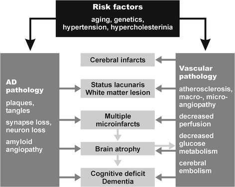
“These factors induce brain atrophy with cognitive and memory impairment (Figure 1) [147], although the complex cascade of these and other noxious factors needs further elucidation.”
§ The overlap between vascular disease and Alzheimer’s disease - lessons from pathology > Review > Pathogenic factors
What the sources say
- Not checked yetcites [147] Jellinger KA, Attems J: Neuropathological evaluation of mixed dementia. J Neurol Sci. 2007, 257: 80-87. source
plaindirect- Asserted: passes — The authors put it forward as true.
- Checkable: passes — It can be checked against evidence.
- Load-bearing: passes — The paper’s conclusions depend on it.
- Based: passes — The paper gives a reason why it holds.
It may state two separate claims as one.
In the paper
“Both hypertension and CAA are associated with an increased prevalence of CVLs [157]”
§ The overlap between vascular disease and Alzheimer’s disease - lessons from pathology > Review > Pathogenic factors
What the sources say
- Not checked yetcites [157] Jellinger KA, Attems J: Prevalence and pathogenic role of cerebrovascular lesions in Alzheimer’s disease. J Neurol Sci. 2005, 229-230: 37-41. source
plaincorrelational- Asserted: passes — The authors put it forward as true.
- Checkable: passes — It can be checked against evidence.
- Load-bearing: unclear — It is unclear whether the paper’s conclusions depend on it.
- Based: passes — The paper gives a reason why it holds.
It may state two separate claims as one.
In the paper
“CVD has been suggested to be an important cause of cognitive impairment in the elderly, both by itself or as a catalyst for the conversion of low-grade AD to overt dementia [186].”
§ The overlap between vascular disease and Alzheimer’s disease - lessons from pathology > Conclusions
What the sources say
- Not checked yetcites [186] Kalaria RN, Kenny RA, Ballard CG, Perry R, Ince P, Polvikoski T: Towards defining the neuropathological substrates of vascular dementia. J Neurol Sci. 2004, 226: 75-80. source
hedgedconditional- Asserted: passes — The authors put it forward as true.
- Checkable: passes — It can be checked against evidence.
- Load-bearing: passes — The paper’s conclusions depend on it.
- Based: passes — The paper gives a reason why it holds.
It may state two separate claims as one.
The authors state this as an assumption.
WMLs contribute to cognitive decline.
“and, thus, are assumed to contribute to cognitive decline [184].”
Claims other papers make about this one
Each is checked against this paper’s own text: does it say what the citing paper says it does?
The cited source says what the claim attributes to it.
In the citing paper
“Studies have consistently found a positive epidemiological correlation between Alzheimer’s disease (AD) and cardiometabolic traits, yet the biological mechanisms behind this correlation is not well understood [1–4].”
From Multi-trait association studies discover pleiotropic loci between Alzheimer’s disease and cardiometabolic traits (2021) · cites this paper as [1]
The cited source says what the claim attributes to it.
In the citing paper
“Studies have consistently found a positive epidemiological correlation between Alzheimer’s disease (AD) and cardiometabolic traits, yet the biological mechanisms behind this correlation is not well understood [1–4].”
From Multi-trait association studies discover pleiotropic loci between Alzheimer’s disease and cardiometabolic traits (2021) · cites this paper as [1]
In the citing paper
“Moreover, the growing recognition of overlapping pathologies (e.g., Lewy body inclusions, TDP-43 pathology, vascular lesions, etc.) among dementia patients indicates a wide range of biological heterogeneity within AD that necessitates a broader approach to scientific investigations of its pathophysiology [37–39].”
From Systems-based proteomics to resolve the biology of Alzheimer’s disease beyond amyloid and tau (2021) · cites this paper as [38]
Self-check
Percentages, sums and reported differences re-computed from the paper’s own numbers.
Each contradiction between two passages listed here was confirmed by an independent check.
Rated minor: it would not touch a result — a value no result rests on, a p-value that stays on the same side of the significance level, or a conflict that is clerical, resolved elsewhere in the paper, or outside its own findings.
In the paper
“Multiple infarcts (61 = 32.5%)”
§ The overlap between vascular disease and Alzheimer’s disease - lessons from pathology > Review > Topographical distribution of cerebrovascular lesions
Reported 61 · recomputed 58
Rated minor: it would not touch a result — a value no result rests on, a p-value that stays on the same side of the significance level, or a conflict that is clerical, resolved elsewhere in the paper, or outside its own findings.
In the paper
§ The overlap between vascular disease and Alzheimer’s disease - lessons from pathology > Review > Topographical distribution of cerebrovascular lesions
“Table: Table 3 Common lesions in AD, VaD, MIX, and aged controls (from[130])”
§ The overlap between vascular disease and Alzheimer’s disease - lessons from pathology > Review > Topographical distribution of cerebrovascular lesions
“The type and average prevalence of CVLs in AD, VaD, mixed dementia, and aged controls is shown in Table 3[147].”
Rated minor: it would not touch a result — a value no result rests on, a p-value that stays on the same side of the significance level, or a conflict that is clerical, resolved elsewhere in the paper, or outside its own findings.
In the paper
§ The overlap between vascular disease and Alzheimer’s disease - lessons from pathology > Review > Cerebrovascular and Alzheimer’s disease pathology in demented and non-demented elderly
“compared to 66.8% in age-matched controls, all showing low Braak stages (P <0.01).”
§ The overlap between vascular disease and Alzheimer’s disease - lessons from pathology > Review > Cerebrovascular and Alzheimer’s disease pathology in demented and non-demented elderly
“Minor and moderate vascular pathology in AD were about twice as frequent as in controls (26.2% vs. 12.2% and 20.9% vs. 11.3%; P <0.01). On the other hand, severe vascular pathology did not significantly differ between both groups (10.2% vs. 12.2%).”
Citations
Cites
Wardlaw, J. M., Smith, E. E., Biessels, G. J., Cordonnier, C., Fazekas, F., Frayne, R., Lindley, R. I., O'Brien, J. T., Barkhof, F., Benavente, O. R., et al. (2013) · The Lancet Neurology · doi:10.1016/s1474-4422(13)70124-8 · Cited by 5,544 · cited 1×
“SVD is a frequent cause of white matter lesions (WMLs; leukoaraiosis) that are increasingly detected by neuroimaging [118-121].”
BackgroundGorelick, P. B., Scuteri, A., Black, S. E., DeCarli, C., Greenberg, S. M., Iadecola, C., Launer, L. J., Laurent, S., Lopez, O. L., Nyenhuis, D., et al. (2011) · Stroke · doi:10.1161/str.0b013e3182299496 · Cited by 3,811 · cited 3×
“Epidemiological studies have shown that AD and CVD share common risk factors such as hypertension during midlife, diabetes mellitus, smoking, apolipoprotein E (ApoE) ε4 isoforms, hypercholesterolemia, homocysteinemia, and, in particular, age [16,30-34].”
BackgroundPantoni, L. (2010) · The Lancet Neurology · doi:10.1016/s1474-4422(10)70104-6 · Cited by 3,405 · cited 1×
“SVD affects small arteries and arterioles and refers to pathological changes similar to atherosclerosis that are termed small vessel arteriosclerosis/atherosclerosis, lipoor fibrohyalinosis, or hypertensive arteriopathy [115].”
BackgroundNeurovascular pathways to neurodegeneration in Alzheimer’s disease and other disorders
Zlokovic, B. V. (2011) · Nature Reviews Neuroscience · doi:10.1038/nrn3114 · Cited by 2,969 · cited 1×
“CVD has been suggested to contribute to AD neuropathological changes including selective brain atrophy and accumulation of abnormal proteins such as Aβ [24,35,84,85].”
BackgroundThe projected effect of risk factor reduction on Alzheimer's disease prevalence
Barnes, D. E., & Yaffe, K. S. (2011) · The Lancet Neurology · doi:10.1016/s1474-4422(11)70072-2 · Cited by 2,846
Mixed brain pathologies account for most dementia cases in community-dwelling older persons
Schneider, J. A., Arvanitakis, Z., Bang, W., & Bennett, D. A. (2007) · Neurology · doi:10.1212/01.wnl.0000271090.28148.24 · Cited by 1,992 · cited 2×
“The burden of vascular and AD-type pathologies are leading and independent causes of dementia in the elderly [4,9-15], suggesting additive or synergistic effects of both types of lesions on cognitive impairment [2,3,5,9,16-29].”
BackgroundThe Pathobiology of Vascular Dementia
Iadecola, C. (2013) · Neuron · doi:10.1016/j.neuron.2013.10.008 · Cited by 1,857 · cited 1×
“The pathophysiology of VaD has been critically reviewed recently [48,144-146].”
BackgroundCerebral microbleeds: a guide to detection and interpretation
Greenberg, S. M., Vernooij, M. W., Cordonnier, C., Viswanathan, A., Al-Shahi Salman, R., Warach, S., Launer, L. J., Van Buchem, M. A., & Breteler, M. M. (2009) · The Lancet Neurology · doi:10.1016/s1474-4422(09)70013-4 · Cited by 1,798
Brain Infarction and the Clinical Expression of Alzheimer Disease<subtitle>The Nun Study</subtitle>
Snowdon, D. A. (1997) · JAMA: The Journal of the American Medical Association · doi:10.1001/jama.1997.03540340047031 · Cited by 1,590
Silent brain infarcts: a systematic review
Vermeer, S. E., Longstreth, W. T., & Koudstaal, P. J. (2007) · The Lancet Neurology · doi:10.1016/s1474-4422(07)70170-9 · Cited by 1,221 · cited 1×
““Silent” lacunar infarcts are frequently detected by MRI or CCT and are not accompanied by any overt clinical symptoms, but double the risk of subsequent stroke and dementia [133].”
BackgroundKivipelto, M., Ngandu, T., Laatikainen, T. K., Winblad, B. G., Soininen, H. S., & Tuomilehto, J. (2006) · The Lancet Neurology · doi:10.1016/s1474-4422(06)70537-3 · Cited by 1,170 · cited 1×
“, atrial fibrillation and congestive heart failure, have also been linked to the pathogenesis and progression of AD and are among the most important modifiable risk factors for AD [35-42].”
BackgroundCerebral microvascular pathology in aging and Alzheimer's disease
Farkas, E., & Luiten, P. G. M. (2001) · Progress in Neurobiology · doi:10.1016/s0301-0082(00)00068-x · Cited by 1,102 · cited 1×
“Vascular disease is thought by many authors to play a major role in the pathogenesis of AD and some even consider AD as being rather a primarily vascular than a neurodegenerative disorder [22,68-74].”
BackgroundClinical diagnosis of cerebral amyloid angiopathy: validation of the Boston criteria
Knudsen, K. A., Rosand, J., Karluk, D., & Greenberg, S. M. (2001) · Neurology · doi:10.1212/wnl.56.4.537 · Cited by 1,015
Prevalence of superficial siderosis in patients with cerebral amyloid angiopathy
Linn, J., Halpin, A., Demaerel, P., Ruhland, J., Giese, A. D., Dichgans, M., van Buchem, M. A., Bruckmann, H., & Greenberg, S. M. (2010) · Neurology · doi:10.1212/wnl.0b013e3181dad605 · Cited by 914
Enlarged perivascular spaces on MRI are a feature of cerebral small vessel disease
Doubal, F. N., MacLullich, A. M. J., Ferguson, K. J., Dennis, M. S., & Wardlaw, J. M. (2010) · Stroke · doi:10.1161/strokeaha.109.564914 · Cited by 904 · cited 1×
“SVD is a frequent cause of white matter lesions (WMLs; leukoaraiosis) that are increasingly detected by neuroimaging [118-121].”
BackgroundNeurovascular mechanisms and blood–brain barrier disorder in Alzheimer’s disease
Bell, R. D., & Zloković, B. V. (2009) · Acta Neuropathologica · doi:10.1007/s00401-009-0522-3 · Cited by 884 · cited 1×
“BBB dysfunctions related to SVD leading to a leakage of plasma proteins into enlarged perivascular spaces [61,128] have been described in WMLs and lacunar stroke [129,130].”
BackgroundAlzheimer Disease as a Vascular Disorder
de la Torre, J. C. (2002) · Stroke · doi:10.1161/01.str.0000014421.15948.67 · Cited by 852 · cited 1×
“Vascular disease is thought by many authors to play a major role in the pathogenesis of AD and some even consider AD as being rather a primarily vascular than a neurodegenerative disorder [22,68-74].”
BackgroundToledo, J. B., Arnold, S. E., Raible, K., Brettschneider, J., Xie, S. X., Grossman, M., Monsell, S. E., Kukull, W. A., & Trojanowski, J. Q. (2013) · Brain · doi:10.1093/brain/awt188 · Cited by 849 · cited 4×
“, lower neuritic Braak stages) [5], similar to earlier studies on respective associations with subcortical vascular pathology [6] and general CVD [1].”
BackgroundResultInfluentialBrain infarction and the clinical expression of Alzheimer disease. The Nun Study.
Snowdon, D. A., Greiner, L. H., Mortimer, J. A., et al. (1997) · PubMed · Cited by 846
Neurovascular mechanisms of Alzheimer's neurodegeneration
Zlokovic, B. V. (2005) · Trends in Neurosciences · doi:10.1016/j.tins.2005.02.001 · Cited by 770 · cited 1×
“CVD has been suggested to contribute to AD neuropathological changes including selective brain atrophy and accumulation of abnormal proteins such as Aβ [24,35,84,85].”
BackgroundBrain Infarction and the Clinical Expression of Alzheimer Disease-Reply
Snowdon, D. A. (1997) · JAMA · doi:10.1001/jama.1997.03550020046024 · Cited by 683
Alzheimer disease and cerebrovascular pathology: an update
Jellinger, K. A. (2002) · Journal of Neural Transmission · doi:10.1007/s007020200068 · Cited by 680 · cited 5×
“, hypertensive vasculopathy), or microvascular degeneration (tortuosity, fibro- and lipohyalinosis,); – Blood–brain barrier (BBB) dysfunction causing white matter lesions (WMLs), microinfarctions, lacunes or lacunar infarcts, and microbleeds [17,87].”
BackgroundInfluentialSelf-citationNeuropathology of Cognitively Normal Elderly
Knopman, D. S., Parisi, J. E., Salviati, A., Floriach-Robert, M., Boeve, B. F., Ivnik, R. J., Smith, G. E., Dickson, D. W., Johnson, K. A., Petersen, L. E., et al. (2003) · Journal of Neuropathology & Experimental Neurology · doi:10.1093/jnen/62.11.1087 · Cited by 674 · cited 1×
“, small or large cerebral infarcts, lacunes, and WMLs, in up to 10% [10,165-167].”
BackgroundCerebral amyloid angiopathy in the elderly
Viswanathan, A., & Greenberg, S. M. (2011) · Annals of Neurology · doi:10.1002/ana.22516 · Cited by 643
Vascular contributions to cognitive impairment and dementia including Alzheimer's disease
Snyder, H. M., Corriveau, R. A., Craft, S., Faber, J. E., Greenberg, S. M., Knopman, D., Lamb, B. T., Montine, T. J., Nedergaard, M., Schaffer, C. B., et al. (2015) · Alzheimer's & Dementia · doi:10.1016/j.jalz.2014.10.008 · Cited by 603 · cited 1×
“The burden of vascular and AD-type pathologies are leading and independent causes of dementia in the elderly [4,9-15], suggesting additive or synergistic effects of both types of lesions on cognitive impairment [2,3,5,9,16-29].”
BackgroundCasserly, I., & Topol, E. J. (2004) · The Lancet · doi:10.1016/s0140-6736(04)15900-x · Cited by 560 · cited 1×
“Epidemiological studies have shown that AD and CVD share common risk factors such as hypertension during midlife, diabetes mellitus, smoking, apolipoprotein E (ApoE) ε4 isoforms, hypercholesterolemia, homocysteinemia, and, in particular, age [16,30-34].”
BackgroundThe overlap between neurodegenerative and vascular factors in the pathogenesis of dementia
Iadecola, C. (2010) · Acta Neuropathologica · doi:10.1007/s00401-010-0718-6 · Cited by 557 · cited 1×
“On the other hand, CVD has been associated with worse cognitive performance in AD and neuropathological studies report that CVD lowers the threshold for dementia in subjects with a pathological diagnosis of AD [5,6,8,9,13,17,23,51,53,81-83].”
BackgroundMild cognitive impairment is related to Alzheimer disease pathology and cerebral infarctions
Bennett, D. A., Schneider, J. A., Bienias, J. L., Evans, D. A., & Wilson, R. S. (2005) · Neurology · doi:10.1212/01.wnl.0000152982.47274.9e · Cited by 537 · cited 1×
“However, AD pathology alone more frequently accounts for dementia than both macroscopic and microscopic infarcts [15] and, in late stages of AD, concomitant SVLs do not significantly influence the overall state and progression of cognitive decline [45,54,76], the severity and extent of AD pathology overwhelming the ra…”
BackgroundWhite matter lesions impair frontal lobe function regardless of their location
Tullberg, M., Fletcher, E., DeCarli, C., Mungas, D., Reed, B. R., Harvey, D. J., Weiner, M. W., Chui, H. C., & Jagust, W. J. (2004) · Neurology · doi:10.1212/01.wnl.0000130530.55104.b5 · Cited by 530 · cited 1×
“They impair frontal functions regardless of their location [175,176] and increase the risk of dementia, particularly in patients with lacunar infarcts [177,178], causing functional network disruption in cognitively-impaired individuals compared with age-matched healthy elderly controls [179,180].”
BackgroundThe role of cerebral ischemia in Alzheimer’s disease
Kalaria, R. N. (2000) · Neurobiology of Aging · doi:10.1016/s0197-4580(00)00125-1 · Cited by 526 · cited 3×
“However, the role of combined cerebrovascular pathology and AD in dementia is still under discussion and data obtained from epidemiological and clinico-pathological studies regarding their relation are controversial [13,17,22,23,52-55].”
BackgroundAlzheimer neuropathologic alterations in aged cognitively normal subjects
Davis, D. G., Schmitt, F. A., Wekstein, D. R., & Markesbery, W. R. (1999) · Journal of Neuropathology and Experimental Neurology · doi:10.1097/00005072-199904000-00008 · Cited by 516 · cited 1×
“, small or large cerebral infarcts, lacunes, and WMLs, in up to 10% [10,165-167].”
BackgroundDiabetes, Alzheimer disease, and vascular dementia
Ahtiluoto, S., Polvikoski, T. M., Peltonen, M., Solomon, A., Tuomilehto, J., Winblad, B. G., Sulkava, R. O., & Kivipelto, M. (2010) · Neurology · doi:10.1212/wnl.0b013e3181f4d7f8 · Cited by 499
Imaging of amyloid burden and distribution in cerebral amyloid angiopathy
Johnson, K. A., Gregas, M., Becker, J. A., Kinnecom, C., Salat, D. H., Moran, E. K., Smith, E. E., Rosand, J., Rentz, D. M., Klunk, W. E., et al. (2007) · Annals of Neurology · doi:10.1002/ana.21164 · Cited by 495 · cited 1×
“In addition to the presence of superficial siderosis, cerebral microbleeds, cortical microinfarcts, and hypointensities in MRI images [106-109], the use of Pittsburgh Compound-B (PiB)positron emission tomography (PET) is useful in detecting CAA intra vitam [110,111], and a significant decrease of both Aβ-40 and Aβ-42…”
BackgroundCerebrovascular disease and threshold for dementia in the early stages of Alzheimer's disease
Esiri, M. M., Nagy, Z., Smith, M. Z., Barnetson, L., & Smith, A. D. (1999) · The Lancet · doi:10.1016/s0140-6736(99)02355-7 · Cited by 493 · cited 3×
“However, the role of combined cerebrovascular pathology and AD in dementia is still under discussion and data obtained from epidemiological and clinico-pathological studies regarding their relation are controversial [13,17,22,23,52-55].”
BackgroundMicrovascular injury and blood–brain barrier leakage in Alzheimer's disease
Zipser, B. D., Johanson, C. E., Gonzalez, L., Berzin, T. M., Tavares, R., Hulette, C. M., Vitek, M. P., Hovanesian, V., & Stopa, E. G. (2007) · Neurobiology of Aging · doi:10.1016/j.neurobiolaging.2006.05.016 · Cited by 487 · cited 1×
“AD has been reported to present frequently together with SVD, microvascular injury, and microscopic CVLs [8,16,47,56-60].”
BackgroundFunctional network disruption in the degenerative dementias
Pievani, M., de Haan, W., Wu, T., Seeley, W. W., & Frisoni, G. B. (2011) · The Lancet Neurology · doi:10.1016/s1474-4422(11)70158-2 · Cited by 481
Overlap Between Pathology of Alzheimer Disease and Vascular Dementia
Kalaria, R. N., & Ballard, C. G. (1999) · Alzheimer Disease & Associated Disorders · doi:10.1097/00002093-199912003-00017 · Cited by 475 · cited 2×
“In a series of 300 autopsy cases of AD, Kalaria and Ballard [148] reported 98% CAA, 100% microvascular degeneration, 31% infarcts of all sizes, and 7% intracerebral hemorrhage, while Olichney [149], in a cohort of 248 autopsy cases of AD, revealed a total of 48% CVLs, with 31% microinfarcts, 12.”
BackgroundInfluentialNeurological Diseases in Relation to the Blood–Brain Barrier
Rosenberg, G. A. (2012) · Journal of Cerebral Blood Flow & Metabolism · doi:10.1038/jcbfm.2011.197 · Cited by 454 · cited 1×
“Mechanisms leading to BBB leakage in aging brains are complex, including oxidative damage and the activation of proteases, matrix metalloproteinases, and cyclooxygenases [131].”
BackgroundPathological correlates of dementia in a longitudinal, population‐based sample of aging
Sonnen, J. A., Larson, E. B., Crane, P. K., Haneuse, S., Li, G., Schellenberg, G. D., Craft, S., Leverenz, J. B., & Montine, T. J. (2007) · Annals of Neurology · doi:10.1002/ana.21208 · Cited by 432 · cited 1×
“The burden of vascular and AD-type pathologies are leading and independent causes of dementia in the elderly [4,9-15], suggesting additive or synergistic effects of both types of lesions on cognitive impairment [2,3,5,9,16-29].”
BackgroundMicroinfarct Pathology, Dementia, and Cognitive Systems
Arvanitakis, Z., Leurgans, S. E., Barnes, L. L., Bennett, D. A., & Schneider, J. A. (2011) · Stroke · doi:10.1161/strokeaha.110.595082 · Cited by 404 · cited 1×
“A recent study assessed CVD in 5,715 autopsy cases of the National Alzheimer’s Coordinating Center (NACC) database, and confirmed previous data on the prevalence of CVD in AD and the additive or interactive deleterious effect of both AD and vascular pathologies on cognition [6,9,47,50,51].”
BackgroundThal, D. R., Ghebremedhin, E., Orantes, M., & Wiestler, O. D. (2003) · Journal of Neuropathology & Experimental Neurology · doi:10.1093/jnen/62.12.1287 · Cited by 384
Kovács, G. G., Milenković, I., Wöehrer, A., Höftberger, R., Gelpí, E., Haberler, C., Hönigschnabl, S., Reiner‐Concin, A., Heinzl, H., Jungwirth, S., et al. (2013) · Acta Neuropathologica · doi:10.1007/s00401-013-1157-y · Cited by 380 · cited 1×
“8% [47], while the majority (24% to 93%) showed mixed pathologies [158,159].”
BackgroundThe Neuropathology of Older Persons with and Without Dementia from Community versus Clinic Cohorts
Schneider, J. A., Aggarwal, N. T., Barnes, L., Boyle, P., & Bennett, D. A. (2009) · Journal of Alzheimer's Disease · doi:10.3233/jad-2009-1227 · Cited by 372 · cited 2×
“, small or large cerebral infarcts, lacunes, and WMLs, in up to 10% [10,165-167].”
BackgroundCerebral amyloid angiopathy and its relationship to Alzheimer’s disease
Thal, D. R., Griffin, W. S. T., de Vos, R. A. I., & Ghebremedhin, E. (2008) · Acta Neuropathologica · doi:10.1007/s00401-008-0366-2 · Cited by 372 · cited 1×
“Due to the location of the underlying vessel disorder, multiple cortical CMIs are often associated with CAA, whereas subcortical microinfarcts are mainly linked to SVD or atherosclerosis-related embolism [135].”
BackgroundReview: sporadic cerebral amyloid angiopathy
Attems, J., Jellinger, K., Thal, D. R., & Van Nostrand, W. (2011) · Neuropathology and Applied Neurobiology · doi:10.1111/j.1365-2990.2010.01137.x · Cited by 371 · cited 2×
“The occipital lobe has been reported to be the site most frequently and severely affected by CAA, followed by either frontal, temporal, or parietal lobes [89,90].”
BackgroundSelf-citationLacunar stroke is associated with diffuse blood-brain barrier dysfunction
Wardlaw, J. M., Doubal, F., Armitage, P., Chappell, F., Carpenter, T., Muñoz Maniega, S., Farrall, A., Sudlow, C., Dennis, M., & Dhillon, B. (2009) · Annals of Neurology · doi:10.1002/ana.21549 · Cited by 364 · cited 1×
“BBB dysfunctions related to SVD leading to a leakage of plasma proteins into enlarged perivascular spaces [61,128] have been described in WMLs and lacunar stroke [129,130].”
BackgroundPathology and pathogenesis of vascular cognitive impairment—a critical update
Jellinger, K. A. (2013) · Frontiers in Aging Neuroscience · doi:10.3389/fnagi.2013.00017 · Cited by 353 · cited 2×
“In contrast to AD and mixed dementia, the prevalence of pure VaD decreased after 80 years of age [145,158].”
BackgroundResultSelf-citationNeuropathological assessment of the lesions of significance in vascular dementia
Esiri, M. M., Wilcock, G. K., & Morris, J. H. (1997) · Journal of Neurology, Neurosurgery & Psychiatry · doi:10.1136/jnnp.63.6.749 · Cited by 347 · cited 2×
“However, the role of combined cerebrovascular pathology and AD in dementia is still under discussion and data obtained from epidemiological and clinico-pathological studies regarding their relation are controversial [13,17,22,23,52-55].”
BackgroundVascular pathology in the aged human brain
Grinberg, L. T., & Thal, D. R. (2010) · Acta Neuropathologica · doi:10.1007/s00401-010-0652-7 · Cited by 345 · cited 2×
“, hypertensive vasculopathy), or microvascular degeneration (tortuosity, fibro- and lipohyalinosis,); – Blood–brain barrier (BBB) dysfunction causing white matter lesions (WMLs), microinfarctions, lacunes or lacunar infarcts, and microbleeds [17,87].”
BackgroundHeterogeneity in age-related white matter changes
Schmidt, R., Schmidt, H., Haybaeck, J., Loitfelder, M., Weis, S., Cavalieri, M., Seiler, S., Enzinger, C., Ropele, S., Erkinjuntti, T., et al. (2011) · Acta Neuropathologica · doi:10.1007/s00401-011-0851-x · Cited by 343 · cited 2×
“WMLs may be caused by both CVD (hypoperfusion) and AD (retrograde degeneration), they progress with age, and they are a considerable risk factor for cognitive impairment [120,173,174].”
BackgroundSelf-citationMRI predictors of cognition in subcortical ischemic vascular disease and Alzheimer's disease
Mungas, D., Jagust, W. J., Reed, B. R., Kramer, J. H., Weiner, M. W., Schuff, N., Norman, D., Mack, W. J., Willis, L., & Chui, H. C. (2001) · Neurology · doi:10.1212/wnl.57.12.2229 · Cited by 341
Contribution of Vascular Risk Factors to the Progression in Alzheimer Disease
Helzner, E., Luchsinger, J. A., Scarmeas, N., Cosentino, S., Brickman, A. M., Glymour, M. M., & Stern, Y. (2009) · Archives of Neurology · doi:10.1001/archneur.66.3.343 · Cited by 338 · cited 1×
“, atrial fibrillation and congestive heart failure, have also been linked to the pathogenesis and progression of AD and are among the most important modifiable risk factors for AD [35-42].”
BackgroundNeuropathologic correlates of white matter hyperintensities
Young, V. G., Halliday, G. M., & Kril, J. J. (2008) · Neurology · doi:10.1212/01.wnl.0000319691.50117.54 · Cited by 336 · cited 2×
“Damage to the vasculature may, in turn, impair the BBB integrity as one mechanism by which WMLs may evolve [124].”
BackgroundSmall Vessel Disease and General Cognitive Function in Nondisabled Elderly
van der Flier, W. M., van Straaten, E. C. W., Barkhof, F., Verdelho, A., Madureira, S., Pantoni, L., Inzitari, D., Erkinjuntti, T., Crisby, M., Waldemar, G., et al. (2005) · Stroke · doi:10.1161/01.str.0000179092.59909.42 · Cited by 324 · cited 1×
“Although WMLs and lacunes may be independently associated with cognitive dysfunction [181,182], WMLs in AD are significantly correlated to cortical and medial temporal lobe atrophy [181-183], and, thus, are assumed to contribute to cognitive decline [184].”
BackgroundMuch of late life cognitive decline is not due to common neurodegenerative pathologies
Boyle, P. A., Wilson, R. S., Yu, L., Barr, A. M., Honer, W. G., Schneider, J. A., & Bennett, D. A. (2013) · Annals of Neurology · doi:10.1002/ana.23964 · Cited by 323 · cited 1×
“However, much of the variation in cognitive decline remains unexplained, suggesting that other important determinants of cognitive decline remain to be identified [156].”
BackgroundVascular factors predict rate of progression in Alzheimer disease
Mielke, M. M., Rosenberg, P. B., Tschanz, J. T., Cook, L., Corcoran, C. M., Hayden, K. M., Norton, M. C., Rabins, P. V., Green, R. C., Welsh-Bohmer, K. A., et al. (2007) · Neurology · doi:10.1212/01.wnl.0000279520.59792.fe · Cited by 311 · cited 1×
“, atrial fibrillation and congestive heart failure, have also been linked to the pathogenesis and progression of AD and are among the most important modifiable risk factors for AD [35-42].”
BackgroundBrain microbleeds and Alzheimer’s disease: innocent observation or key player?
Cordonnier, C., & van der Flier, W. M. (2011) · Brain · doi:10.1093/brain/awq321 · Cited by 310
Evidence that Alzheimer’s disease is a microvascular disorder: the role of constitutive nitric oxide
de la Torre, J. C., & Stefano, G. B. (2000) · Brain Research Reviews · doi:10.1016/s0165-0173(00)00043-6 · Cited by 307 · cited 1×
“Vascular disease is thought by many authors to play a major role in the pathogenesis of AD and some even consider AD as being rather a primarily vascular than a neurodegenerative disorder [22,68-74].”
BackgroundAmyloid PET imaging in Alzheimer’s disease: a comparison of three radiotracers
Initiative, T. A. D. N., Landau, S. M., Thomas, B. A., Thurfjell, L., Schmidt, M. E., Margolin, R. A., Mintun, M. A., Pontecorvo, M. J., Baker, S. L., & Jagust, W. J. (2014) · European Journal of Nuclear Medicine and Molecular Imaging · doi:10.1007/s00259-014-2753-3 · Cited by 306 · cited 1×
“) [188-190] will enable the identification of AD and CVD patients in clinical and research settings.”
BackgroundRelation of cerebral infarctions to dementia and cognitive function in older persons
Schneider, J. A., Wilson, R. S., Cochran, E. J., Bienias, J. L., Arnold, S. E., Evans, D. A., & Bennett, D. A. (2003) · Neurology · doi:10.1212/01.wnl.0000055863.87435.b2 · Cited by 305 · cited 1×
“The burden of vascular and AD-type pathologies are leading and independent causes of dementia in the elderly [4,9-15], suggesting additive or synergistic effects of both types of lesions on cognitive impairment [2,3,5,9,16-29].”
BackgroundNelson, P. T., Jicha, G. A., Schmitt, F. A., Liu, H., Davis, D. G., Mendiondo, M. S., Abner, E. L., & Markesbery, W. R. (2007) · Journal of Neuropathology and Experimental Neurology · doi:10.1097/nen.0b013e31815c5efb · Cited by 292 · cited 1×
“These data add further evidence for AD pathology (mainly neurofibrillary tangles and neuritic plaques) being the main morphological substrate of clinical dementia [51,79,80].”
BackgroundTowards defining the neuropathological substrates of vascular dementia
Kalaria, R. N., Kenny, R. A., Ballard, C. G., Perry, R., Ince, P., & Polvikoski, T. (2004) · Journal of the Neurological Sciences · doi:10.1016/j.jns.2004.09.019 · Cited by 291 · cited 1×
“Conclusions CVD has been suggested to be an important cause of cognitive impairment in the elderly, both by itself or as a catalyst for the conversion of low-grade AD to overt dementia [186].”
BackgroundSchrag, M., McAuley, G. A., Pomakian, J., Jiffry, A., Tung, S., Mueller, C., Vinters, H. V., Haacke, E. M., Holshouser, B. A., Kido, D. K., et al. (2009) · Acta Neuropathologica · doi:10.1007/s00401-009-0615-z · Cited by 290 · cited 1×
“In addition to the presence of superficial siderosis, cerebral microbleeds, cortical microinfarcts, and hypointensities in MRI images [106-109], the use of Pittsburgh Compound-B (PiB)positron emission tomography (PET) is useful in detecting CAA intra vitam [110,111], and a significant decrease of both Aβ-40 and Aβ-42…”
BackgroundNeuropathological evaluation of mixed dementia
Jellinger, K. A., & Attems, J. (2007) · Journal of the Neurological Sciences · doi:10.1016/j.jns.2007.01.045 · Cited by 289 · cited 2×
“These factors induce brain atrophy with cognitive and memory impairment (Figure 1) [147], although the complex cascade of these and other noxious factors needs further elucidation.”
BackgroundSelf-citationAtherosclerosis and AD: Analysis of data from the US National Alzheimer’s Coordinating Center
Honig, L. S., Kukull, W., & Mayeux, R. (2005) · Neurology · doi:10.1212/01.wnl.0000150886.50187.30 · Cited by 284
Topakian, R., Barrick, T. R., Howe, F. A., & Markus, H. S. (2010) · Journal of Neurology, Neurosurgery & Psychiatry · doi:10.1136/jnnp.2009.172072 · Cited by 281 · cited 1×
“BBB dysfunctions related to SVD leading to a leakage of plasma proteins into enlarged perivascular spaces [61,128] have been described in WMLs and lacunar stroke [129,130].”
BackgroundCerebrovascular atherosclerosis correlates with Alzheimer pathology in neurodegenerative dementias
Yarchoan, M. M., Xie, S. X., Kling, M. A., Toledo, J. B., Wolk, D. A., Lee, E. B., Deerlin, V. M. V., Lee, V. M. Y., Trojanowski, J. Q., & Arnold, S. E. (2012) · Brain · doi:10.1093/brain/aws271 · Cited by 268 · cited 1×
“atherosclerosis in the circle of Willis has been specifically linked to AD [138-140], and the presence of largevessel CVD was strongly associated with an increased frequency of neuritic plaques, suggesting a common etiology or a reciprocal regulation for atherosclerosis and AD [138,141].”
BackgroundCerebral Microinfarcts: A Systematic Review of Neuropathological Studies
Brundel, M., de Bresser, J., van Dillen, J. J., Kappelle, L. J., & Biessels, G. J. (2012) · Journal of Cerebral Blood Flow & Metabolism · doi:10.1038/jcbfm.2011.200 · Cited by 258
Ihara, M., Polvikoski, T. M., Hall, R., Slade, J. Y., Perry, R. H., Oakley, A. E., Englund, E. M., O'Brien, J. T., Ince, P. G., & Kalaria, R. N. (2010) · Acta Neuropathologica · doi:10.1007/s00401-009-0635-8 · Cited by 251 · cited 1×
“WMLs may be caused by both CVD (hypoperfusion) and AD (retrograde degeneration), they progress with age, and they are a considerable risk factor for cognitive impairment [120,173,174].”
BackgroundCognitive impact of subcortical vascular and Alzheimer’s disease pathology
Chui, H. C., Zarow, C., Mack, W. J., Ellis, W. G., Zheng, L., Jagust, W. J., Mungas, D., Reed, B. R., Kramer, J. H., DeCarli, C. C., et al. (2006) · Annals of Neurology · doi:10.1002/ana.21009 · Cited by 247 · cited 4×
“A recent study assessed CVD in 5,715 autopsy cases of the National Alzheimer’s Coordinating Center (NACC) database, and confirmed previous data on the prevalence of CVD in AD and the additive or interactive deleterious effect of both AD and vascular pathologies on cognition [6,9,47,50,51].”
BackgroundResultInfluentialVascular pathologies and cognition in a population-based cohort of elderly people
Fernando, M. S., & Ince, P. G. (2004) · Journal of the Neurological Sciences · doi:10.1016/j.jns.2004.09.004 · Cited by 239 · cited 1×
“The proportion of multiple CVL was higher in the demented group, while only 21% of clinically-demented patients showed “pure” AD pathology at post-mortem, indicating that most patients had mixed disease [152].”
ResultAtherosclerosis, dementia, and Alzheimer disease in the Baltimore Longitudinal Study of aging cohort
Dolan, H., Crain, B. J., Troncoso, J. C., Resnick, S. M., Zonderman, A. B., & O’Brien, R. (2010) · Annals of Neurology · doi:10.1002/ana.22055 · Cited by 235 · cited 2×
“Assessment of 175 autopsy cases in the Baltimore Longitudinal Study of Aging cohort found no relationship between the degree of atherosclerosis in the aorta, heart, and intracranial vessels and the degree of AD pathology, while the presence of intracranial atherosclerosis significantly increased the odds of dementia,…”
Beach, T. G., Wilson, J. R., Sue, L. I., Newell, A., Poston, M., Cisneros, R., Pandya, Y., Esh, C. L., Connor, D. J., Sabbagh, M. N., et al. (2006) · Acta Neuropathologica · doi:10.1007/s00401-006-0136-y · Cited by 232 · cited 1×
“atherosclerosis in the circle of Willis has been specifically linked to AD [138-140], and the presence of largevessel CVD was strongly associated with an increased frequency of neuritic plaques, suggesting a common etiology or a reciprocal regulation for atherosclerosis and AD [138,141].”
BackgroundAD lesions and infarcts in demented and non‐demented Japanese‐American men
Petrovitch, H., Ross, G. W., Steinhorn, S. C., Abbott, R. D., Markesbery, W., Davis, D., Nelson, J., Hardman, J., Masaki, K., Vogt, M. R., et al. (2005) · Annals of Neurology · doi:10.1002/ana.20318 · Cited by 230
Charidimou, A., Meegahage, R., Fox, Z., Peeters, A., Vandermeeren, Y., Laloux, P., Baron, J.-C., Jager, H. R., & Werring, D. J. (2013) · Journal of Neurology, Neurosurgery & Psychiatry · doi:10.1136/jnnp-2012-304434 · Cited by 225 · cited 2×
“Correlations of these criteria with post-mortem neuropathological findings indicate that the diagnosis of probable CAA-related hemorrhage can be made intra vitam with high accuracy [102-105].”
BackgroundArima, H., Tzourio, C., Anderson, C. S., Woodward, M., Bousser, M., MacMahon, S. W., Neal, B. C., & Chalmers, J. P. (2010) · Stroke · doi:10.1161/strokeaha.109.563932 · Cited by 223 · cited 1×
“Of note, the majority of cases with CAA-related ICH had hypertension, suggesting that hypertension is an important additional causal factor in CAA-related ICHs [95,96].”
BackgroundVascular Dementia in a Population-Based Autopsy Study
Knopman, D. S., Parisi, J. E., Boeve, B. F., Cha, R. H., Apaydın, H., Salviati, A., Edland, S. D., & Rocca, W. A. (2003) · Archives of Neurology · doi:10.1001/archneur.60.4.569 · Cited by 222 · cited 1×
“Criteria for pure VaD using imaging results (Mayo Clinic criteria) showed 75% sensitivity and 81% specificity [151].”
MethodRelation of neuropathology to cognition in persons without cognitive impairment
Bennett, D. A., Wilson, R. S., Boyle, P. A., Buchman, A. S., & Schneider, J. A. (2012) · Annals of Neurology · doi:10.1002/ana.23654 · Cited by 220 · cited 1×
“The burden of vascular and AD-type pathologies are leading and independent causes of dementia in the elderly [4,9-15], suggesting additive or synergistic effects of both types of lesions on cognitive impairment [2,3,5,9,16-29].”
BackgroundEffect of infarcts on dementia in the Baltimore longitudinal study of aging
Troncoso, J. C., Zonderman, A. B., Resnick, S. M., Crain, B., Pletnikova, O., & O'Brien, R. J. (2008) · Annals of Neurology · doi:10.1002/ana.21413 · Cited by 219 · cited 2×
“However, AD pathology alone more frequently accounts for dementia than both macroscopic and microscopic infarcts [15] and, in late stages of AD, concomitant SVLs do not significantly influence the overall state and progression of cognitive decline [45,54,76], the severity and extent of AD pathology overwhelming the ra…”
BackgroundVascular determinants of cholinergic deficits in Alzheimer disease and vascular dementia.
Román, G. C., & Kalaria, R. N. (2006) · Neurobiology of Aging · doi:10.1016/j.neurobiolaging.2005.10.004 · Cited by 213 · cited 1×
“Microvascular changes in the aged brain and in AD induce impairment of cerebral perfusion, in particular decrease of regional blood flow, reduction of glucose transport and utilization, loss of vascular innervation with special impact on the cholinergic and transmitter deficits in AD [171], impairment of neurovascular…”
BackgroundCerebral infarcts in patients with autopsy-proven Alzheimer's disease
Heyman, A., Fillenbaum, G. G., Welsh-Bohmer, K. A., Gearing, M., Mirra, S. S., Mohs, R. C., Peterson, B. L., & Pieper, C. F. (1998) · Neurology · doi:10.1212/wnl.51.1.159 · Cited by 206 · cited 1×
“On the other hand, CVD has been associated with worse cognitive performance in AD and neuropathological studies report that CVD lowers the threshold for dementia in subjects with a pathological diagnosis of AD [5,6,8,9,13,17,23,51,53,81-83].”
BackgroundCortical Microinfarcts and Demyelination Significantly Affect Cognition in Brain Aging
Kövari, E., Gold, G., Herrmann, F. R., Canuto, A., Hof, P. R., Michel, J.-P., Bouras, C., & Giannakopoulos, P. (2004) · Stroke · doi:10.1161/01.str.0000110791.51378.4e · Cited by 202 · cited 1×
“Further, the neuropathological evaluation of focal and white matter gliosis may have no clinical validity [185].”
BackgroundCerebrospinal fluid amyloid β40 is decreased in cerebral amyloid angiopathy
Verbeek, M. M., Kremer, B. P. H., Rikkert, M. O., Van Domburg, P. H. M. F., Skehan, M. E., & Greenberg, S. M. (2009) · Annals of Neurology · doi:10.1002/ana.21694 · Cited by 202 · cited 1×
“In addition to the presence of superficial siderosis, cerebral microbleeds, cortical microinfarcts, and hypointensities in MRI images [106-109], the use of Pittsburgh Compound-B (PiB)positron emission tomography (PET) is useful in detecting CAA intra vitam [110,111], and a significant decrease of both Aβ-40 and Aβ-42…”
BackgroundWhite, L. (2009) · Journal of Alzheimer's Disease · doi:10.3233/jad-2009-1178 · Cited by 193 · cited 1×
“There are considerable differences in the pathological burden in relation to age of onset of dementia, suggesting that late onset is associated with increased vascular pathology and lower AD burden [161,162].”
BackgroundBrayne, C., the Cambridge City over-75s Cohort (CC75C) study neuropathology collaboration, Richardson, K., Matthews, F. E., Fleming, J., Hunter, S., Xuereb, J. H., Paykel, E., Mukaetova-Ladinska, E. B., Huppert, F. A., et al. (2009) · Journal of Alzheimer's Disease · doi:10.3233/jad-2009-1182 · Cited by 191
Attems, J., Jellinger, K. A., & Lintner, F. (2005) · Acta Neuropathologica · doi:10.1007/s00401-005-1064-y · Cited by 188 · cited 1×
“The occipital lobe has been reported to be the site most frequently and severely affected by CAA, followed by either frontal, temporal, or parietal lobes [89,90].”
BackgroundSelf-citationCerebrovascular lesions induce transient -amyloid deposition
Garcia-Alloza, M., Gregory, J., Kuchibhotla, K. V., Fine, S., Wei, Y., Ayata, C., Frosch, M. P., Greenberg, S. M., & Bacskai, B. J. (2011) · Brain · doi:10.1093/brain/awr300 · Cited by 187 · cited 1×
“SVD-induced ApoE leakage was associated with AD and accumulation of β-amyloid (Aβ) in perivascular astrocytes [61] and transient induction of Aβ deposition [62].”
BackgroundGregoire, S. M., Charidimou, A., Gadapa, N., Dolan, E., Antoun, N. M., Peeters, A. P., Vandermeeren, Y., Laloux, P., Baron, J., Jäger, H. R., et al. (2011) · Brain · doi:10.1093/brain/awr172 · Cited by 187 · cited 1×
“Of note, the majority of cases with CAA-related ICH had hypertension, suggesting that hypertension is an important additional causal factor in CAA-related ICHs [95,96].”
BackgroundDegenerative and vascular lesions of the brain have synergistic effects in dementia of the elderly
Zekry, D., Duyckaerts, C. J., Moulias, R., Belmin, J., Geoffre, C., Herrmann, F. R., & Hauw, J. (2002) · Acta Neuropathologica · doi:10.1007/s00401-001-0493-5 · Cited by 184 · cited 4×
“However, the role of combined cerebrovascular pathology and AD in dementia is still under discussion and data obtained from epidemiological and clinico-pathological studies regarding their relation are controversial [13,17,22,23,52-55].”
BackgroundInfluentialDuering, M., Csanadi, E., Gesierich, B., Jouvent, E., Herve, D., Seiler, S., Belaroussi, B., Ropele, S., Schmidt, R., Chabriat, H., et al. (2013) · Brain · doi:10.1093/brain/awt184 · Cited by 183 · cited 1×
“SVD is a frequent cause of white matter lesions (WMLs; leukoaraiosis) that are increasingly detected by neuroimaging [118-121].”
BackgroundHawkes, C. A., Sullivan, P. M., Hands, S. L., Weller, R. O., Nicoll, J. A. R., & Carare, R. O. (2012) · PLoS ONE · doi:10.1371/journal.pone.0041636 · Cited by 182
Effects of White Matter Lesions and Lacunes on Cortical Function
Reed, B., Eberling, J. L., Mungas, D., Weiner, M. W., Kramer, J. H., & Jagust, W. J. (2004) · Archives of Neurology · doi:10.1001/archneur.61.10.1545 · Cited by 182 · cited 1×
“They impair frontal functions regardless of their location [175,176] and increase the risk of dementia, particularly in patients with lacunar infarcts [177,178], causing functional network disruption in cognitively-impaired individuals compared with age-matched healthy elderly controls [179,180].”
BackgroundIn vivo evaluation of a novel tau imaging tracer for Alzheimer’s disease
Villemagne, V. L., Furumoto, S., Fodero‐Tavoletti, M. T., Mulligan, R. S., Hodges, J. R., Harada, R., Yates, P. A., Piguet, O., Pejoska, S., Doré, V., et al. (2014) · European Journal of Nuclear Medicine and Molecular Imaging · doi:10.1007/s00259-013-2681-7 · Cited by 173 · cited 1×
“) [188-190] will enable the identification of AD and CVD patients in clinical and research settings.”
BackgroundWirth, M., Villeneuve, S., Haase, C. M., Madison, C. M., Oh, H., Landau, S. M., Rabinovici, G. D., & Jagust, W. J. (2013) · JAMA Neurology · doi:10.1001/jamaneurol.2013.4013 · Cited by 171 · cited 1×
“7 years) confirmed that a substantial number harbor neurodegeneration without Aβ burden, but association of neurodegenerative lesions with CVD can emerge through non-Aβ pathways within regions most affected by AD [170].”
BackgroundMicroinfarcts, brain atrophy, and cognitive function: The Honolulu Asia Aging Study Autopsy Study
Launer, L. J., Hughes, T. M., & White, L. R. (2011) · Annals of Neurology · doi:10.1002/ana.22520 · Cited by 171 · cited 1×
“AD has been reported to present frequently together with SVD, microvascular injury, and microscopic CVLs [8,16,47,56-60].”
BackgroundProgression of white matter lesions and hemorrhages in cerebral amyloid angiopathy
Chen, Y. W., Gurol, M. E., Rosand, J. M., Viswanathan, A., Rakich, S. M., Groover, T. R., Greenberg, S. M., & Smith, E. E. (2006) · Neurology · doi:10.1212/01.wnl.0000223613.57229.24 · Cited by 167 · cited 3×
“CAA may cause lobar intracerebral hemorrhages (ICH) and microbleeds [91]; it is indeed considered a risk factor for non-traumatic ICHs in the elderly and is present in up to 20% of all cases with ICH [92].”
BackgroundThe enigma of vascular cognitive disorder and vascular dementia
Jellinger, K. A. (2007) · Acta Neuropathologica · doi:10.1007/s00401-006-0185-2 · Cited by 166 · cited 4×
“4%, Table 2), suggesting different pathogenic mechanisms between these types of disorders [2,3].”
BackgroundInfluentialSelf-citationPark, J., Seo, S. W., Kim, C., Kim, G. H., Noh, H. J., Kim, S. T., Kwak, K., Yoon, U., Lee, J. M., Lee, J. W., et al. (2013) · Annals of Neurology · doi:10.1002/ana.23845 · Cited by 164 · cited 1×
“Deep cerebral microbleeds (CMB) are mainly linked to subcortical SVD, while both subcortical SVD and CAA interact to increase the risk of lobar CMBs [122,123].”
Background. Identification of Alzheimer and vascular lesion thresholds for mixed dementia.
Gold, G., Giannakopoulos, P., Herrmann, F. R., Bouras, C., & Kovari, E. (2007) · Brain · doi:10.1093/brain/awm228 · Cited by 163 · cited 1×
“However, AD pathology alone more frequently accounts for dementia than both macroscopic and microscopic infarcts [15] and, in late stages of AD, concomitant SVLs do not significantly influence the overall state and progression of cognitive decline [45,54,76], the severity and extent of AD pathology overwhelming the ra…”
BackgroundA Population-Based Clinicopathological Study in the Oldest-Old: The 90+ Study
M. Corrada, M., J. Berlau, D., & H. Kawas, C. (2012) · Current Alzheimer Research · doi:10.2174/156720512801322537 · Cited by 162 · cited 1×
“However, in a 90+ study, there was extensive overlap in pathology among those with and without dementia; 22% of demented subjects did not have significant pathology to account for their cognitive impairment [163].”
BackgroundKester, M. I., Goos, J. D. C., Teunissen, C. E., Benedictus, M. R., Bouwman, F. H., Wattjes, M. P., Barkhof, F., Scheltens, P., & van der Flier, W. M. (2014) · JAMA Neurology · doi:10.1001/jamaneurol.2014.754 · Cited by 161 · cited 1×
“Modern CSF biomarkers may support a direct relationship between SVD and AD pathology [195], although in the Alzheimer Disease Neuroimaging Initiative that is focused on AD, no interactions were noted between vascular risk factors and AD biomarkers [26].”
BackgroundCapizzano, A. A. (2004) · Journal of Neurology Neurosurgery & Psychiatry · doi:10.1136/jnnp.2003.019273 · Cited by 160
Vascular burden and Alzheimer disease pathologic progression
Lo, R. Y., Jagust, W. J., Weiner, M., Aisen, P., Weiner, M., Aisen, P., Petersen, R., Jack, C. R., Jagust, W., Trojanowki, J. Q., et al. (2012) · Neurology · doi:10.1212/wnl.0b013e31826c1b9d · Cited by 156 · cited 2×
“Modern CSF biomarkers may support a direct relationship between SVD and AD pathology [195], although in the Alzheimer Disease Neuroimaging Initiative that is focused on AD, no interactions were noted between vascular risk factors and AD biomarkers [26].”
BackgroundCerebral Microinfarcts Associated with Severe Cerebral β‐Amyloid Angiopathy
Soontornniyomkij, V., Lynch, M. D., Mermash, S., Pomakian, J., Badkoobehi, H., Clare, R., & Vinters, H. V. (2009) · Brain Pathology · doi:10.1111/j.1750-3639.2009.00322.x · Cited by 156 · cited 1×
“CAA has been suggested to cause cortical microinfarcts [98,99], while others did not confirm such an association [100].”
ResultCortical superficial siderosis and intracerebral hemorrhage risk in cerebral amyloid angiopathy
Charidimou, A., Peeters, A. P., Jäger, R., Fox, Z., Vandermeeren, Y., Laloux, P., Baron, J.-C., & Werring, D. J. (2013) · Neurology · doi:10.1212/01.wnl.0000435298.80023.7a · Cited by 155 · cited 1×
“Correlations of these criteria with post-mortem neuropathological findings indicate that the diagnosis of probable CAA-related hemorrhage can be made intra vitam with high accuracy [102-105].”
BackgroundWhite, L., Small, B. J., Petrovitch, H., Ross, G. W., Masaki, K., Abbott, R. D., Hardman, J., Davis, D., Nelson, J., & Markesbery, W. (2005) · Journal of Geriatric Psychiatry and Neurology · doi:10.1177/0891988705281872 · Cited by 155 · cited 1×
“There are considerable differences in the pathological burden in relation to age of onset of dementia, suggesting that late onset is associated with increased vascular pathology and lower AD burden [161,162].”
BackgroundMeng, X.-F., Yu, J.-T., Wang, H.-F., Tan, M.-S., Wang, C., Tan, C.-C., & Tan, L. (2014) · Journal of Alzheimer s Disease · doi:10.3233/jad-140954 · Cited by 150 · cited 1×
“Epidemiological studies have shown that AD and CVD share common risk factors such as hypertension during midlife, diabetes mellitus, smoking, apolipoprotein E (ApoE) ε4 isoforms, hypercholesterolemia, homocysteinemia, and, in particular, age [16,30-34].”
BackgroundAPOE-ε2 and APOE-ε4 Correlate With Increased Amyloid Accumulation in Cerebral Vasculature
Nelson, P. T., Pious, N. M., Jicha, G. A., Wilcock, D. M., Fardo, D. W., Estus, S., & Rebeck, G. W. (2013) · Journal of Neuropathology & Experimental Neurology · doi:10.1097/nen.0b013e31829a25b9 · Cited by 150
Morphologic diagnosis of “vascular dementia” — A critical update
Jellinger, K. A. (2008) · Journal of the Neurological Sciences · doi:10.1016/j.jns.2008.03.006 · Cited by 150 · cited 1×
“Epidemiological studies have shown that AD and CVD share common risk factors such as hypertension during midlife, diabetes mellitus, smoking, apolipoprotein E (ApoE) ε4 isoforms, hypercholesterolemia, homocysteinemia, and, in particular, age [16,30-34].”
BackgroundSelf-citationIntracranial atherosclerosis as a contributing factor to Alzheimer's disease dementia
Roher, A. E., Tyas, S. L., Maarouf, C. L., Daugs, I. D., Kokjohn, T. A., Emmerling, M. R., Garami, Z. F., Bëlohlávek, M., Sabbagh, M. N., Sue, L. I., et al. (2011) · Alzheimer s & Dementia · doi:10.1016/j.jalz.2010.08.228 · Cited by 148 · cited 1×
“atherosclerosis in the circle of Willis has been specifically linked to AD [138-140], and the presence of largevessel CVD was strongly associated with an increased frequency of neuritic plaques, suggesting a common etiology or a reciprocal regulation for atherosclerosis and AD [138,141].”
BackgroundJellinger, K. A. (2007) · Alzheimer s & Dementia · doi:10.1016/j.jalz.2006.09.002 · Cited by 148 · cited 4×
“4%, Table 2), suggesting different pathogenic mechanisms between these types of disorders [2,3].”
BackgroundInfluentialSelf-citationPrevalence and pathogenic role of cerebrovascular lesions in Alzheimer disease
Jellinger, K. A., & Attems, J. (2004) · Journal of the Neurological Sciences · doi:10.1016/j.jns.2004.11.018 · Cited by 147 · cited 2×
“Both hypertension and CAA are associated with an increased prevalence of CVLs [157], and both human and experimental studies in transgenic mice overexpressing amyloid precursor protein suggest that cerebrovascular effects of Aβ render the aged brain more vulnerable to ischemic injury [172].”
BackgroundSelf-citationLewis, H. D., Beher, D., Cookson, N., Oakley, A. E., Piggott, M. A., Morris, C. M., Jaros, E., Perry, R., Ince, P. G., Kenny, R. A., et al. (2006) · Neuropathology and Applied Neurobiology · doi:10.1111/j.1365-2990.2006.00696.x · Cited by 143 · cited 1×
“It was also suggested that more Aβ accumulates with age in brains of vascular dementia (VaD) subjects compared to elderly without CVD [66].”
BackgroundVascular disease and dementias: Paradigm shifts to drive research in new directions
Kling, M. A., Trojanowski, J. Q., Wolk, D. A., Lee, V. M. ‐., & Arnold, S. E. (2012) · Alzheimer s & Dementia · doi:10.1016/j.jalz.2012.02.007 · Cited by 137 · cited 2×
“Therefore, an integrating rather than a strictly taxonomic approach (instead of discriminating AD, VaD, and other diseases) to elucidate specific pathophysiological mechanisms that contribute to dementia phenotypes and neuropathological causes has been proposed [37].”
BackgroundCerebral amyloid PET imaging in Alzheimer’s disease
Jack, C. R., Barrio, J. R., & Kepe, V. (2013) · Acta Neuropathologica · doi:10.1007/s00401-013-1185-7 · Cited by 137 · cited 1×
“However, recent evidence comparing PiB-PET with postmortem or biopsy results raised doubts about this method as representative of Aβ loads in the living brain [191,192] and PiB-positivity was observed in 55% of non-demented subjects over 80 [193].”
ResultHomocysteine and Cognitive Performance in the Framingham Offspring Study: Age Is Important
Elias, M. F., Sullivan, L. M., D'Agostino, R. B., Elias, P. K., Jacques, P. F., Selhub, J., Seshadri, S., Au, R., Beiser, A., & Wolf, P. A. (2005) · American Journal of Epidemiology · doi:10.1093/aje/kwi259 · Cited by 136 · cited 1×
“Epidemiological studies have shown that AD and CVD share common risk factors such as hypertension during midlife, diabetes mellitus, smoking, apolipoprotein E (ApoE) ε4 isoforms, hypercholesterolemia, homocysteinemia, and, in particular, age [16,30-34].”
BackgroundCerebrovascular Disease Pathology and Parkinsonian Signs in Old Age
Buchman, A. S., Leurgans, S. E., Nag, S., Bennett, D. A., & Schneider, J. A. (2011) · Stroke · doi:10.1161/strokeaha.111.623462 · Cited by 133
Toledo, J. B., Toledo, E., Weiner, M. W., Jack, C. R., Jagust, W., Lee, V. M. ‐., Shaw, L. M. J., & Trojanowski, J. Q. (2012) · Alzheimer s & Dementia · doi:10.1016/j.jalz.2011.08.008 · Cited by 130
Prevalence and Impact of Cerebrovascular Lesions in Alzheimer and Lewy Body Diseases
Jellinger, K. A. (2010) · Neurodegenerative Diseases · doi:10.1159/000285518 · Cited by 129 · cited 2×
“Both the incidence and severity of CVLs increased with higher neuritic Braak stages as was reported in a previous study [12].”
BackgroundResultSelf-citationSmallwood, A., Oulhaj, A., Joachim, C., Christie, S., Sloan, C., Smith, A. D., & Esiri, M. (2012) · Neuropathology and Applied Neurobiology · doi:10.1111/j.1365-2990.2011.01221.x · Cited by 127 · cited 1×
“However, the role of combined cerebrovascular pathology and AD in dementia is still under discussion and data obtained from epidemiological and clinico-pathological studies regarding their relation are controversial [13,17,22,23,52-55].”
BackgroundPrevalence and mechanisms of cortical superficial siderosis in cerebral amyloid angiopathy
Charidimou, A., Jäger, H. R., Fox, Z. V., Peeters, A. P., Vandermeeren, Y., Laloux, P., Baron, J., & Werring, D. J. (2013) · Neurology · doi:10.1212/wnl.0b013e3182a08f2c · Cited by 126 · cited 2×
“Correlations of these criteria with post-mortem neuropathological findings indicate that the diagnosis of probable CAA-related hemorrhage can be made intra vitam with high accuracy [102-105].”
Backgroundvan der Flier, W. M. (2005) · Journal of Neurology Neurosurgery & Psychiatry · doi:10.1136/jnnp.2005.064998 · Cited by 124 · cited 1×
“Although WMLs and lacunes may be independently associated with cognitive dysfunction [181,182], WMLs in AD are significantly correlated to cortical and medial temporal lobe atrophy [181-183], and, thus, are assumed to contribute to cognitive decline [184].”
BackgroundIncidence of cerebrovascular lesions in Alzheimer's disease: a postmortem study
Jellinger, K. A., & Attems, J. (2003) · Acta Neuropathologica · doi:10.1007/s00401-002-0634-5 · Cited by 121 · cited 1×
“2% of AD cases, out of which 26% showed severe degrees [150].”
BackgroundSelf-citationDu, A., Schuff, N. W., Chao, L. L., Kornak, J., Ezekiel, F., Jagust, W. J., Kramer, J. H., Reed, B., Miller, B. L., Norman, D. M., et al. (2004) · Neurobiology of Aging · doi:10.1016/j.neurobiolaging.2004.05.002 · Cited by 119
Decline in intelligence is associated with progression in white matter hyperintensity volume
Garde, E. (2005) · Journal of Neurology, Neurosurgery & Psychiatry · doi:10.1136/jnnp.2004.055905 · Cited by 118 · cited 1×
“Both hypertension and CAA are associated with an increased prevalence of CVLs [157], and both human and experimental studies in transgenic mice overexpressing amyloid precursor protein suggest that cerebrovascular effects of Aβ render the aged brain more vulnerable to ischemic injury [172].”
BackgroundAD brain pathology: Vascular origins?
Launer, L. J., Petrovitch, H., Ross, G. W., Markesbery, W., & White, L. R. (2008) · Neurobiology of Aging · doi:10.1016/j.neurobiolaging.2007.03.008 · Cited by 117 · cited 1×
“The burden of vascular and AD-type pathologies are leading and independent causes of dementia in the elderly [4,9-15], suggesting additive or synergistic effects of both types of lesions on cognitive impairment [2,3,5,9,16-29].”
BackgroundContribution of vascular pathology to the clinical expression of dementia
Altschul, D., Dickson, D. W., Lipton, R. B., Katz, M. J., Derby, C. A., Lee, S. H., Wang, C., & Verghese, J. (2008) · Neurobiology of Aging · doi:10.1016/j.neurobiolaging.2008.09.011 · Cited by 117 · cited 2×
“The burden of vascular and AD-type pathologies are leading and independent causes of dementia in the elderly [4,9-15], suggesting additive or synergistic effects of both types of lesions on cognitive impairment [2,3,5,9,16-29].”
BackgroundEffect of Vascular Lesions on Cognition in Alzheimer's Disease: A Community‐Based Study
Riekse, R. G., Leverenz, J. B., McCormick, W. C., Bowen, J. D., Teri, L. L., Nochlin, D., Simpson, K., Eugenio, C. J., Larson, E. B., & Tsuang, D. W. (2004) · Journal of the American Geriatrics Society · doi:10.1111/j.1532-5415.2004.52405.x · Cited by 109 · cited 1×
“On the other hand, CVD has been associated with worse cognitive performance in AD and neuropathological studies report that CVD lowers the threshold for dementia in subjects with a pathological diagnosis of AD [5,6,8,9,13,17,23,51,53,81-83].”
BackgroundThe vascular lesions in vascular and mixed dementia: the weight of functional neuroanatomy
Zekry, D., Duyckaerts, C. J., Belmin, J., Geoffre, C., Herrmann, F. R., Moulias, R., & Hauw, J. (2002) · Neurobiology of Aging · doi:10.1016/s0197-4580(02)00066-0 · Cited by 108 · cited 1×
“However, AD pathology alone more frequently accounts for dementia than both macroscopic and microscopic infarcts [15] and, in late stages of AD, concomitant SVLs do not significantly influence the overall state and progression of cognitive decline [45,54,76], the severity and extent of AD pathology overwhelming the ra…”
BackgroundSerrano‐Pozo, A., Qian, J., Monsell, S. E., Frosch, M. P., Betensky, R. A., & Hyman, B. T. (2013) · Journal of Neuropathology & Experimental Neurology · doi:10.1097/nen.0000000000000016 · Cited by 106 · cited 1×
“Thus, concomitant CVD strongly correlated with cognitive impairment in this sample selected to represent the AD pathology continuum, confirming the uncertainty of AD clinico-pathological correlations based only on neurofibrillary tangles and Aβplaques [155].”
BackgroundKövari, E., Herrmann, F. R., Hof, P. R., & Bouras, C. (2012) · Neuropathology and Applied Neurobiology · doi:10.1111/nan.12003 · Cited by 105 · cited 1×
“CAA has been suggested to cause cortical microinfarcts [98,99], while others did not confirm such an association [100].”
ResultEffect of White Matter Changes on Cognitive Impairment in Patients With Lacunar Infarcts
Wen, H. M., Mok, V., Fan, Y. H., Lam, W. W. M., Tang, W. K., Wong, A., Huang, R. X., & Wong, K. S. (2004) · Stroke · doi:10.1161/01.str.0000133686.29320.58 · Cited by 99 · cited 1×
“They impair frontal functions regardless of their location [175,176] and increase the risk of dementia, particularly in patients with lacunar infarcts [177,178], causing functional network disruption in cognitively-impaired individuals compared with age-matched healthy elderly controls [179,180].”
BackgroundIn vivo assessment of amyloid‐β deposition in nondemented very elderly subjects
Mathis, C. A., Kuller, L. H., Klunk, W. E., Snitz, B. E., Price, J. C., Weissfeld, L. A., Rosario, B. L., Lopresti, B. J., Saxton, J. A., Aizenstein, H. J., et al. (2013) · Annals of Neurology · doi:10.1002/ana.23797 · Cited by 98
The impact of cerebrovascular lesions in Alzheimer disease
Jellinger, K. A., & Mitter-Ferstl, E. (2003) · Journal of Neurology · doi:10.1007/s00415-003-0142-0 · Cited by 98
Self-citationDetection of isolated cerebrovascular β‐amyloid with pittsburgh compound B
Greenberg, S. M., Grabowski, T. J., Gurol, M. E., Skehan, M. E., Nandigam, R. N. K., Becker, J. A., Garcı́a-Alloza, M., Prada, C. M., Frosch, M. P., Rosand, J. M., et al. (2008) · Annals of Neurology · doi:10.1002/ana.21528 · Cited by 97 · cited 1×
“In addition to the presence of superficial siderosis, cerebral microbleeds, cortical microinfarcts, and hypointensities in MRI images [106-109], the use of Pittsburgh Compound-B (PiB)positron emission tomography (PET) is useful in detecting CAA intra vitam [110,111], and a significant decrease of both Aβ-40 and Aβ-42…”
BackgroundViggars, A. P., Wharton, S. B., Simpson, J. E., Matthews, F. E., Brayne, C., Savva, G. M., Garwood, C. J., Drew, D., Shaw, P. J., & Ince, P. G. (2011) · Neuroscience Letters · doi:10.1016/j.neulet.2011.09.049 · Cited by 96 · cited 1×
“Evidence of early increase of BBB changes and their progression with severity of AD-type pathology suggest that BBB dysfunction contributes to damage in the aging brain [132].”
BackgroundGiannakopoulos, P., Gold, G., Kövari, E., von Gunten, A., Imhof, A., Bouras, C., & Hof, P. R. (2006) · Acta Neuropathologica · doi:10.1007/s00401-006-0144-y · Cited by 96 · cited 1×
“Since 50% to 84% of the brains of persons who die aged 80 to 90+ show appreciable cerebrovascular lesions (CVL) [1], a specific problem is their impact in relation to AD pathology [2-8].”
BackgroundVascular risk factors in dementia
Schmidt, R., Schmidt, H., & Fazekas, F. (2000) · Journal of Neurology · doi:10.1007/s004150050021 · Cited by 95 · cited 1×
“Vascular disease is thought by many authors to play a major role in the pathogenesis of AD and some even consider AD as being rather a primarily vascular than a neurodegenerative disorder [22,68-74].”
BackgroundLee, J. H., Olichney, J., Hansen, L. A., Hofstetter, C. R., & Thal, L. J. (2000) · Archives of Neurology · doi:10.1001/archneur.57.10.1474 · Cited by 95 · cited 2×
“However, AD pathology alone more frequently accounts for dementia than both macroscopic and microscopic infarcts [15] and, in late stages of AD, concomitant SVLs do not significantly influence the overall state and progression of cognitive decline [45,54,76], the severity and extent of AD pathology overwhelming the ra…”
BackgroundDiffusion-Weighted Imaging and Cognition in the Leukoariosis and Disability in the Elderly Study
Schmidt, R., Ropele, S., Ferro, J. M., Madureira, S., Verdelho, A., Petrovic, K. E., Gouw, A. A., van der Flier, W. M., Enzinger, C., Pantoni, L., et al. (2010) · Stroke · doi:10.1161/strokeaha.109.576629 · Cited by 95
Cognitive reserve, cortical plasticity and resistance to Alzheimer's disease
Esiri, M. M., & Chance, S. A. (2012) · Alzheimer's Research & Therapy · doi:10.1186/alzrt105 · Cited by 95
Recent Updates on Subcortical Ischemic Vascular Dementia
Roh, J. H., & Lee, J.-H. (2014) · Journal of Stroke · doi:10.5853/jos.2014.16.1.18 · Cited by 93 · cited 1×
“The recent development of in vivo amyloid imaging enables further pathological breakdown of SVD into pure forms and mixed dementia based on the absence or presence of amyloid pathology in the brain [194].”
BackgroundWhite matter loss in healthy ageing: A postmortem analysis
Piguet, O., Double, K. L., Kril, J. J., Harasty, J., Macdonald, V., McRitchie, D. A., & Halliday, G. M. (2008) · Neurobiology of Aging · doi:10.1016/j.neurobiolaging.2007.10.015 · Cited by 90
Vascular abnormalities: the insidious pathogenesis of Alzheimer’s disease☆
Shi, J., Perry, G. L., Smith, M. A., & Friedland, R. P. (2000) · Neurobiology of Aging · doi:10.1016/s0197-4580(00)00119-6 · Cited by 89 · cited 1×
“Vascular disease is thought by many authors to play a major role in the pathogenesis of AD and some even consider AD as being rather a primarily vascular than a neurodegenerative disorder [22,68-74].”
BackgroundFrontal lobe white matter hyperintensities and neurofibrillary pathology in the oldest old
Polvikoski, T. M., van Straaten, E. C. W., Barkhof, F., Sulkava, R., Aronen, H. J., Niinistö, L., Oinas, M., Scheltens, P., Erkinjuntti, T., & Kalaria, R. N. (2010) · Neurology · doi:10.1212/wnl.0b013e318200d6f9 · Cited by 88
Amyloid-β Positron Emission Tomography Imaging Probes: A Critical Review
Kepe, V., Moghbel, M. C., Långström, B., Zaidi, H., Vinters, H. V., Huang, S., Satyamurthy, N., Doudet, D. J., Mishani, E., Cohen, R. M., et al. (2013) · Journal of Alzheimer s Disease · doi:10.3233/jad-130485 · Cited by 87
Neuroanatomical localisation and clinical correlates of white matter lesions in the elderly
Artéro, S. (2004) · Journal of Neurology Neurosurgery & Psychiatry · doi:10.1136/jnnp.2003.023713 · Cited by 87 · cited 1×
“WMLs may be caused by both CVD (hypoperfusion) and AD (retrograde degeneration), they progress with age, and they are a considerable risk factor for cognitive impairment [120,173,174].”
BackgroundUnexpectedly low prevalence of intracerebral hemorrhages in sporadic cerebral amyloid angiopathy
Attems, J., Lauda, F., & Jellinger, K. A. (2008) · Journal of Neurology · doi:10.1007/s00415-008-0674-4 · Cited by 87 · cited 1×
“However, in a large autopsy cohort, the prevalence of ICH was similar in cases with and without CAA (around 5%) [93,94].”
ResultSelf-citationCurrent state and future directions of neurochemical biomarkers for Alzheimer's disease
de Jong, D., Kremer, B. P. H., Rikkert, M. G. M. O., & Verbeek, M. M. (2007) · Clinical Chemistry and Laboratory Medicine (CCLM) · doi:10.1515/cclm.2007.320 · Cited by 84 · cited 1×
“In addition to the presence of superficial siderosis, cerebral microbleeds, cortical microinfarcts, and hypointensities in MRI images [106-109], the use of Pittsburgh Compound-B (PiB)positron emission tomography (PET) is useful in detecting CAA intra vitam [110,111], and a significant decrease of both Aβ-40 and Aβ-42…”
BackgroundJudith Miklossy, A. (2003) · Neurological Research · doi:10.1179/016164103101202048 · Cited by 82 · cited 1×
“Cerebral hypoperfusioninducing cortical microinfarcts may further aggravate cognitive decline in AD [75].”
BackgroundA Review of the Major Vascular Risk Factors Related to Alzheimer's Disease
Polidori, M. C., Pientka, L., & Mecocci, P. (2012) · Journal of Alzheimer s Disease · doi:10.3233/jad-2012-120871 · Cited by 80 · cited 1×
“, atrial fibrillation and congestive heart failure, have also been linked to the pathogenesis and progression of AD and are among the most important modifiable risk factors for AD [35-42].”
BackgroundUtter, S., Tamboli, I. Y., Walter, J., Upadhaya, A. R., Birkenmeier, G., Pietrzik, C. U., Ghebremedhin, E., & Thal, D. R. (2008) · Journal of Neuropathology & Experimental Neurology · doi:10.1097/nen.0b013e3181836a71 · Cited by 80 · cited 3×
“SVD-induced ApoE leakage was associated with AD and accumulation of β-amyloid (Aβ) in perivascular astrocytes [61] and transient induction of Aβ deposition [62].”
BackgroundQiu, C., Xu, W., Winblad, B. G., & Fratiglioni, L. (2010) · Journal of Alzheimer s Disease · doi:10.3233/jad-2010-1361 · Cited by 79 · cited 1×
“, atrial fibrillation and congestive heart failure, have also been linked to the pathogenesis and progression of AD and are among the most important modifiable risk factors for AD [35-42].”
BackgroundRenard, D., Castelnovo, G., Wacongne, A., et al. (2012) · Journal of Neurology · doi:10.1007/s00415-012-6520-8 · Cited by 77 · cited 1×
“In addition to the presence of superficial siderosis, cerebral microbleeds, cortical microinfarcts, and hypointensities in MRI images [106-109], the use of Pittsburgh Compound-B (PiB)positron emission tomography (PET) is useful in detecting CAA intra vitam [110,111], and a significant decrease of both Aβ-40 and Aβ-42…”
BackgroundCerebral Amyloid Angiopathy: A Common Cause of Cerebral Hemorrhage
Pezzini, A., Zotto, E. D., Volonghi, I., Giossi, A., Costa, P., & Padovani, A. (2009) · Current Medicinal Chemistry · doi:10.2174/092986709788682047 · Cited by 77 · cited 5×
“The progression of WMLs in subjects with CAA has been associated with incident lobar ICHs [97].”
BackgroundInfluentialSelected Findings from the Religious Orders Study and Rush Memory and Aging Project
Bennett, D. A., Wilson, R. S., Arvanitakis, Z., Boyle, P. A., de Toledo-Morrell, L., & Schneider, J. A. (2012) · Journal of Alzheimer's Disease · doi:10.3233/jad-2012-129007 · Cited by 69 · cited 3×
“These data add further evidence for AD pathology (mainly neurofibrillary tangles and neuritic plaques) being the main morphological substrate of clinical dementia [51,79,80].”
BackgroundAre cerebrovascular factors involved in Alzheimer’s disease?
Kudo, T., Imaizumi, K., Tanimukai, H., Katayama, T., Sato, N., Nakamura, Y., Tanaka, T., Kashiwagi, Y., Jinno, Y., Tohyama, M., et al. (2000) · Neurobiology of Aging · doi:10.1016/s0197-4580(00)00129-9 · Cited by 66 · cited 1×
“Vascular disease is thought by many authors to play a major role in the pathogenesis of AD and some even consider AD as being rather a primarily vascular than a neurodegenerative disorder [22,68-74].”
BackgroundCortical microinfarcts in Alzheimer's disease and subcortical vascular dementia
Okamoto, Y., Ihara, M., Fujita, Y., Ito, H., Takahashi, R., & Tomimoto, H. (2009) · Neuroreport · doi:10.1097/wnr.0b013e32832d2e6a · Cited by 66 · cited 1×
“CAA has been suggested to cause cortical microinfarcts [98,99], while others did not confirm such an association [100].”
ResultCapillary CAA and perivascular Aβ-deposition: Two distinct features of Alzheimer's disease pathology
Attems, J., Yamaguchi, H., Saido, T. C., & Thal, D. R. (2010) · Journal of the Neurological Sciences · doi:10.1016/j.jns.2010.08.030 · Cited by 63 · cited 1×
“In addition, pericapillary Aβ refers to Aβ depositions in the glia limitans and adjacent neuropil, whereas in capillary CAA Aβ depositions are present in the capillary wall [88].”
BackgroundSelf-citationRichardson, K., Stephan, B. C. M., Ince, P. G., Brayne, C., & Matthews, F. E. (2012) · Current Alzheimer Research · doi:10.2174/156720512801322654 · Cited by 63 · cited 1×
“In the Medical Research Council Cognitive Function and Ageing Study, vascular risk factors were not associated with an increased burden of AD pathology at death in old age, whereas cerebral small vessel disease (SVD) and cardiovascular disease were interrelated [43].”
BackgroundInverse relation between Braak stage and cerebrovascular pathology in Alzheimer predominant dementia
Goulding, J. M. R., Signorini, D. F., Chatterjee, S., Nicoll, J. A. R., Stewart, J., Morris, R. G., & Lammie, G. A. (1999) · Journal of Neurology Neurosurgery & Psychiatry · doi:10.1136/jnnp.67.5.654 · Cited by 62
Prevalence and Pathology of Vascular Dementia in the Oldest-Old1
Jellinger, K. A., & Attems, J. (2010) · Journal of Alzheimer s Disease · doi:10.3233/jad-2010-100603 · Cited by 61 · cited 4×
“6% to 57%) and mixed dementia (2% to 86%) [47].”
BackgroundInfluentialSelf-citationAssociation of silent lacunar infarct with brain atrophy and cognitive impairment
Thong, J. Y. J., Hilal, S., Wang, Y., Soon, H. W., Dong, Y., Collinson, S. L., Anh, T. T., Ikram, M. K., Wong, T. Y., Venketasubramanian, N., et al. (2013) · Journal of Neurology Neurosurgery & Psychiatry · doi:10.1136/jnnp-2013-305310 · Cited by 60 · cited 1×
“They have been shown to be associated with atrophy in multiple subcortical structures, ventricular enlargement, and widespread cortical thinning, supporting the assumption of a vascular contribution to neurodegeneration and cognitive impairment [134].”
BackgroundOlichney, J., Ellis, R. J., Katzman, R., Sabbagh, M. N., & Hansen, L. (1997) · Annals of the New York Academy of Sciences · doi:10.1111/j.1749-6632.1997.tb48511.x · Cited by 57 · cited 2×
“In a series of 300 autopsy cases of AD, Kalaria and Ballard [148] reported 98% CAA, 100% microvascular degeneration, 31% infarcts of all sizes, and 7% intracerebral hemorrhage, while Olichney [149], in a cohort of 248 autopsy cases of AD, revealed a total of 48% CVLs, with 31% microinfarcts, 12.”
BackgroundInfluentialAltered default-mode network activation in mild cognitive impairment compared with healthy aging
Vogelaere, F. D., Santens, P., Achten, E., Boon, P., & Vingerhoets, G. (2012) · Neuroradiology · doi:10.1007/s00234-012-1036-6 · Cited by 57 · cited 1×
“They impair frontal functions regardless of their location [175,176] and increase the risk of dementia, particularly in patients with lacunar infarcts [177,178], causing functional network disruption in cognitively-impaired individuals compared with age-matched healthy elderly controls [179,180].”
BackgroundAdvances in molecular genetics and pathology of cerebrovascular disorders
Kalaria, R. N. (2001) · Trends in Neurosciences · doi:10.1016/s0166-2236(00)01836-1 · Cited by 56 · cited 1×
“AD has been reported to present frequently together with SVD, microvascular injury, and microscopic CVLs [8,16,47,56-60].”
BackgroundNonstroke Cardiovascular Disease and Risk of Alzheimer Disease and Dementia
Eriksson, U. K., Bennet, A. M., Gatz, M., Dickman, P. W., & Pedersen, N. L. (2010) · Alzheimer Disease & Associated Disorders · doi:10.1097/wad.0b013e3181d1b99b · Cited by 53
Sporadic cerebral amyloid angiopathy is not a frequent cause of spontaneous brain hemorrhage
Jellinger, K. A., Lauda, F., & Attems, J. (2007) · European Journal of Neurology · doi:10.1111/j.1468-1331.2007.01880.x · Cited by 52 · cited 1×
“However, in a large autopsy cohort, the prevalence of ICH was similar in cases with and without CAA (around 5%) [93,94].”
ResultSelf-citationResearch and practice in Alzheimer's disease 2002.
Vellas, B., & Fitten, L. J. (2002) · PubMed · Cited by 51
Current pathophysiological concepts in cerebral small vessel disease
Rincón, F., & Wright, C. B. (2014) · Frontiers in Aging Neuroscience · doi:10.3389/fnagi.2014.00024 · Cited by 50
Neuropathology and general autopsy findings in nondemented aged subjects
Jellinger, K. A., & Attems, J. (2012) · Clinical Neuropathology · doi:10.5414/np300418 · Cited by 48 · cited 1×
“A lacunar state in basal ganglia and/or white matter was observed in 73%, hippocampal sclerosis in 3%, and mixed cerebral pathologies in 6%, whereas only 9% were free of CVLs [169].”
BackgroundSelf-citationSmall Vascular and Alzheimer Disease-Related Pathologic Determinants of Dementia in the Oldest-Old
Sinka, L., Kövari, E., Gold, G., Hof, P. R., Herrmann, F. R., Bouras, C., & Giannakopoulos, P. (2010) · Journal of Neuropathology & Experimental Neurology · doi:10.1097/nen.0b013e3181ffc3b9 · Cited by 44 · cited 1×
“AD has been reported to present frequently together with SVD, microvascular injury, and microscopic CVLs [8,16,47,56-60].”
BackgroundEducation Differences in Life Expectancy With Cognitive Impairment
Matthews, F. E., Jagger, C., Miller, L. L., Brayne, C., & CFAS, M. (2009) · The Journals of Gerontology Series A · doi:10.1093/gerona/gln003 · Cited by 43 · cited 1×
“Moderate to severe CAA is considered to be an independent risk factor for cognitive impairment [101].”
BackgroundEsiri, M. M., Joachim, C., Sloan, C., Christie, S., Agacinski, G., Bridges, L. R., Wilcock, G. K., & Smith, A. D. (2014) · Alzheimer Disease & Associated Disorders · doi:10.1097/wad.0b013e31829b72f1 · Cited by 42 · cited 5×
“AD has been reported to present frequently together with SVD, microvascular injury, and microscopic CVLs [8,16,47,56-60].”
BackgroundResultInfluentialStephan, B. C. M., Matthews, F. E., Ma, B., Terrera, G. M., Hunter, S., Davis, D. H. J., McKeith, I. G., Foster, G., Ince, P. G., & Brayne, C. (2012) · Journal of Alzheimer s Disease · doi:10.3233/jad-2011-110518 · Cited by 40 · cited 1×
“The burden of vascular and AD-type pathologies are leading and independent causes of dementia in the elderly [4,9-15], suggesting additive or synergistic effects of both types of lesions on cognitive impairment [2,3,5,9,16-29].”
BackgroundJellinger, K. A. (2001) · Archives of Neurology · doi:10.1001/archneur.58.3.520 · Cited by 37 · cited 2×
“However, AD pathology alone more frequently accounts for dementia than both macroscopic and microscopic infarcts [15] and, in late stages of AD, concomitant SVLs do not significantly influence the overall state and progression of cognitive decline [45,54,76], the severity and extent of AD pathology overwhelming the ra…”
BackgroundSelf-citationEvaluation of Selection Bias in an Incident-Based Dementia Autopsy Case Series
Tsuang, D., Simpson, K. L., Li, G., Barnhart, R. L., Edland, S. D., Bowen, J., McCormick, W., Teri, L., Nochlin, D., Larson, E. B., et al. (2005) · Alzheimer Disease & Associated Disorders · doi:10.1097/01.wad.0000165507.67993.47 · Cited by 37 · cited 1×
“A specific caveat in this respect is the effect of sample selection in incident-bases dementia autopsy series [164].”
BackgroundMicrovascular Burden and Alzheimer-Type Lesions Across the Age Spectrum
Costanza, A., Xekardaki, A., Kövari, E., Gold, G., Bouras, C., & Giannakopoulos, P. (2012) · Journal of Alzheimer's Disease · doi:10.3233/jad-2012-120835 · Cited by 36 · cited 2×
“Together with cortical microinfarcts, WMLs may contribute to the progression of cognitive impairment, but do not necessarily interact with AD pathology to increase the likelihood of dementia beyond their additive effect [20].”
BackgroundMalykhin, N. V., Vahidy, S., Michielse, S., Coupland, N., Camicioli, R. M., Seres, P., & Carter, R. (2011) · Brain Structure and Function · doi:10.1007/s00429-011-0321-1 · Cited by 36 · cited 1×
“They impair frontal functions regardless of their location [175,176] and increase the risk of dementia, particularly in patients with lacunar infarcts [177,178], causing functional network disruption in cognitively-impaired individuals compared with age-matched healthy elderly controls [179,180].”
BackgroundLuoto, T., Haikonen, S., Haapasalo, H., Goebeler, S., Huhtala, H., Erkinjuntti, T., & Karhunen, P. (2009) · European Neurology · doi:10.1159/000222779 · Cited by 34 · cited 1×
“Others, however, saw no direct association between large-vessel cerebral atherosclerosis and AD pathology [142], suggesting that atherosclerosis of the intracranial vessels is an independent and important risk factor for dementia due to potentially reversible pathways unrelated to AD pathology and stroke [143].”
BackgroundCerebral Cortical Microinfarcts at 7Tesla MRI in Patients with Early Alzheimer's Disease
van Veluw, S. J., on behalf of the Utrecht Vascular Cognitive Impairment study group, Heringa, S. M., Kuijf, H. J., Koek, H. L., Luijten, P. R., & Biessels, G. J. (2014) · Journal of Alzheimer s Disease · doi:10.3233/jad-131040 · Cited by 32 · cited 1×
“A systemic review of CMIs reported frequencies of 43% in patients with AD and 24% in non-demented older adults [136], while a 7-Tesla MRI study revealed CMI occurrence in 55% of early AD and 45% of non-demented age-matched controls [137].”
BackgroundPathogenesis and Treatment of Vascular Cognitive Impairment
Jellinger, K. A. (2014) · Neurodegenerative Disease Management · doi:10.2217/nmt.14.37 · Cited by 30 · cited 1×
“The pathophysiology of VaD has been critically reviewed recently [48,144-146].”
BackgroundSelf-citationCerebrovascular Pathology and Amyloid Plaque Formation in Alzheimer’s Disease
Lee, C.-W., Shih, Y., & Kuo, Y. (2014) · Current Alzheimer Research · doi:10.2174/1567205010666131119234308 · Cited by 28 · cited 1×
“CVD has been shown to induce Aβ deposition, which may by itself cause CVD, in particular micro-vascular degeneration [63].”
BackgroundLate and early onset dementia: What is the role of vascular factors? A retrospective study
Carotenuto, A., Rea, R., Colucci, L., Ziello, A. R., Molino, I., Carpi, S., Traini, E., Amenta, F., & Fasanaro, A. M. (2012) · Journal of the Neurological Sciences · doi:10.1016/j.jns.2012.07.066 · Cited by 27 · cited 1×
“Cerebrovascular lesions are found in the majority of lateonset AD and only in half of early-onset AD cases [160].”
BackgroundRisk factors and neurodegenerative mechanisms in stroke related dementia.
Kalaria, R. N. (2012) · PubMed · Cited by 27
McAleese, K. E., Firbank, M., Hunter, D., Sun, L., Hall, R., Neal, J., Mann, D. M. A., Esiri, M. M., Jellinger, K. A., O'Brien, J. T., et al. (2012) · Neuropathology and Applied Neurobiology · doi:10.1111/j.1365-2990.2012.01310.x · Cited by 24 · cited 1×
“Routine histological assessment may underrate mild to moderate subcortical vascular lesions, but MRI imaging of fixed post-mortem brains reliably reflects subcortical vascular pathology of the white matter [126,127].”
Self-citationMolecular Imaging of Alzheimer Disease Pathology
Kantarci, K. (2014) · American Journal of Neuroradiology · doi:10.3174/ajnr.a3847 · Cited by 24 · cited 1×
“) [188-190] will enable the identification of AD and CVD patients in clinical and research settings.”
BackgroundBurhan, A. M., Bartha, R., Bocti, C., BORRIE, M. J., Laforce, R. J., Rosa‐Neto, P., & Soucy, J. (2013) · Alzheimer s Research & Therapy · doi:10.1186/alzrt200 · Cited by 24
Ortner, M., Kurz, A., Alexopoulos, P., Auer, F., Diehl-Schmid, J., Drzezga, A., Förster, S., Förstl, H., Perneczky, R., Sorg, C., et al. (2015) · Biological Psychiatry · doi:10.1016/j.biopsych.2014.01.019 · Cited by 17 · cited 1×
“In AD neither Aβ load nor metabolic deficit are dependent on the age of disease onset, but patients with late-onset AD show a significantly higher amount of SVD that influences the association between metabolic deficit and clinical symptoms [117].”
BackgroundO'Brien, T. J., Wadley, V. G., Nicholas, A. P., et al. (2009) · Archives of Clinical Neuropsychology · doi:10.1093/arclin/acp029 · Cited by 17 · cited 1×
“The burden of vascular and AD-type pathologies are leading and independent causes of dementia in the elderly [4,9-15], suggesting additive or synergistic effects of both types of lesions on cognitive impairment [2,3,5,9,16-29].”
BackgroundCerebrovascular lesions in elderly Japanese patients with Alzheimer's disease
Nagata, K., Takano, D., Yamazaki, T., Maeda, T., Satoh, Y., Nakase, T., & Ikeda, Y. (2012) · Journal of the Neurological Sciences · doi:10.1016/j.jns.2012.07.001 · Cited by 16 · cited 1×
“AD has been reported to present frequently together with SVD, microvascular injury, and microscopic CVLs [8,16,47,56-60].”
BackgroundChallenges in the Neuropathological Diagnosis of Dementias
Jellinger, K. A. (2012) · International Journal of Neuropathology · doi:10.7726/ijn.2013.1002 · Cited by 15 · cited 2×
“The pathophysiology of VaD has been critically reviewed recently [48,144-146].”
BackgroundResultSelf-citationAmyloid and tau in the brain in sporadic Alzheimer’s disease: defining the chicken and the egg
Hawkes, C. A., Carare, R. O., & Weller, R. O. (2014) · Acta Neuropathologica · doi:10.1007/s00401-014-1243-9 · Cited by 14 · cited 1×
“In addition, aging, per se, has an effect on cerebral arteries in relation to AD since such age related changes may impair the drainage of soluble Aβ out of the brain, which in turn leads to Aβ accumulation in vessel walls and brain parenchyma associated with perturbation of cerebral perfusion and loss of homeostasis…”
BackgroundA physiologic biomarker for cerebral amyloid angiopathy
Gurol, M. E., & Greenberg, S. M. (2013) · Neurology · doi:10.1212/01.wnl.0000435303.10519.15 · Cited by 7 · cited 1×
“In addition to the presence of superficial siderosis, cerebral microbleeds, cortical microinfarcts, and hypointensities in MRI images [106-109], the use of Pittsburgh Compound-B (PiB)positron emission tomography (PET) is useful in detecting CAA intra vitam [110,111], and a significant decrease of both Aβ-40 and Aβ-42…”
BackgroundHulette, C. M., Ervin, J. F., Edmonds, Y., Antoine, S., Stewart, N., Szymanski, M. H., Hayden, K. M., Pieper, C. F., Burke, J. R., & Welsh-Bohmer, K. A. (2009) · Journal of Neuropathology & Experimental Neurology · doi:10.1097/nen.0b013e31819e6334 · Cited by 6 · cited 1×
“Activity of smooth muscle actin (SMA) was reduced in the brains of patients with late stage AD, while increased arteriolar SMA expression together with frequent Aβ plaques observed in the brains of non-demented subjects suggests that increased SMA expression might represent a physiological response to neurodegeneratio…”
Background
Research works printed but not linked: 2
- Crystal H, Dickson D: Cerebral infarcts in patients with autopsy proven Alzheimer’s disease. Neurobiol Aging. 2002, 23: 207.
- Jentoft M, Parisi J, Dickson D, Johnson K, Boeve B, Knopman D, Petersen R: Neuropathologic findings in 32 nondemented elderly subjects (abs.). J Neuropathol Exp Neurol. 2011, 70: 531.
Cited by
Sorted by citations
The Cellular Phase of Alzheimer’s Disease
De Strooper, B., & Karran, E. (2016) · Cell · doi:10.1016/j.cell.2015.12.056 · Cited by 1,990
Impact of multiple pathologies on the threshold for clinically overt dementia
Kapasi, A., DeCarli, C., & Schneider, J. A. (2017) · Acta Neuropathologica · doi:10.1007/s00401-017-1717-7 · Cited by 739
“Other common pathologies presented in persons with mixed pathologies are Lewy bodies [7, 131].”
BackgroundIadecola, C., Yaffe, K., Biller, J., Bratzke, L. C., Faraci, F. M., Gorelick, P. B., Gulati, M., Kamel, H., Knopman, D. S., Launer, L. J., et al. (2016) · Hypertension · doi:10.1161/hyp.0000000000000053 · Cited by 746
“Although dementia caused by AD and vascular dementia have traditionally been considered distinct nosological entities, increasing evidence indicates that these conditions often coexist.(8,9) Thus, the neuropathological hallmarks of AD, amyloid plaques and neurofibrillary tangles (NFTs), are frequently associated with…”
Blood-based biomarkers for Alzheimer disease: mapping the road to the clinic
Hampel, H., O’Bryant, S. E., Molinuevo, J. L., Zetterberg, H., Masters, C. L., Lista, S., Kiddle, S. J., Batrla, R., & Blennow, K. (2018) · Nature Reviews Neurology · doi:10.1038/s41582-018-0079-7 · Cited by 716
A Quarter Century of APOE and Alzheimer’s Disease: Progress to Date and the Path Forward
Belloy, M. E., Napolioni, V., & Greicius, M. D. (2019) · Neuron · doi:10.1016/j.neuron.2019.01.056 · Cited by 570
“To complicate matters further, cerebrovascular disease and AD often co-occur, particularly in older patients (Attems and Jellinger, 2014; Grinberg et al., 2010).”
Metadata
- Authors
- Attems, J., & Jellinger, K. A.
- Affiliations
- Newcastle University (GB)
- Medical University of Vienna (AT)
- Corresponding author
- Attems, Johannes
- Venue
- BMC Medicine
- Published
- 2014-11-11
- Volume
- 12
- Issue
- 1
- Pages
- 206-206
- ISSN
- 1741-7015 (ISSN-L)
- PMC
- PMC4226890
- OpenAlex
- W2043522411
- Cited by
- 739
- References
- 196
- Citations by year
- 2026: 55 · 2025: 79 · 2024: 73 · 2023: 66 · 2022: 61 · 2021: 71 · 2020: 91 · 2019: 70 · 2018: 53 · 2017: 44 · 2016: 53 · 2015: 19 · 2014: 3
- FWCI
- 5.49
- Type
- journal article
- Language
- en
- Open access
- gold
- doi.org
- Locations
- BMC Medicine (publishedVersion, cc-by)
- Europe PMC (PubMed Central) (publishedVersion, cc-by)
- PubMed Central (publishedVersion, cc-by)
- PubMed (publishedVersion)
- Newcastle University ePrints (Newcastle Univesity) (cc-by)
- Funders
- Alzheimer's Society
- National Institute for Health and Care Research
- Dunhill Medical Trust (R173/1110)
- Newcastle University (G0400074)
- Newcastle upon Tyne Hospitals NHS Foundation Trust
- Medical Research Council (G0400074)
- Topics
- Health Sciences › Medicine › Psychiatry and Mental health › Dementia and Cognitive Impairment Research
- Life Sciences › Neuroscience › Neurology › Neurological Disease Mechanisms and Treatments
- Life Sciences › Neuroscience › Cellular and Molecular Neuroscience › Alzheimer's disease research and treatments
- Keywords
- Alzheimer's disease, cerebrovascular disease, cerebral amyloid angiopathy, cerebral small vessel disease, vascular dementia, mixed dementia, cognitive decline, dementia progression, subcortical infarcts
- MeSH terms
- Aging, Alzheimer Disease, Cerebrovascular Disorders, Humans
About these checks
Registry, retraction, Cites and statistics results are computed, not judged, with one exception: a printed reference that no index lists is read once by a model before it counts as a citation error. Claims are extracted by a model and kept only when their quote appears verbatim in the text; each cited source is then checked, with a model’s help, for whether it says what the claim relies on. Those verdicts can be wrong — read the claim’s quote and the cited source before relying on one.
A language model drew the claims from the paper on 2026-10-10, under version 3 of the claim definition (model deepseek-v4.1-flash, prompt extract-v3).
A second model labelled each claim on 2026-10-10 (model jev-1.13-20260917, definition revision gold-guideline-v0.2).
Statistics and arithmetic are recomputed, not judged.
Confidence bands are shown only for fields calibrated on our annotated test set. None is calibrated yet, so no band is shown.
What counts as a claim ›How we measure confidence ›How we check this ›
Are you an author, or do you see a problem with this page? Request a review
