

The DOI matches its Crossref record; 195 of the 196 references it cites were found in a registry or an index; none of the 57 sources its claims cite is checked for support yet.
The overlap between vascular disease and Alzheimer’s disease - lessons from pathology
Open accessAbstract
Recent epidemiological and clinico-pathological data indicate considerable overlap between cerebrovascular disease (CVD) and Alzheimer's disease (AD) and suggest additive or synergistic effects of both pathologies on cognitive decline. The most frequent vascular pathologies in the aging brain and in AD are cerebral amyloid angiopathy and small vessel disease. Up to 84% of aged subjects show morphological substrates of CVD in addition to AD pathology. AD brains with minor CVD, similar to pure vascular dementia, show subcortical vascular lesions in about two-thirds, while in mixed type dementia (AD plus vascular dementia), multiple larger infarcts are more frequent. Small infarcts in patients with full-blown AD have no impact on cognitive decline but are overwhelmed by the severity of Alzheimer pathology, while in early stages of AD, cerebrovascular lesions may influence and promote cognitive impairment, lowering the threshold for clinically overt dementia. Further studies are warranted to elucidate the many hitherto unanswered questions regarding the overlap between CVD and AD as well as the impact of both CVD and AD pathologies on the development and progression of dementia.
Overview
AI snapshot
Written from the paper’s full text.
TL;DR
This review synthesizes pathological and clinicopathological evidence that cerebrovascular disease (CVD) and Alzheimer’s disease (AD) frequently overlap, with additive or synergistic effects on cognitive decline. It emphasizes that vascular lesions may lower the threshold for dementia in early AD, while severe AD pathology often overwhelms the cognitive impact of small vascular lesions.
Key findings
- Up to 84% of aged subjects show morphological substrates of CVD in addition to AD pathology; cerebral amyloid angiopathy (CAA) is present in 82% to 98% of AD patients, and small vessel disease (SVD) is common in both aging and AD.
- In pure vascular dementia (VaD), about 68% of cases show lacunar/strategic subcortical infarcts and 32.5% show multiple large cortico-subcortical infarcts; in mixed dementia (AD + severe CVD), 56.6% show multiple large/lobar or cortico-subcortical lesions and 43.4% show small subcortical lacunar, micro-infarcts, or strategic infarcts.
- Small infarcts in full-blown AD have no impact on cognitive decline and are overwhelmed by Alzheimer pathology, whereas in early AD, cerebrovascular lesions may promote cognitive impairment and lower the threshold for clinically overt dementia.
- Converging autopsy, amyloid PET, functional MRI, and CSF biomarker evidence indicates that AD and CVD exert additive rather than interactive adverse effects on cognitive health, though interactions between vascular factors and amyloidosis/tauopathy remain unresolved.
Methods
- Narrative review of epidemiological, clinico-pathological, and autopsy studies on the overlap between CVD and AD, including data from large cohorts such as the National Alzheimer’s Coordinating Center (NACC) database and the Rush Memory and Aging Project/Religious Orders Study.
- The paper summarizes prevalence and topographical distribution of cerebrovascular lesions across AD, VaD, mixed dementia, and aged controls, using tables from prior autopsy series (e.g., VaD total 188; mixed dementia n = 83; NACC 5,715 autopsy cases).
- Main analysis is qualitative synthesis of reported frequencies, odds ratios, and clinicopathological correlations; no new primary data or systematic meta-analysis are presented.
Limitations
- The paper states that further studies are warranted to elucidate unanswered questions about CVD–AD overlap and that there is still a lack of consensus on incorporating vascular disease into clinical and neuropathological classification schemes for dementias.
- It notes that sample selection may strongly bias results: community samples tend to show greater cerebrovascular pathology than hospital-based samples, and mixed AD/CVD prevalence was higher in the community-based RUSH Memory and Aging Project (44%) than in the RUSH Religious Order Study (28%).
- The text also raises doubts about PiB-PET as representative of Aβ loads in the living brain and states that differentiation of mixed AD/CVD with CSF biomarkers may be difficult; evident from the text, this is a narrative review without a systematic search or quantitative pooling, so findings depend on heterogeneous autopsy cohorts and diagnostic criteria.
Model-written summary. Check it against the paper before relying on it.
Integrity signals
Registry
Cites
Statistics
Figures
Verification
Claims the paper makes
51 Not checked yet · 12 Own interpretation · 6 No basis given · 1 Own result
The paper states this without a source or data behind it here, so there is nothing to check it against. The quote shows it is in the paper.
In the paper
“In AD brains with minor CVD the majority of CVLs are lacunar infarcts in basal ganglia and white matter, and multiple micro-infarcts.”
§ The overlap between vascular disease and Alzheimer’s disease - lessons from pathology > Review > Topographical distribution of cerebrovascular lesions
plain- Asserted: passes — The authors put it forward as true.
- Checkable: passes — It can be checked against evidence.
- Load-bearing: passes — The paper’s conclusions depend on it.
- Based: passes — The paper gives a reason why it holds.
The paper states this without a source or data behind it here, so there is nothing to check it against. The quote shows it is in the paper.
In the paper
“while in mixed type dementia (AD plus vascular dementia), multiple larger infarcts are more frequent.”
§ The overlap between vascular disease and Alzheimer’s disease - lessons from pathology > Abstract
plaincorrelational- Asserted: passes — The authors put it forward as true.
- Checkable: passes — It can be checked against evidence.
- Load-bearing: unclear — It is unclear whether the paper’s conclusions depend on it.
- Based: passes — The paper gives a reason why it holds.
A result the paper reports from its own data — it cites no other source for it, so there is none to check. The quote shows it is in the paper.
In the paper
“By contrast, mixed dementia (AD + severe CVD), according to our experience, is more frequently characterized by large or lobar infarcts, and multiple cortico-subcortical lesions (56.6%) than small subcortical lacunar infarcts, micro-infarcts, or strategic infarcts (43.4%, Table 2)”
§ The overlap between vascular disease and Alzheimer’s disease - lessons from pathology > Review > Topographical distribution of cerebrovascular lesions
hedgedcorrelational- Asserted: passes — The authors put it forward as true.
- Checkable: passes — It can be checked against evidence.
- Load-bearing: passes — The paper’s conclusions depend on it.
- Based: passes — The paper gives a reason why it holds.
The paper states this without a source or data behind it here, so there is nothing to check it against. The quote shows it is in the paper.
In the paper
“AD brains with minor CVD, similar to pure vascular dementia, show subcortical vascular lesions in about two-thirds”
§ The overlap between vascular disease and Alzheimer’s disease - lessons from pathology > Abstract
plaincorrelational- Asserted: passes — The authors put it forward as true.
- Checkable: passes — It can be checked against evidence.
- Load-bearing: unclear — It is unclear whether the paper’s conclusions depend on it.
- Based: passes — The paper gives a reason why it holds.
In the paper
“Up to 84% of aged subjects show morphological substrates of CVD in addition to AD pathology.”
§ The overlap between vascular disease and Alzheimer’s disease - lessons from pathology > Abstract
What the sources say
- Not checked yetcites [1] Petrovitch H, Ross GW, Steinhorn SC, Abbott RD, Markesberry W, Davis DG, Nelson J, Hardman J, Masaki KH, Vogt MR, Launer LJ, White LR: AD lesions and infarcts in demented and no-demented Japanese-American men. Ann Neurol. 2005, 57: 98-103. source
hedgedcorrelational- Asserted: passes — The authors put it forward as true.
- Checkable: passes — It can be checked against evidence.
- Load-bearing: passes — The paper’s conclusions depend on it.
- Based: passes — The paper gives a reason why it holds.
None of the sources cited for this claim have been checked yet.
In the paper
“These data add further evidence for AD pathology (mainly neurofibrillary tangles and neuritic plaques) being the main morphological substrate of clinical dementia [51],[79],[80].”
§ The overlap between vascular disease and Alzheimer’s disease - lessons from pathology > Review > Coincidence between cerebrovascular disease and Alzheimer’s disease
What the sources say
- Not checked yetcites [51] Bennett DA, Wilson RS, Arvanitakis Z, Boyle PA, de Toledo-Morrell L, Schneider JA: Selected findings from the religious orders study and rush memory and aging project. J Alzheimers Dis. 2013, 33: S397-S403. source
- Not checked yetcites [79] Nelson PT, Jicha GA, Schmitt FA, Liu H, Davis DG, Mendiondo MS, Abner EL, Markesbery WR: Clinicopathologic correlations in a large Alzheimer disease center autopsy cohort: neuritic plaques and neurofibrillary tangles “do count” when staging disease severity. J Neuropathol Exp Neurol. 2007, 66: 1136-1146. source
- Not checked yetcites [80] Nelson PT, Pious NM, Jicha GA, Wilcock DM, Fardo DW, Estus S, Rebeck GW: APOE-epsilon2 and APOE-epsilon4 correlate with increased amyloid accumulation in cerebral vasculature. J Neuropathol Exp Neurol. 2013, 72: 708-715. source
plaindirect- Asserted: passes — The authors put it forward as true.
- Checkable: passes — It can be checked against evidence.
- Load-bearing: passes — The paper’s conclusions depend on it.
- Based: passes — The paper gives a reason why it holds.
A conclusion the paper draws from its own results — it cites no other source for it, so there is none to check. The quote shows it is in the paper.
In the paper
“while small CVLs alone, seen in 10% to 50% of aged cognitively unimpaired controls, are not likely to account for a single cause of dementia.”
§ The overlap between vascular disease and Alzheimer’s disease - lessons from pathology > Review > Topographical distribution of cerebrovascular lesions
hedged- Asserted: passes — The authors put it forward as true.
- Checkable: passes — It can be checked against evidence.
- Load-bearing: passes — The paper’s conclusions depend on it.
- Based: passes — The paper gives a reason why it holds.
In the paper
“However, AD pathology alone more frequently accounts for dementia than both macroscopic and microscopic infarcts [15]”
§ The overlap between vascular disease and Alzheimer’s disease - lessons from pathology > Review > Coincidence between cerebrovascular disease and Alzheimer’s disease
What the sources say
- Not checked yetcites [15] Troncoso JC, Zonderman AB, Resnick SM, Crain B, Pletnikova O, O’Brien RJ: Effect of infarcts on dementia in the Baltimore longitudinal study of aging. Ann Neurol. 2008, 64: 168-176. source
plaindirect- Asserted: passes — The authors put it forward as true.
- Checkable: passes — It can be checked against evidence.
- Load-bearing: passes — The paper’s conclusions depend on it.
- Based: passes — The paper gives a reason why it holds.
A conclusion the paper draws from its own results — it cites no other source for it, so there is none to check. The quote shows it is in the paper.
In the paper
“while in early stages of AD, cerebrovascular lesions may influence and promote cognitive impairment, lowering the threshold for clinically overt dementia.”
§ The overlap between vascular disease and Alzheimer’s disease - lessons from pathology > Abstract
hedgedconditional- Asserted: passes — The authors put it forward as true.
- Checkable: passes — It can be checked against evidence.
- Load-bearing: passes — The paper’s conclusions depend on it.
- Based: passes — The paper gives a reason why it holds.
A conclusion the paper draws from its own results — it cites no other source for it, so there is none to check. The quote shows it is in the paper.
In the paper
“The combination of two or more pathological processes may influence the severity of cognitive deficits, unmasking preclinical dementia due to mild AD lesions”
§ The overlap between vascular disease and Alzheimer’s disease - lessons from pathology > Review > Topographical distribution of cerebrovascular lesions
hedgedconditional- Asserted: passes — The authors put it forward as true.
- Checkable: passes — It can be checked against evidence.
- Load-bearing: passes — The paper’s conclusions depend on it.
- Based: passes — The paper gives a reason why it holds.
- It may state two separate claims as one. 7
- Probably not a substantial claim. 1
A conclusion the paper draws from its own results — it cites no other source for it, so there is none to check. The quote shows it is in the paper.
In the paper
“Recent epidemiological and clinico-pathological data indicate considerable overlap between cerebrovascular disease (CVD) and Alzheimer’s disease (AD) and suggest additive or synergistic effects of both pathologies on cognitive decline.”
§ The overlap between vascular disease and Alzheimer’s disease - lessons from pathology > Abstract
hedgedcorrelational- Asserted: passes — The authors put it forward as true.
- Checkable: passes — It can be checked against evidence.
- Load-bearing: passes — The paper’s conclusions depend on it.
- Based: passes — The paper gives a reason why it holds.
It may state two separate claims as one.
In the paper
“There are considerable differences in the pathological burden in relation to age of onset of dementia, suggesting that late onset is associated with increased vascular pathology and lower AD burden [161],[162].”
§ The overlap between vascular disease and Alzheimer’s disease - lessons from pathology > Review > Cerebrovascular and Alzheimer’s disease pathology in demented and non-demented elderly
What the sources say
- Not checked yetcites [161],[162]
hedgedcorrelational- Asserted: passes — The authors put it forward as true.
- Checkable: passes — It can be checked against evidence.
- Load-bearing: passes — The paper’s conclusions depend on it.
- Based: passes — The paper gives a reason why it holds.
It may state two separate claims as one.
In the paper
“A lacunar state in basal ganglia and/or white matter was observed in 73%, hippocampal sclerosis in 3%, and mixed cerebral pathologies in 6%, whereas only 9% were free of CVLs [169].”
§ The overlap between vascular disease and Alzheimer’s disease - lessons from pathology > Review > Cerebrovascular and Alzheimer’s disease pathology in demented and non-demented elderly
What the sources say
- Not checked yetcites [169] Jellinger KA, Attems J: Neuropathology and general autopsy findings in nondemented aged subjects. Clin Neuropathol. 2012, 31: 87-98. source
plain- Asserted: passes — The authors put it forward as true.
- Checkable: passes — It can be checked against evidence.
- Load-bearing: fails — The paper’s conclusions may not depend on it.
- Based: passes — The paper gives a reason why it holds.
Probably not a substantial claim. The paper’s conclusions may not depend on it.
In the paper
“The pathogenic chain of these and other deleterious effects, in a vicious circle, finally produces either structural cerebral disintegration (lacunes, infarcts, WMLs) with compromised neuronal metabolism, mitochondrial deficiency, oxidative stress, protein degradation, failure promoting cytoskeletal lesions with deposition of Aβ, and formation of neuritic lesions (e.g., neurofibrillary tangles).”
§ The overlap between vascular disease and Alzheimer’s disease - lessons from pathology > Review > Pathogenic factors
What the sources say
- Not checked yetcites [147] Jellinger KA, Attems J: Neuropathological evaluation of mixed dementia. J Neurol Sci. 2007, 257: 80-87. source
plaindirect- Asserted: passes — The authors put it forward as true.
- Checkable: passes — It can be checked against evidence.
- Load-bearing: passes — The paper’s conclusions depend on it.
- Based: passes — The paper gives a reason why it holds.
It may state two separate claims as one.
In the paper
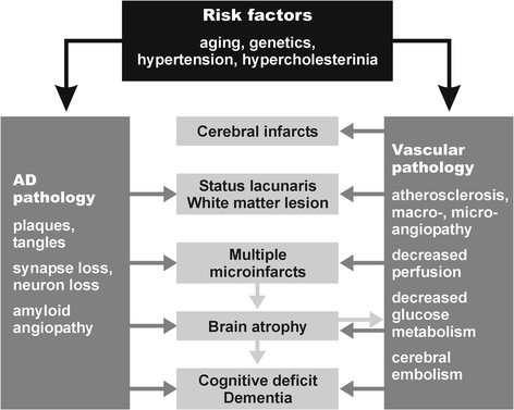
“Microvascular changes in the aged brain and in AD induce impairment of cerebral perfusion, in particular decrease of regional blood flow, reduction of glucose transport and utilization, loss of vascular innervation with special impact on the cholinergic and transmitter deficits in AD [171], impairment of neurovascular regulation, ultrastructural changes in capillaries and basement membranes due to”
§ The overlap between vascular disease and Alzheimer’s disease - lessons from pathology > Review > Pathogenic factors
What the sources say
- Not checked yetcites [171] Roman GC, Kalaria RN: Vascular determinants of cholinergic deficits in Alzheimer disease and vascular dementia. Neurobiol Aging. 2006, 27: 1769-1785. source
plaindirect- Asserted: passes — The authors put it forward as true.
- Checkable: passes — It can be checked against evidence.
- Load-bearing: passes — The paper’s conclusions depend on it.
- Based: passes — The paper gives a reason why it holds.
It may state two separate claims as one.
In the paper
“These factors induce brain atrophy with cognitive and memory impairment (Figure 1) [147], although the complex cascade of these and other noxious factors needs further elucidation.”
§ The overlap between vascular disease and Alzheimer’s disease - lessons from pathology > Review > Pathogenic factors
What the sources say
- Not checked yetcites [147] Jellinger KA, Attems J: Neuropathological evaluation of mixed dementia. J Neurol Sci. 2007, 257: 80-87. source
plaindirect- Asserted: passes — The authors put it forward as true.
- Checkable: passes — It can be checked against evidence.
- Load-bearing: passes — The paper’s conclusions depend on it.
- Based: passes — The paper gives a reason why it holds.
It may state two separate claims as one.
In the paper
“Both hypertension and CAA are associated with an increased prevalence of CVLs [157]”
§ The overlap between vascular disease and Alzheimer’s disease - lessons from pathology > Review > Pathogenic factors
What the sources say
- Not checked yetcites [157] Jellinger KA, Attems J: Prevalence and pathogenic role of cerebrovascular lesions in Alzheimer’s disease. J Neurol Sci. 2005, 229-230: 37-41. source
plaincorrelational- Asserted: passes — The authors put it forward as true.
- Checkable: passes — It can be checked against evidence.
- Load-bearing: unclear — It is unclear whether the paper’s conclusions depend on it.
- Based: passes — The paper gives a reason why it holds.
It may state two separate claims as one.
In the paper
“CVD has been suggested to be an important cause of cognitive impairment in the elderly, both by itself or as a catalyst for the conversion of low-grade AD to overt dementia [186].”
§ The overlap between vascular disease and Alzheimer’s disease - lessons from pathology > Conclusions
What the sources say
- Not checked yetcites [186] Kalaria RN, Kenny RA, Ballard CG, Perry R, Ince P, Polvikoski T: Towards defining the neuropathological substrates of vascular dementia. J Neurol Sci. 2004, 226: 75-80. source
hedgedconditional- Asserted: passes — The authors put it forward as true.
- Checkable: passes — It can be checked against evidence.
- Load-bearing: passes — The paper’s conclusions depend on it.
- Based: passes — The paper gives a reason why it holds.
It may state two separate claims as one.
The authors state this as an assumption.
WMLs contribute to cognitive decline.
“and, thus, are assumed to contribute to cognitive decline [184].”
Claims other papers make about this one
Each is checked against this paper’s own text: does it say what the citing paper says it does?
The cited source says what the claim attributes to it.
In the citing paper
“Studies have consistently found a positive epidemiological correlation between Alzheimer’s disease (AD) and cardiometabolic traits, yet the biological mechanisms behind this correlation is not well understood [1–4].”
From Multi-trait association studies discover pleiotropic loci between Alzheimer’s disease and cardiometabolic traits (2021) · cites this paper as [1]
The cited source says what the claim attributes to it.
In the citing paper
“Studies have consistently found a positive epidemiological correlation between Alzheimer’s disease (AD) and cardiometabolic traits, yet the biological mechanisms behind this correlation is not well understood [1–4].”
From Multi-trait association studies discover pleiotropic loci between Alzheimer’s disease and cardiometabolic traits (2021) · cites this paper as [1]
In the citing paper
“Moreover, the growing recognition of overlapping pathologies (e.g., Lewy body inclusions, TDP-43 pathology, vascular lesions, etc.) among dementia patients indicates a wide range of biological heterogeneity within AD that necessitates a broader approach to scientific investigations of its pathophysiology [37–39].”
From Systems-based proteomics to resolve the biology of Alzheimer’s disease beyond amyloid and tau (2021) · cites this paper as [38]
Self-check
Percentages, sums and reported differences re-computed from the paper’s own numbers.
Each contradiction between two passages listed here was confirmed by an independent check.
Rated minor: it would not touch a result — a value no result rests on, a p-value that stays on the same side of the significance level, or a conflict that is clerical, resolved elsewhere in the paper, or outside its own findings.
In the paper
“Multiple infarcts (61 = 32.5%)”
§ The overlap between vascular disease and Alzheimer’s disease - lessons from pathology > Review > Topographical distribution of cerebrovascular lesions
Reported 61 · recomputed 58
Rated minor: it would not touch a result — a value no result rests on, a p-value that stays on the same side of the significance level, or a conflict that is clerical, resolved elsewhere in the paper, or outside its own findings.
In the paper
§ The overlap between vascular disease and Alzheimer’s disease - lessons from pathology > Review > Topographical distribution of cerebrovascular lesions
“Table: Table 3 Common lesions in AD, VaD, MIX, and aged controls (from[130])”
§ The overlap between vascular disease and Alzheimer’s disease - lessons from pathology > Review > Topographical distribution of cerebrovascular lesions
“The type and average prevalence of CVLs in AD, VaD, mixed dementia, and aged controls is shown in Table 3[147].”
Rated minor: it would not touch a result — a value no result rests on, a p-value that stays on the same side of the significance level, or a conflict that is clerical, resolved elsewhere in the paper, or outside its own findings.
In the paper
§ The overlap between vascular disease and Alzheimer’s disease - lessons from pathology > Review > Cerebrovascular and Alzheimer’s disease pathology in demented and non-demented elderly
“compared to 66.8% in age-matched controls, all showing low Braak stages (P <0.01).”
§ The overlap between vascular disease and Alzheimer’s disease - lessons from pathology > Review > Cerebrovascular and Alzheimer’s disease pathology in demented and non-demented elderly
“Minor and moderate vascular pathology in AD were about twice as frequent as in controls (26.2% vs. 12.2% and 20.9% vs. 11.3%; P <0.01). On the other hand, severe vascular pathology did not significantly differ between both groups (10.2% vs. 12.2%).”
Citations
Cites
Wardlaw, J. M., Smith, E. E., Biessels, G. J., Cordonnier, C., Fazekas, F., Frayne, R., Lindley, R. I., O'Brien, J. T., Barkhof, F., Benavente, O. R., et al. (2013) · The Lancet Neurology · doi:10.1016/s1474-4422(13)70124-8 · Cited by 5,544 · cited 1×
“SVD is a frequent cause of white matter lesions (WMLs; leukoaraiosis) that are increasingly detected by neuroimaging [118-121].”
BackgroundGorelick, P. B., Scuteri, A., Black, S. E., DeCarli, C., Greenberg, S. M., Iadecola, C., Launer, L. J., Laurent, S., Lopez, O. L., Nyenhuis, D., et al. (2011) · Stroke · doi:10.1161/str.0b013e3182299496 · Cited by 3,811 · cited 3×
“Epidemiological studies have shown that AD and CVD share common risk factors such as hypertension during midlife, diabetes mellitus, smoking, apolipoprotein E (ApoE) ε4 isoforms, hypercholesterolemia, homocysteinemia, and, in particular, age [16,30-34].”
BackgroundPantoni, L. (2010) · The Lancet Neurology · doi:10.1016/s1474-4422(10)70104-6 · Cited by 3,405 · cited 1×
“SVD affects small arteries and arterioles and refers to pathological changes similar to atherosclerosis that are termed small vessel arteriosclerosis/atherosclerosis, lipoor fibrohyalinosis, or hypertensive arteriopathy [115].”
BackgroundNeurovascular pathways to neurodegeneration in Alzheimer’s disease and other disorders
Zlokovic, B. V. (2011) · Nature Reviews Neuroscience · doi:10.1038/nrn3114 · Cited by 2,969 · cited 1×
“CVD has been suggested to contribute to AD neuropathological changes including selective brain atrophy and accumulation of abnormal proteins such as Aβ [24,35,84,85].”
BackgroundThe projected effect of risk factor reduction on Alzheimer's disease prevalence
Barnes, D. E., & Yaffe, K. S. (2011) · The Lancet Neurology · doi:10.1016/s1474-4422(11)70072-2 · Cited by 2,846
Mixed brain pathologies account for most dementia cases in community-dwelling older persons
Schneider, J. A., Arvanitakis, Z., Bang, W., & Bennett, D. A. (2007) · Neurology · doi:10.1212/01.wnl.0000271090.28148.24 · Cited by 1,992 · cited 2×
“The burden of vascular and AD-type pathologies are leading and independent causes of dementia in the elderly [4,9-15], suggesting additive or synergistic effects of both types of lesions on cognitive impairment [2,3,5,9,16-29].”
BackgroundThe Pathobiology of Vascular Dementia
Iadecola, C. (2013) · Neuron · doi:10.1016/j.neuron.2013.10.008 · Cited by 1,857 · cited 1×
“The pathophysiology of VaD has been critically reviewed recently [48,144-146].”
BackgroundCerebral microbleeds: a guide to detection and interpretation
Greenberg, S. M., Vernooij, M. W., Cordonnier, C., Viswanathan, A., Al-Shahi Salman, R., Warach, S., Launer, L. J., Van Buchem, M. A., & Breteler, M. M. (2009) · The Lancet Neurology · doi:10.1016/s1474-4422(09)70013-4 · Cited by 1,798
Brain Infarction and the Clinical Expression of Alzheimer Disease<subtitle>The Nun Study</subtitle>
Snowdon, D. A. (1997) · JAMA: The Journal of the American Medical Association · doi:10.1001/jama.1997.03540340047031 · Cited by 1,590
Silent brain infarcts: a systematic review
Vermeer, S. E., Longstreth, W. T., & Koudstaal, P. J. (2007) · The Lancet Neurology · doi:10.1016/s1474-4422(07)70170-9 · Cited by 1,221 · cited 1×
““Silent” lacunar infarcts are frequently detected by MRI or CCT and are not accompanied by any overt clinical symptoms, but double the risk of subsequent stroke and dementia [133].”
Background
Research works printed but not linked: 2
- Crystal H, Dickson D: Cerebral infarcts in patients with autopsy proven Alzheimer’s disease. Neurobiol Aging. 2002, 23: 207.
- Jentoft M, Parisi J, Dickson D, Johnson K, Boeve B, Knopman D, Petersen R: Neuropathologic findings in 32 nondemented elderly subjects (abs.). J Neuropathol Exp Neurol. 2011, 70: 531.
Cited by
Sorted by citations
The Cellular Phase of Alzheimer’s Disease
De Strooper, B., & Karran, E. (2016) · Cell · doi:10.1016/j.cell.2015.12.056 · Cited by 1,990
Impact of multiple pathologies on the threshold for clinically overt dementia
Kapasi, A., DeCarli, C., & Schneider, J. A. (2017) · Acta Neuropathologica · doi:10.1007/s00401-017-1717-7 · Cited by 739
“Other common pathologies presented in persons with mixed pathologies are Lewy bodies [7, 131].”
BackgroundIadecola, C., Yaffe, K., Biller, J., Bratzke, L. C., Faraci, F. M., Gorelick, P. B., Gulati, M., Kamel, H., Knopman, D. S., Launer, L. J., et al. (2016) · Hypertension · doi:10.1161/hyp.0000000000000053 · Cited by 746
“Although dementia caused by AD and vascular dementia have traditionally been considered distinct nosological entities, increasing evidence indicates that these conditions often coexist.(8,9) Thus, the neuropathological hallmarks of AD, amyloid plaques and neurofibrillary tangles (NFTs), are frequently associated with…”
Blood-based biomarkers for Alzheimer disease: mapping the road to the clinic
Hampel, H., O’Bryant, S. E., Molinuevo, J. L., Zetterberg, H., Masters, C. L., Lista, S., Kiddle, S. J., Batrla, R., & Blennow, K. (2018) · Nature Reviews Neurology · doi:10.1038/s41582-018-0079-7 · Cited by 716
A Quarter Century of APOE and Alzheimer’s Disease: Progress to Date and the Path Forward
Belloy, M. E., Napolioni, V., & Greicius, M. D. (2019) · Neuron · doi:10.1016/j.neuron.2019.01.056 · Cited by 570
“To complicate matters further, cerebrovascular disease and AD often co-occur, particularly in older patients (Attems and Jellinger, 2014; Grinberg et al., 2010).”
High Cortisol and the Risk of Dementia and Alzheimer’s Disease: A Review of the Literature
Ouanes, S., & Popp, J. (2019) · Frontiers in Aging Neuroscience · doi:10.3389/fnagi.2019.00043 · Cited by 503
Dominguez, L. J., Veronese, N., Vernuccio, L., Catanese, G., Inzerillo, F., Salemi, G., & Barbagallo, M. (2021) · Nutrients · doi:10.3390/nu13114080 · Cited by 433
“Specifically, vascular disease risk factors, more so in midlife, are associated with brain accumulation of beta-amyloid [71,72].”
BackgroundAssociation of Intensive vs Standard Blood Pressure Control With Cerebral White Matter Lesions
Nasrallah, I. M., Pajewski, N. M., Auchus, A. P., Chelune, G. J., Cheung, A. K., Cleveland, M. L., Coker, L. H., Crowe, M. G., Cushman, W. C., Cutler, J. A., et al. (2019) · JAMA · doi:10.1001/jama.2019.10551 · Cited by 433
Global Prevalence of Young-Onset Dementia: A Systematic Review and Meta-analysis
Hendriks, S., Peetoom, K., Bakker, C., van der Flier, W. M., Papma, J. M., Koopmans, R., Verhey, F. R. J., de Vugt, M., Köhler, S., Young-Onset Dementia Epidemiology Study Group, et al. (2021) · JAMA Neurology · doi:10.1001/jamaneurol.2021.2161 · Cited by 409
A major role for cardiovascular burden in age-related cognitive decline
Qiu, C., & Fratiglioni, L. (2015) · Nature Reviews Cardiology · doi:10.1038/nrcardio.2014.223 · Cited by 422
Traffic-related air pollution and dementia incidence in northern Sweden: a longitudinal study
Oudin, A., Forsberg, B., Adolfsson, A. N., Lind, N., Modig, L., Nordin, M., Nordin, S., Adolfsson, R., & Nilsson, L.-G. (2016) · Environmental Health Perspectives · doi:10.1289/ehp.1408322 · Cited by 358
Bennett, R. E., Robbins, A. B., Hu, M., Cao, X., Betensky, R. A., Clark, T., Das, S., & Hyman, B. T. (2018) · Proceedings of the National Academy of Sciences · doi:10.1073/pnas.1710329115 · Cited by 299
“Mounting evidence indicates that, as assessed in life by MRI scans or at autopsy by neuropathological evaluation, “pure” AD is substantially less common than mixed dementias, in which tangles and plaques are accompanied by vascular changes (1, 2).”
BackgroundCerebrovascular disease in ageing and Alzheimer’s disease
Love, S., & Miners, J. S. (2016) · Acta Neuropathologica · doi:10.1007/s00401-015-1522-0 · Cited by 299
“ture was reviewed in detail by Attems and Jellinger [8].”
MethodZhong, B.-L., Chen, S.-L., Tu, X., & Conwell, Y. (2017) · The Journals of Gerontology Series B: Psychological Sciences and Social Sciences · doi:10.1093/geronb/gbw037 · Cited by 259
“…vessel disease, leading to increases in cerebral amyloid plaques, neurofibrillary tangles, and microinfarcts, the most common pathological correlates of late-life dementia (Attems & Jellinger, 2014; G. Cheng, Huang, Deng, & Wang, 2012; Faraco & Iadecola, 2013; Pregelj, 2008; Sharp et al., 2011).”
Rafnsson, S. B., Orrell, M., d’Orsi, E., Hogervorst, E., & Steptoe, A. (2020) · The Journals of Gerontology: Series B · doi:10.1093/geronb/gbx087 · Cited by 262
“Cardiovascular risk factors promote atherosclerotic small vessel disease and the neurofibrillary tangles characteristic of Alzheimer’s disease (Attems & Jellinger, 2014).”
BackgroundThe Vascular Impairment of Cognition Classification Consensus Study
Skrobot, O. A., O'Brien, J., Black, S., Chen, C., DeCarli, C., Erkinjuntti, T., Ford, G. A., Kalaria, R. N., Pantoni, L., Pasquier, F., et al. (2017) · Alzheimer's & Dementia · doi:10.1016/j.jalz.2016.10.007 · Cited by 242
“CVD commonly contributes to many forms of dementia, including AD[26-28], and may be targeted with some success [29], although further research into possible associations and causal relationships is needed.”
Self-citationA natural experiment on the effect of herpes zoster vaccination on dementia
Eyting, M., Xie, M., Michalik, F., Heß, S., Chung, S., & Geldsetzer, P. (2025) · Nature · doi:10.1038/s41586-025-08800-x · Cited by 273
Corriveau, R. A., Bosetti, F., Emr, M., Gladman, J. T., Koenig, J. I., Moy, C. S., Pahigiannis, K., Waddy, S. P., & Koroshetz, W. (2016) · Cellular and Molecular Neurobiology · doi:10.1007/s10571-016-0334-7 · Cited by 240
“1) (Breteler 2000a, b; Gorelick et al. 2011; Iadecola 2013; Attems and Jellinger 2014).”
Peters, R., Ee, N., Peters, J., Beckett, N., Booth, A., Rockwood, K., & Anstey, K. J. (2019) · Therapeutic Advances in Chronic Disease · doi:10.1177/2040622319880392 · Cited by 213
Cognitive impact of COVID-19: looking beyond the short term
Miners, J. S., Kehoe, P. G., & Love, S. (2020) · Alzheimer s Research & Therapy · doi:10.1186/s13195-020-00744-w · Cited by 261
The Neuroprotective Effects of Exercise: Maintaining a Healthy Brain Throughout Aging
Vecchio, L. M., Meng, Y., Xhima, K., Lipsman, N., Hamani, C., & Aubert, I. (2018) · Brain Plasticity · doi:10.3233/bpl-180069 · Cited by 244
Emerging Concepts in Vascular Dementia: A Review
Bir, S. C., Khan, M. W., Javalkar, V., Toledo, E. G., & Kelley, R. E. (2021) · Journal of Stroke and Cerebrovascular Diseases · doi:10.1016/j.jstrokecerebrovasdis.2021.105864 · Cited by 247
Insulin Resistance as a Link between Amyloid-Beta and Tau Pathologies in Alzheimer’s Disease
Mullins, R. J., Diehl, T. C., Chia, C. W., & Kapogiannis, D. (2017) · Frontiers in Aging Neuroscience · doi:10.3389/fnagi.2017.00118 · Cited by 227
“…infarcts, leukoaraiosis (seen on magnetic resonance imaging (MRI) as white matter hyperintensities), cerebral amyloid angiopathy (CAA), microaneurysms, and small and large vessel ischemic/hemorrhagic stroke (Breteler, 2000; Gorelick et al., 2011; Attems and Jellinger, 2014; Corriveau et al., 2016).”
BackgroundKapasi, A., & Schneider, J. A. (2016) · Biochimica et Biophysica Acta (BBA) - Molecular Basis of Disease · doi:10.1016/j.bbadis.2015.12.023 · Cited by 221
Matěj, R., Tesař, A., & Rusina, R. (2019) · Clinical Biochemistry · doi:10.1016/j.clinbiochem.2019.08.005 · Cited by 164
Multimorbidity burden and dementia risk in older adults: The role of inflammation and genetics
Grande, G., Marengoni, A., Vetrano, D. L., Roso‐Llorach, A., Rizzuto, D., Zucchelli, A., Qiu, C., Fratiglioni, L., & Calderón‐Larrañaga, A. (2021) · Alzheimer s & Dementia · doi:10.1002/alz.12237 · Cited by 164
Challenges of multimorbidity of the aging brain: a critical update
Jellinger, K. A., & Attems, J. (2015) · Journal of Neural Transmission · doi:10.1007/s00702-014-1288-x · Cited by 154
Self-citationA global view of the genetic basis of Alzheimer disease
Reitz, C., Pericak‐Vance, M. A., Foroud, T., & Mayeux, R. P. (2023) · Nature Reviews Neurology · doi:10.1038/s41582-023-00789-z · Cited by 181
Millan, M. J. (2017) · Progress in Neurobiology · doi:10.1016/j.pneurobio.2017.03.004 · Cited by 151
Blood pressure lowering and prevention of dementia: an individual patient data meta-analysis
Peters, R., Xu, Y., Fitzgerald, O., Aung, H. L., Beckett, N., Bulpitt, C., Chalmers, J., Forette, F., Gong, J., Harris, K., et al. (2022) · European Heart Journal · doi:10.1093/eurheartj/ehac584 · Cited by 142
de la Torre, J. (2018) · Journal of Alzheimer s Disease · doi:10.3233/jad-180004 · Cited by 161
“Not only do both dementias share nearly identical symptoms and signs, but both show comparable microstructural changes, negative impact on cognitive function and a link to more than two dozen vascular risk factors [60, 61].”
Alzheimer's Disease–Related Dementias Summit 2016: National research priorities
Corriveau, R. A., Koroshetz, W. J., Gladman, J. T., Jeon, S., Babcock, D., Bennett, D. A., Carmichael, S. T., Dickinson, S. L.-J., Dickson, D. W., Emr, M., et al. (2017) · Neurology · doi:10.1212/wnl.0000000000004717 · Cited by 143
Post-mortem assessment in vascular dementia: advances and aspirations
McAleese, K. E., Alafuzoff, I. I., Charidimou, A., Reuck, J. D., Grinberg, L. T., Hainsworth, A. H., Hortobágyi, T., Ince, P. G., Jellinger, K. A., Gao, J., et al. (2016) · BMC Medicine · doi:10.1186/s12916-016-0676-5 · Cited by 144
“interact with pathophysiological processes in AD either independently of each other or through additive or synergistic effects on cognitive decline [39, 40].”
BackgroundSelf-citationDissecting the genetic relationship between cardiovascular risk factors and Alzheimer’s disease
Broce, I. J., Tan, C. H., Fan, C. C., Jansen, I., Savage, J. E., Witoelar, A., Wen, N., Hess, C. P., Dillon, W. P., Glastonbury, C. M., et al. (2019) · Acta Neuropathologica · doi:10.1007/s00401-018-1928-6 · Cited by 140
Dementia risk and prevention by targeting modifiable vascular risk factors
Tariq, S., & Barber, P. A. (2017) · Journal of Neurochemistry · doi:10.1111/jnc.14132 · Cited by 140
Whole-Brain Vasculature Reconstruction at the Single Capillary Level
Giovanna, A. P. D., Tibo, A., Silvestri, L., Müllenbroich, M. C., Costantini, I., Mascaro, A. L. A., Sacconi, L., Frasconi, P., & Pavone, F. S. (2018) · Scientific Reports · doi:10.1038/s41598-018-30533-3 · Cited by 135
Villabona-Rueda, A. F., Erice, C., Pardo, C. A., & Stins, M. F. (2019) · Frontiers in Cellular Neuroscience · doi:10.3389/fncel.2019.00405 · Cited by 130
“In addition, differences in vascular pathologies between different lobes and WM versus GM are reported, with the occipital lobe more severely affected by cerebral amyloid angiopathies, followed by frontal-, temporal-, and parietal lobes (Vinters et al., 1996; Attems and Jellinger, 2014).”
BackgroundGannon, O. J., Robison, L. S., Salinero, A. E., Abi-Ghanem, C., Mansour, F. M., Kelly, R. D., Tyagi, A., Brawley, R. R., Ogg, J. D., & Zuloaga, K. L. (2022) · Journal of Neuroinflammation · doi:10.1186/s12974-022-02466-2 · Cited by 127
Update on Vascular Cognitive Impairment Associated with Subcortical Small-Vessel Disease
Wallin, A., Román, G. C., Esiri, M. M., Kettunen, P., Svensson, J., Paraskevas, G. P., & Kapaki, E. N. (2018) · Journal of Alzheimer s Disease · doi:10.3233/jad-170803 · Cited by 133
“may be present in 40–80% of VCI patients [11, 12].”
BackgroundCzakó, C. N., Kovács, T., Ungvari, Z. I., Csiszár, A., Yabluchanskiy, A., Conley, S. M., Csípő, T., Lipécz, Á., Horváth, H., Sándor, G. L., et al. (2020) · GeroScience · doi:10.1007/s11357-020-00252-7 · Cited by 130
CSF/serum albumin ratio in dementias: a cross-sectional study on 1861 patients
Skillbäck, T. B., Delsing, L., Synnergren, J., Mattsson, N., Janelidze, S., Nägga, K., Kilander, L., Hicks, R., Wimo, A. B., Winblad, B. G., et al. (2017) · Neurobiology of Aging · doi:10.1016/j.neurobiolaging.2017.06.028 · Cited by 127
“Taken together, these results might stem from contaminant cerebral vascular pathology and a relative rarity of biochemically pure AD, but could also indicate abundance of WM lesions (Attems and Jellinger, 2014, McAleese et al., 2016).”
Modular machine learning for Alzheimer's disease classification from retinal vasculature
Tian, J., Smith, G. E., Guo, H. W., Liu, B., Pan, Z., Wang, Z., Xiong, S., & Fang, R. (2021) · Scientific Reports · doi:10.1038/s41598-020-80312-2 · Cited by 131
Systems-based proteomics to resolve the biology of Alzheimer’s disease beyond amyloid and tau
Rayaprolu, S., Higginbotham, L., Bagchi, P., Watson, C. M., Zhang, T., Levey, A. I., Rangaraju, S., & Seyfried, N. T. (2021) · Neuropsychopharmacology · doi:10.1038/s41386-020-00840-3 · Cited by 118
Peri-Infarct Upregulation of the Oxytocin Receptor in Vascular Dementia
McKay, E. C., Beck, J. S., Khoo, S. K., Dykema, K. J., Cottingham, S. L., Winn, M. E., Paulson, H. L., Lieberman, A. P., & Counts, S. (2019) · Journal of Neuropathology & Experimental Neurology · doi:10.1093/jnen/nlz023 · Cited by 121
“Fisher concluded that stroke alone could lead to dementia (21), the high rate of mixed pathology dementias from both AD and vascular damage has confounded efforts to combat VaD resulting from stroke or cerebrovascular disease alone (6, 22).”
BackgroundLi, Y., Choi, W. J., Wei, W., Song, S., Zhang, Q., Liu, J., & Wang, R. K. (2018) · Neurobiology of Aging · doi:10.1016/j.neurobiolaging.2018.06.017 · Cited by 119
“Although there is no consensus reached regarding the causal relationship between a decreased cerebral blood flow (CBF) and decline of neuronal function, there is an increasing prevalence of coincident cerebrovascular deficiency and cognitive dysfunction with aging that is also well recognized in Alzheimer’s disease (A…”
BackgroundThe blood brain barrier in Alzheimer’s disease
Chakraborty, A., de Wit, N. M., van der Flier, W. M., & de Vries, H. E. (2017) · Vascular Pharmacology · doi:10.1016/j.vph.2016.11.008 · Cited by 118
Amyloid-beta and tau protein beyond Alzheimer’s disease
Abyadeh, M., Gupta, V., Paulo, J. A., Mahmoudabad, A. G., Shadfar, S., Mirshahvaladi, S., Gupta, V. B., Nguyen, C. T. O., Finkelstein, D. I., You, Y., et al. (2023) · Neural Regeneration Research · doi:10.4103/1673-5374.386406 · Cited by 126
“There is strong evidence indicating a causal association between CVDs and dementias; and individuals with CVDs are at higher risk of developing AD (Attems and Jellinger, 2014; Santos et al., 2017; Tini et al., 2020).”
BackgroundHDL from an Alzheimer's disease perspective
Button, E. B., Robert, J., Caffrey, T. M., Fan, J., Zhao, W., & Wellington, C. L. (2019) · Current Opinion in Lipidology · doi:10.1097/mol.0000000000000604 · Cited by 114
Pharmacological interventions for apathy in Alzheimer's disease
Ruthirakuhan, M. T., Herrmann, N., Abraham, E. H., Chan, S., & Lanctôt, K. L. (2018) · Cochrane Database of Systematic Reviews · doi:10.1002/14651858.cd012197.pub2 · Cited by 113
Robert, J., Button, E. B., Yuen, B., Gilmour, M., Kang, K., Bahrabadi, A., Stukas, S. K., Zhao, W., Kulić, I., & Wellington, C. L. (2017) · eLife · doi:10.7554/elife.29595 · Cited by 118
“…following equation: Papp=(dQ/dt)*(1/A*C0*60) where dQ/dt is the amount of FITC-dextran transported per minute (ng/min), A is the surface area of the tissue (cm [Attems and Jellinger, 2014]), C0 is the initial concentration of FITC-Dextran (ng/ml) and 60 is the conversion from minutes to seconds.”
Zamolodchikov, D., Renné, T., & Strickland, S. (2015) · Journal of Thrombosis and Haemostasis · doi:10.1111/jth.13209 · Cited by 113
Ji, F., Pasternak, O., Liu, S., Loke, Y. M., Choo, B. L., Hilal, S., Xu, X., Ikram, M. K., Venketasubramanian, N., Chen, C., et al. (2017) · Alzheimer s Research & Therapy · doi:10.1186/s13195-017-0292-4 · Cited by 109
“White matter hyperintensity (WMH), which is associated with increased water content and vascular changes [3], serves as an important clinical diagnostic criterion by which to identify CeVD status [4].”
BackgroundMcAleese, K. E., Colloby, S. J., Thomas, A. J., Al‐Sarraj, S., Ansorge, O., Neal, J., Roncaroli, F., Love, S., Francis, P. T., & Attems, J. (2021) · Alzheimer s & Dementia · doi:10.1002/alz.12291 · Cited by 110
Self-citationEndothelial Mitochondrial Dysfunction in Cerebral Amyloid Angiopathy and Alzheimer’s Disease
Parodi‐Rullán, R. M., Sone, J. Y., & Fossati, S. (2019) · Journal of Alzheimer s Disease · doi:10.3233/jad-190357 · Cited by 110
Chong, J. R., Ashton, N. J., Karikari, T. K., Tanaka, T., Schöll, M., Zetterberg, H., Blennow, K., Chen, C., & Lai, M. K. P. (2021) · Journal of Neurology Neurosurgery & Psychiatry · doi:10.1136/jnnp-2021-327370 · Cited by 111
Mullane, K., & Williams, M. J. (2020) · Biochemical Pharmacology · doi:10.1016/j.bcp.2020.113945 · Cited by 109
Plasma hydrogen sulfide: A biomarker of Alzheimer's disease and related dementias
Disbrow, E. A., Stokes, K. Y., Ledbetter, C. R., Patterson, J. C., Kelley, R. E., Pardue, S., Reekes, T. H., Larmeu, L., Batra, V., Yuan, S. S., et al. (2021) · Alzheimer s & Dementia · doi:10.1002/alz.12305 · Cited by 100
Further understanding the connection between Alzheimer's disease and Down syndrome
Snyder, H. M., Bain, L. J., Brickman, A. M., Carrillo, M. C., Esbensen, A. J., Espinosa, J. M., Fernandez, F., Fortea, J., Hartley, S. L., Head, E., et al. (2020) · Alzheimer's & Dementia · doi:10.1002/alz.12112 · Cited by 105
Toward precision medicine in Alzheimer’s disease
Reitz, C. (2016) · Annals of Translational Medicine · doi:10.21037/atm.2016.03.05 · Cited by 100
“Adding to this complexity is frequent clinical and pathological overlap with other pathologies in particular Lewy Body disease (LBD) and cerebrovascular disease (4-6).”
BackgroundCerebral small vessel disease and Alzheimer’s disease
Cai, Z., Wang, C., He, W., Tu, H., Tang, Z., Xiao, M., & Yan, L. (2015) · Clinical Interventions in Aging · doi:10.2147/cia.s90871 · Cited by 102
“relationship between AD and cognitive impairment and VaD has been reported while CVD and stroke are related to high risk of both cognitive impairment and AD.(139,140) The manifestation of CVD in imaging is extremely similar to AD in the elderly, whereas imaging markers of subcortical vascular diseases (leukoaraiosis,…”
Tsartsalis, S., Sleven, H., Fancy, N., Wessely, F., Smith, A. M., Willumsen, N., Cheung, T. K. D., Rokicki, M. J., Chau, V., Ifie, E., et al. (2024) · Nature Communications · doi:10.1038/s41467-024-46630-z · Cited by 95
“In the 3 rd paragraph of the “Quality control, dataset integration, dimension reduction and clustering” section, we briefly describe how cell type identification was performed based on the full of co-expressed genes that distinguish each cluster.”
mTOR signaling and Alzheimer's disease: What we know and where we are?
Davoody, S., Asgari Taei, A., Khodabakhsh, P., & Dargahi, L. (2024) · CNS Neuroscience & Therapeutics · doi:10.1111/cns.14463 · Cited by 96
“128 mTOR signaling through specific mechanisms active in endothelial and smooth muscle cells of vasculature regulates vascular function acutely and chronically.”
BackgroundVascular cognitive impairment and dementia: Mechanisms, treatment, and future directions
Mok, V., Cai, Y., & Markus, H. S. (2024) · International Journal of Stroke · doi:10.1177/17474930241279888 · Cited by 120
“53,54 In addition, preclinical and clinical evidence showed that SVD is an early initiating event in the development of A β .”
BackgroundA Life-Long Approach to Physical Activity for Brain Health
Macpherson, H., Teo, W.-P., Schneider, L. A., & Smith, A. E. (2017) · Frontiers in Aging Neuroscience · doi:10.3389/fnagi.2017.00147 · Cited by 104
“…restricts blood flow, disrupts the integrity of the cerebral vessels and likely has further cascading effects on brain perfusion, resulting in disruption to homeostasis, increasing inflammation, neuronal dysfunction and cell death as well as inhibiting neuroplasticity (Attems and Jellinger, 2014).”
BackgroundInfluentialVascular risk factors and Alzheimer’s disease
O'Brien, J. T., & Markus, H. S. (2014) · BMC Medicine · doi:10.1186/s12916-014-0218-y · Cited by 99
“Jellinger and Attems [9] provide a detailed account of”
BackgroundThree-dimensional mapping of neurofibrillary tangle burden in the human medial temporal lobe
Yushkevich, P. A., Muñoz, M., Martin, M. M. I. D. O., Ittyerah, R., Lim, S., Ravikumar, S., Bedard, M. L., Pickup, S., Liu, W., Wang, J., et al. (2021) · Brain · doi:10.1093/brain/awab262 · Cited by 96
Blood Pressure Variability and Progression of Clinical Alzheimer Disease
de Heus, R. A. A., Olde Rikkert, M. G. M., Tully, P. J., Lawlor, B. A., Claassen, J. A. H. R., & for the NILVAD Study Group (2019) · Hypertension · doi:10.1161/hypertensionaha.119.13664 · Cited by 88
Review: Vascular dementia: clinicopathologic and genetic considerations
Vinters, H. V., Zarow, C., Borys, E., Whitman, J. D., Tung, S., Ellis, W. G., Zheng, L., & Chui, H. C. (2018) · Neuropathology and Applied Neurobiology · doi:10.1111/nan.12472 · Cited by 90
Motoric cognitive risk syndrome: Integration of two early harbingers of dementia in older adults
Semba, R. D., Tian, Q., Carlson, M. C., Xue, Q., & Ferrucci, L. (2020) · Ageing Research Reviews · doi:10.1016/j.arr.2020.101022 · Cited by 88
“Recent studies show AD is a multifactorial and heterogeneous disease with multiple contributors, including cerebrovascular disease (Schneider et al., 2007; Toledo et al., 2013; Attems and Jellinger, 2014; Sweeney et al., 2019; De Rueck et al., 2018).”
BackgroundAPP as a Protective Factor in Acute Neuronal Insults
Hefter, D., & Draguhn, A. (2017) · Frontiers in Molecular Neuroscience · doi:10.3389/fnmol.2017.00022 · Cited by 97
“These common pathways may then result in overlapping histopathological findings (de la Torre, 2002; Pluta et al., 2009, 2012; Attems and Jellinger, 2014).”
BackgroundWhite matter hyperintensities and CSF Alzheimer disease biomarkers in preclinical Alzheimer disease
Soldan, A., Pettigrew, C. A., Zhu, Y., Wang, M., Moghekar, A., Gottesman, R. F., Singh, B., Martinez, O., Fletcher, E. M., DeCarli, C. S., et al. (2019) · Neurology · doi:10.1212/wnl.0000000000008864 · Cited by 82
“Alzheimer disease (AD) and cerebrovascular disease frequently co-occur among older adults.(1) It remains unclear, however, whether these 2 pathologies independently affect the risk of cognitive impairment, or whether they have synergistic effects on cognitive decline.”
BackgroundAndrogens’ effects on cerebrovascular function in health and disease
Abi‐Ghanem, C., Robison, L. S., & Zuloaga, K. L. (2020) · Biology of Sex Differences · doi:10.1186/s13293-020-00309-4 · Cited by 89
“Moreover, VCID is comorbid in up to 80% of AD patients, resulting in multi-etiology dementia [109, 110].”
BackgroundCognitive disorder and dementia in type 2 diabetes mellitus
Ortíz, G. G., Huerta, M., González-Usigli, H., Torres-Sánchez, E. D., Delgado-Lara, D. L. C., Pacheco-Moisés, F. P., Mireles-Ramírez, M. A., Torres-Mendoza, B. M., Moreno-Cih, R. I., & Velázquez-Brizuela, I. E. (2022) · World Journal of Diabetes · doi:10.4239/wjd.v13.i4.319 · Cited by 93
Mehta, R. I., & Schneider, J. A. (2021) · Current Opinion in Neurology · doi:10.1097/wco.0000000000000912 · Cited by 87
“Moreover, novel insights into resilience factors [48 & ,49 && ,50 && ] and brain–heart [23], brain–gut [51 && ,52 && ], brain– kidney [53 && ], and brain–lung connections [54 && ] also suggest the existence of broader neuroimmune and brain–body connections in manifestations of disease.”
BackgroundResultInfluentialWalker, L. C. (2020) · Free Neuropathology · doi:10.17879/freeneuropathology-2020-3025 · Cited by 113
Bertram, S., Brixius, K., & Brinkmann, C. K. (2016) · Endocrine · doi:10.1007/s12020-016-0976-8 · Cited by 89
Microvascular Dysfunction Is Associated With Worse Cognitive Performance
Rensma, S. P., van Sloten, T. T., Houben, A. J. H. M., Köhler, S., van Boxtel, M. P. J., Berendschot, T. T. J. M., Jansen, J. F. A., Verhey, F. R. J., Kroon, A. A., Koster, A., et al. (2019) · Hypertension · doi:10.1161/hypertensionaha.119.13023 · Cited by 85
Slot, R. E. R., van Harten, A. C., Kester, M. I., Jongbloed, W., Bouwman, F. H., Teunissen, C. E., Scheltens, P., Veerhuis, R., & van der Flier, W. M. (2016) · Journal of Alzheimer s Disease · doi:10.3233/jad-151068 · Cited by 82
Potential health benefits of (poly)phenols derived from fruit and 100% fruit juice
Ho, K. K. H. Y., Ferruzzi, M. G., & Wightman, J. D. (2019) · Nutrition Reviews · doi:10.1093/nutrit/nuz041 · Cited by 81
Brain Aging Among Racially and Ethnically Diverse Middle-Aged and Older Adults
Turney, I. C., Lao, P. J., Rentería, M. A., Igwe, K. C., Berroa, J., Rivera, A. M., Benavides, A., Morales, C. D., Rizvi, B. M., Schupf, N., et al. (2022) · JAMA Neurology · doi:10.1001/jamaneurol.2022.3919 · Cited by 87
Associations of the metabolic syndrome and its components with cognitive impairment in older adults
Feinkohl, I., Janke, J., Hadzidiakos, D. A., Slooter, A. J. C., Winterer, G., Spies, C., & Pischon, T. (2019) · BMC Geriatrics · doi:10.1186/s12877-019-1073-7 · Cited by 82
Semba, R. D. (2020) · Advances in Nutrition · doi:10.1093/advances/nmaa024 · Cited by 78
Brain amyloid and vascular risk are related to distinct white matter hyperintensity patterns
Pålhaugen, L., Sudre, C. H., Tecelao, S., Nakling, A., Almdahl, I. S., Kalheim, L. F., Cardoso, M. J., Johnsen, S. H., Rongve, A., Aarsland, D., et al. (2021) · Journal of Cerebral Blood Flow & Metabolism · doi:10.1177/0271678x20957604 · Cited by 79
“None of the guidelines for preclinical or predementia AD include WMHs in their biomarker models,24,32 and extensive WMH burden is an exclusion criterion in several recent clinical trials in predementia AD.”
BackgroundInfluentialMacklin, L. N., Griffith, C. M., Cai, Y., Rose, G. M., Yan, X., & Patrylo, P. R. (2016) · Experimental Gerontology · doi:10.1016/j.exger.2016.12.019 · Cited by 86
“…altered metabolic capacity (Mosconi et al., 2008; Struble et al., 2010), 3) changes in neurovascularization (Scheibel et al., 1989; Cai et al., 2010; Attems and Jellinger, 2014; Li et al., 2015; Snyder et al., 2015; Xue et al., 2015), 4) changes in cholinergic innervation (Coyle et al., 1983;…”
Systemic infection exacerbates cerebrovascular dysfunction in Alzheimer’s disease
Asby, D. J., Boche, D., Allan, S. M., Love, S., & Miners, J. S. (2021) · Brain · doi:10.1093/brain/awab094 · Cited by 83
Wu, A., Sharrett, A. R., Gottesman, R. F., Power, M. C., Mosley, T. H., Jack, C. R., Knopman, D. S., Windham, B. G., Gross, A. L., & Coresh, J. (2019) · JAMA Network Open · doi:10.1001/jamanetworkopen.2019.3359 · Cited by 71
Cerebrovascular plasticity: Processes that lead to changes in the architecture of brain microvessels
Bogorad, M. I., DeStefano, J. G., Linville, R. M., Wong, A. D., & Searson, P. C. (2019) · Journal of Cerebral Blood Flow & Metabolism · doi:10.1177/0271678x19855875 · Cited by 79
Siedliński, M., Carnevale, L., Xu, X., Carnevale, D., Εvangelou, Ε., Caulfield, M. J., Maffia, P., Wardlaw, J. M., Samani, N. J., Tomaszewski, M., et al. (2023) · European Heart Journal · doi:10.1093/eurheartj/ehad101 · Cited by 75
Neuroinflammatory Signaling in the Pathogenesis of Alzheimer’s Disease
Uddin, M. S., Kabir, M. T., Jalouli, M., Rahman, M. A., Jeandet, P., Behl, T., Alexiou, A. T., Albadrani, G. M., Abdel‐Daim, M. M., Perveen, A., et al. (2021) · Current Neuropharmacology · doi:10.2174/1570159x19666210826130210 · Cited by 76
“Numerous neuropathological, clinical, and epidemiological studies have revealed that vascular pathology is a crucial risk factor for AD development [187, 188].”
BackgroundAn MRI measure of degenerative and cerebrovascular pathology in Alzheimer disease
Brickman, A. M., Tosto, G., Gutierrez, J., Andrews, H., Gu, Y., Narkhede, A., Rizvi, B. M., Guzmán, V., Manly, J. J., Vonsattel, J. P., et al. (2018) · Neurology · doi:10.1212/wnl.0000000000006310 · Cited by 73
“On MRI, LOAD is manifest by medial temporal lobe and cortical atrophy, but white matter hyperintensities (WMHs) and brain infarcts are also frequent.(4,6) Structural MRI biomarker studies of LOAD often treat cerebrovascular disease and atrophy as distinct entities, but they coexist more frequently than not and the ons…”
BackgroundDisentangling the effects of Alzheimer’s and small vessel disease on white matter fibre tracts
Dewenter, A., Jacob, M. A., Cai, M., Gesierich, B. G., Hager, P., Kopczak, A., Biel, D., Ewers, M., Tuladhar, A. M., de Leeuw, F., et al. (2022) · Brain · doi:10.1093/brain/awac265 · Cited by 72
“As expected from epidemiological and histopathology studies, 6,7 concomitant SVD was found in the Alzheimer’s disease sample, with the largest difference in WMH burden between the A+T– group and matched A–”
Cerebral white matter lesions – associations with Aβ isoforms and amyloid PET
van Westen, D., Lindqvist, D., Blennow, K., Minthon, L., Nägga, K., Stomrud, E., Zetterberg, H., & Hansson, O. (2016) · Scientific Reports · doi:10.1038/srep20709 · Cited by 66
Beishon, L., Batterham, A. P., Quinn, T. J., Nelson, C. P., Panerai, R. B., Robinson, T. G., & Haunton, V. J. (2019) · Cochrane Database of Systematic Reviews · doi:10.1002/14651858.cd013282.pub2 · Cited by 77
“As knowledge and understanding has evolved, it has become increasingly diOicult to distinguish between these dementia subtypes, as there is considerable clinical and pathological overlap between them (Attems 2014; Mandal 2006).”
BackgroundInfluentialYu, W., Li, Y., Hu, J. P., Wu, J., & Huang, Y. (2022) · Journal of Clinical Medicine · doi:10.3390/jcm11164742 · Cited by 75
“Moreover, the pathological evidence of cerebral hypoperfusion was observed in both VCID and AD, mainly inflammation, trophic uncoupling, BBB disruption and WMLs, but with distinctions in the distribution and associated pathologies [7].”
BackgroundGallart‐Palau, X., Lee, B. S. T., Adav, S. S., Qian, J., Serra, A., Park, J. E., Lai, M. K. P., Chen, C., Kalaria, R. N., & Sze, S. K. (2016) · Molecular Brain · doi:10.1186/s13041-016-0205-7 · Cited by 72
“Affected individuals display a complex brain pathology characterized by senile plaques with microinfarcts, as well as a poorly understood degeneration of the small vessels that supply blood to the brain [4–7].”
BackgroundInteraction between cerebrovascular disease and Alzheimer pathology
Saito, S., & Ihara, M. (2016) · Current Opinion in Psychiatry · doi:10.1097/yco.0000000000000239 · Cited by 69
“© 2016 Wolters Kluwer cerebrovascular disease (CVD) has long been noted [4] and mixed Alzheimer’s disease/CVD pathology is commonly seen in patients with clinically diagnosed Alzheimer’s disease [5 && ].”
BackgroundSpecific Patterns of White Matter Alterations Help Distinguishing Alzheimer's and Vascular Dementia
Palesi, F., Rinaldis, A. D., Vitali, P., Castellazzi, G., Casiraghi, L., Germani, G., Bernini, S., Anzalone, N., Ramusino, M. C., Denaro, F. M., et al. (2018) · Frontiers in Neuroscience · doi:10.3389/fnins.2018.00274 · Cited by 71
“In older subjects and in the presence of cerebral amyloid angiopathy, AD patients can also present a certain degree of cerebrovascular lesions contributing to a mixed type of dementia (Attems and Jellinger, 2014).”
BackgroundZeng, F., Liu, Y., Huang, W., Qing, H., Kadowaki, T., Kashiwazaki, H., Ni, J., & Wu, Z. (2020) · Journal of Neurochemistry · doi:10.1111/jnc.15096 · Cited by 72
“Scale bars in b: 50 μm; c: 10 μm as cerebrovascular-related amyloidogenesis has been found in over 80% of AD patients (Attems & Jellinger, 2014; Jellinger & Attems, 2005) and this condition positively correlates with a cognitive decline (Thal, Griffin, de Vos, & Ghebremedhin, 2008; Weller, Boche, &…”
BackgroundHerpes Zoster Vaccination and Dementia Occurrence
Pomirchy, M., Bommer, C., Pradella, F., Michalik, F., Peters, R. K., & Geldsetzer, P. (2025) · JAMA · doi:10.1001/jama.2025.5013 · Cited by 74
McCaddon, A., & Miller, J. W. (2015) · Nutrition Reviews · doi:10.1093/nutrit/nuv022 · Cited by 69
Metadata
- Authors
- Attems, J., & Jellinger, K. A.
- Affiliations
- Newcastle University (GB)
- Medical University of Vienna (AT)
- Corresponding author
- Attems, Johannes
- Venue
- BMC Medicine
- Published
- 2014-11-11
- Volume
- 12
- Issue
- 1
- Pages
- 206-206
- ISSN
- 1741-7015 (ISSN-L)
- PMC
- PMC4226890
- OpenAlex
- W2043522411
- Cited by
- 739
- References
- 196
- Citations by year
- 2026: 55 · 2025: 79 · 2024: 73 · 2023: 66 · 2022: 61 · 2021: 71 · 2020: 91 · 2019: 70 · 2018: 53 · 2017: 44 · 2016: 53 · 2015: 19 · 2014: 3
- FWCI
- 5.49
- Type
- journal article
- Language
- en
- Open access
- gold
- doi.org
- Locations
- BMC Medicine (publishedVersion, cc-by)
- Europe PMC (PubMed Central) (publishedVersion, cc-by)
- PubMed Central (publishedVersion, cc-by)
- PubMed (publishedVersion)
- Newcastle University ePrints (Newcastle Univesity) (cc-by)
- Funders
- Alzheimer's Society
- National Institute for Health and Care Research
- Dunhill Medical Trust (R173/1110)
- Newcastle University (G0400074)
- Newcastle upon Tyne Hospitals NHS Foundation Trust
- Medical Research Council (G0400074)
- Topics
- Health Sciences › Medicine › Psychiatry and Mental health › Dementia and Cognitive Impairment Research
- Life Sciences › Neuroscience › Neurology › Neurological Disease Mechanisms and Treatments
- Life Sciences › Neuroscience › Cellular and Molecular Neuroscience › Alzheimer's disease research and treatments
- Keywords
- Alzheimer's disease, cerebrovascular disease, cerebral amyloid angiopathy, cerebral small vessel disease, vascular dementia, mixed dementia, cognitive decline, dementia progression, subcortical infarcts
- MeSH terms
- Aging, Alzheimer Disease, Cerebrovascular Disorders, Humans
About these checks
Registry, retraction, Cites and statistics results are computed, not judged, with one exception: a printed reference that no index lists is read once by a model before it counts as a citation error. Claims are extracted by a model and kept only when their quote appears verbatim in the text; each cited source is then checked, with a model’s help, for whether it says what the claim relies on. Those verdicts can be wrong — read the claim’s quote and the cited source before relying on one.
A language model drew the claims from the paper on 2026-10-10, under version 3 of the claim definition (model deepseek-v4.1-flash, prompt extract-v3).
A second model labelled each claim on 2026-10-10 (model jev-1.13-20260917, definition revision gold-guideline-v0.2).
Statistics and arithmetic are recomputed, not judged.
Confidence bands are shown only for fields calibrated on our annotated test set. None is calibrated yet, so no band is shown.
What counts as a claim ›How we measure confidence ›How we check this ›
Are you an author, or do you see a problem with this page? Request a review
