

The DOI matches its Crossref record; 195 of the 196 references it cites were found in a registry or an index; none of the 57 sources its claims cite is checked for support yet.
The overlap between vascular disease and Alzheimer’s disease - lessons from pathology
Open accessAbstract
Recent epidemiological and clinico-pathological data indicate considerable overlap between cerebrovascular disease (CVD) and Alzheimer's disease (AD) and suggest additive or synergistic effects of both pathologies on cognitive decline. The most frequent vascular pathologies in the aging brain and in AD are cerebral amyloid angiopathy and small vessel disease. Up to 84% of aged subjects show morphological substrates of CVD in addition to AD pathology. AD brains with minor CVD, similar to pure vascular dementia, show subcortical vascular lesions in about two-thirds, while in mixed type dementia (AD plus vascular dementia), multiple larger infarcts are more frequent. Small infarcts in patients with full-blown AD have no impact on cognitive decline but are overwhelmed by the severity of Alzheimer pathology, while in early stages of AD, cerebrovascular lesions may influence and promote cognitive impairment, lowering the threshold for clinically overt dementia. Further studies are warranted to elucidate the many hitherto unanswered questions regarding the overlap between CVD and AD as well as the impact of both CVD and AD pathologies on the development and progression of dementia.
Overview
AI snapshot
Written from the paper’s full text.
TL;DR
This review synthesizes pathological and clinicopathological evidence that cerebrovascular disease (CVD) and Alzheimer’s disease (AD) frequently overlap, with additive or synergistic effects on cognitive decline. It emphasizes that vascular lesions may lower the threshold for dementia in early AD, while severe AD pathology often overwhelms the cognitive impact of small vascular lesions.
Key findings
- Up to 84% of aged subjects show morphological substrates of CVD in addition to AD pathology; cerebral amyloid angiopathy (CAA) is present in 82% to 98% of AD patients, and small vessel disease (SVD) is common in both aging and AD.
- In pure vascular dementia (VaD), about 68% of cases show lacunar/strategic subcortical infarcts and 32.5% show multiple large cortico-subcortical infarcts; in mixed dementia (AD + severe CVD), 56.6% show multiple large/lobar or cortico-subcortical lesions and 43.4% show small subcortical lacunar, micro-infarcts, or strategic infarcts.
- Small infarcts in full-blown AD have no impact on cognitive decline and are overwhelmed by Alzheimer pathology, whereas in early AD, cerebrovascular lesions may promote cognitive impairment and lower the threshold for clinically overt dementia.
- Converging autopsy, amyloid PET, functional MRI, and CSF biomarker evidence indicates that AD and CVD exert additive rather than interactive adverse effects on cognitive health, though interactions between vascular factors and amyloidosis/tauopathy remain unresolved.
Methods
- Narrative review of epidemiological, clinico-pathological, and autopsy studies on the overlap between CVD and AD, including data from large cohorts such as the National Alzheimer’s Coordinating Center (NACC) database and the Rush Memory and Aging Project/Religious Orders Study.
- The paper summarizes prevalence and topographical distribution of cerebrovascular lesions across AD, VaD, mixed dementia, and aged controls, using tables from prior autopsy series (e.g., VaD total 188; mixed dementia n = 83; NACC 5,715 autopsy cases).
- Main analysis is qualitative synthesis of reported frequencies, odds ratios, and clinicopathological correlations; no new primary data or systematic meta-analysis are presented.
Limitations
- The paper states that further studies are warranted to elucidate unanswered questions about CVD–AD overlap and that there is still a lack of consensus on incorporating vascular disease into clinical and neuropathological classification schemes for dementias.
- It notes that sample selection may strongly bias results: community samples tend to show greater cerebrovascular pathology than hospital-based samples, and mixed AD/CVD prevalence was higher in the community-based RUSH Memory and Aging Project (44%) than in the RUSH Religious Order Study (28%).
- The text also raises doubts about PiB-PET as representative of Aβ loads in the living brain and states that differentiation of mixed AD/CVD with CSF biomarkers may be difficult; evident from the text, this is a narrative review without a systematic search or quantitative pooling, so findings depend on heterogeneous autopsy cohorts and diagnostic criteria.
Model-written summary. Check it against the paper before relying on it.
Integrity signals
Registry
Cites
Statistics
Figures
Verification
Claims the paper makes
51 Not checked yet · 12 Own interpretation · 6 No basis given · 1 Own result
The paper states this without a source or data behind it here, so there is nothing to check it against. The quote shows it is in the paper.
In the paper
“In AD brains with minor CVD the majority of CVLs are lacunar infarcts in basal ganglia and white matter, and multiple micro-infarcts.”
§ The overlap between vascular disease and Alzheimer’s disease - lessons from pathology > Review > Topographical distribution of cerebrovascular lesions
plain- Asserted: passes — The authors put it forward as true.
- Checkable: passes — It can be checked against evidence.
- Load-bearing: passes — The paper’s conclusions depend on it.
- Based: passes — The paper gives a reason why it holds.
The paper states this without a source or data behind it here, so there is nothing to check it against. The quote shows it is in the paper.
In the paper
“while in mixed type dementia (AD plus vascular dementia), multiple larger infarcts are more frequent.”
§ The overlap between vascular disease and Alzheimer’s disease - lessons from pathology > Abstract
plaincorrelational- Asserted: passes — The authors put it forward as true.
- Checkable: passes — It can be checked against evidence.
- Load-bearing: unclear — It is unclear whether the paper’s conclusions depend on it.
- Based: passes — The paper gives a reason why it holds.
A result the paper reports from its own data — it cites no other source for it, so there is none to check. The quote shows it is in the paper.
In the paper
“By contrast, mixed dementia (AD + severe CVD), according to our experience, is more frequently characterized by large or lobar infarcts, and multiple cortico-subcortical lesions (56.6%) than small subcortical lacunar infarcts, micro-infarcts, or strategic infarcts (43.4%, Table 2)”
§ The overlap between vascular disease and Alzheimer’s disease - lessons from pathology > Review > Topographical distribution of cerebrovascular lesions
hedgedcorrelational- Asserted: passes — The authors put it forward as true.
- Checkable: passes — It can be checked against evidence.
- Load-bearing: passes — The paper’s conclusions depend on it.
- Based: passes — The paper gives a reason why it holds.
The paper states this without a source or data behind it here, so there is nothing to check it against. The quote shows it is in the paper.
In the paper
“AD brains with minor CVD, similar to pure vascular dementia, show subcortical vascular lesions in about two-thirds”
§ The overlap between vascular disease and Alzheimer’s disease - lessons from pathology > Abstract
plaincorrelational- Asserted: passes — The authors put it forward as true.
- Checkable: passes — It can be checked against evidence.
- Load-bearing: unclear — It is unclear whether the paper’s conclusions depend on it.
- Based: passes — The paper gives a reason why it holds.
In the paper
“Up to 84% of aged subjects show morphological substrates of CVD in addition to AD pathology.”
§ The overlap between vascular disease and Alzheimer’s disease - lessons from pathology > Abstract
What the sources say
- Not checked yetcites [1] Petrovitch H, Ross GW, Steinhorn SC, Abbott RD, Markesberry W, Davis DG, Nelson J, Hardman J, Masaki KH, Vogt MR, Launer LJ, White LR: AD lesions and infarcts in demented and no-demented Japanese-American men. Ann Neurol. 2005, 57: 98-103. source
hedgedcorrelational- Asserted: passes — The authors put it forward as true.
- Checkable: passes — It can be checked against evidence.
- Load-bearing: passes — The paper’s conclusions depend on it.
- Based: passes — The paper gives a reason why it holds.
None of the sources cited for this claim have been checked yet.
In the paper
“These data add further evidence for AD pathology (mainly neurofibrillary tangles and neuritic plaques) being the main morphological substrate of clinical dementia [51],[79],[80].”
§ The overlap between vascular disease and Alzheimer’s disease - lessons from pathology > Review > Coincidence between cerebrovascular disease and Alzheimer’s disease
What the sources say
- Not checked yetcites [51] Bennett DA, Wilson RS, Arvanitakis Z, Boyle PA, de Toledo-Morrell L, Schneider JA: Selected findings from the religious orders study and rush memory and aging project. J Alzheimers Dis. 2013, 33: S397-S403. source
- Not checked yetcites [79] Nelson PT, Jicha GA, Schmitt FA, Liu H, Davis DG, Mendiondo MS, Abner EL, Markesbery WR: Clinicopathologic correlations in a large Alzheimer disease center autopsy cohort: neuritic plaques and neurofibrillary tangles “do count” when staging disease severity. J Neuropathol Exp Neurol. 2007, 66: 1136-1146. source
- Not checked yetcites [80] Nelson PT, Pious NM, Jicha GA, Wilcock DM, Fardo DW, Estus S, Rebeck GW: APOE-epsilon2 and APOE-epsilon4 correlate with increased amyloid accumulation in cerebral vasculature. J Neuropathol Exp Neurol. 2013, 72: 708-715. source
plaindirect- Asserted: passes — The authors put it forward as true.
- Checkable: passes — It can be checked against evidence.
- Load-bearing: passes — The paper’s conclusions depend on it.
- Based: passes — The paper gives a reason why it holds.
A conclusion the paper draws from its own results — it cites no other source for it, so there is none to check. The quote shows it is in the paper.
In the paper
“while small CVLs alone, seen in 10% to 50% of aged cognitively unimpaired controls, are not likely to account for a single cause of dementia.”
§ The overlap between vascular disease and Alzheimer’s disease - lessons from pathology > Review > Topographical distribution of cerebrovascular lesions
hedged- Asserted: passes — The authors put it forward as true.
- Checkable: passes — It can be checked against evidence.
- Load-bearing: passes — The paper’s conclusions depend on it.
- Based: passes — The paper gives a reason why it holds.
In the paper
“However, AD pathology alone more frequently accounts for dementia than both macroscopic and microscopic infarcts [15]”
§ The overlap between vascular disease and Alzheimer’s disease - lessons from pathology > Review > Coincidence between cerebrovascular disease and Alzheimer’s disease
What the sources say
- Not checked yetcites [15] Troncoso JC, Zonderman AB, Resnick SM, Crain B, Pletnikova O, O’Brien RJ: Effect of infarcts on dementia in the Baltimore longitudinal study of aging. Ann Neurol. 2008, 64: 168-176. source
plaindirect- Asserted: passes — The authors put it forward as true.
- Checkable: passes — It can be checked against evidence.
- Load-bearing: passes — The paper’s conclusions depend on it.
- Based: passes — The paper gives a reason why it holds.
A conclusion the paper draws from its own results — it cites no other source for it, so there is none to check. The quote shows it is in the paper.
In the paper
“while in early stages of AD, cerebrovascular lesions may influence and promote cognitive impairment, lowering the threshold for clinically overt dementia.”
§ The overlap between vascular disease and Alzheimer’s disease - lessons from pathology > Abstract
hedgedconditional- Asserted: passes — The authors put it forward as true.
- Checkable: passes — It can be checked against evidence.
- Load-bearing: passes — The paper’s conclusions depend on it.
- Based: passes — The paper gives a reason why it holds.
A conclusion the paper draws from its own results — it cites no other source for it, so there is none to check. The quote shows it is in the paper.
In the paper
“The combination of two or more pathological processes may influence the severity of cognitive deficits, unmasking preclinical dementia due to mild AD lesions”
§ The overlap between vascular disease and Alzheimer’s disease - lessons from pathology > Review > Topographical distribution of cerebrovascular lesions
hedgedconditional- Asserted: passes — The authors put it forward as true.
- Checkable: passes — It can be checked against evidence.
- Load-bearing: passes — The paper’s conclusions depend on it.
- Based: passes — The paper gives a reason why it holds.
- It may state two separate claims as one. 7
- Probably not a substantial claim. 1
A conclusion the paper draws from its own results — it cites no other source for it, so there is none to check. The quote shows it is in the paper.
In the paper
“Recent epidemiological and clinico-pathological data indicate considerable overlap between cerebrovascular disease (CVD) and Alzheimer’s disease (AD) and suggest additive or synergistic effects of both pathologies on cognitive decline.”
§ The overlap between vascular disease and Alzheimer’s disease - lessons from pathology > Abstract
hedgedcorrelational- Asserted: passes — The authors put it forward as true.
- Checkable: passes — It can be checked against evidence.
- Load-bearing: passes — The paper’s conclusions depend on it.
- Based: passes — The paper gives a reason why it holds.
It may state two separate claims as one.
In the paper
“There are considerable differences in the pathological burden in relation to age of onset of dementia, suggesting that late onset is associated with increased vascular pathology and lower AD burden [161],[162].”
§ The overlap between vascular disease and Alzheimer’s disease - lessons from pathology > Review > Cerebrovascular and Alzheimer’s disease pathology in demented and non-demented elderly
What the sources say
- Not checked yetcites [161],[162]
hedgedcorrelational- Asserted: passes — The authors put it forward as true.
- Checkable: passes — It can be checked against evidence.
- Load-bearing: passes — The paper’s conclusions depend on it.
- Based: passes — The paper gives a reason why it holds.
It may state two separate claims as one.
In the paper
“A lacunar state in basal ganglia and/or white matter was observed in 73%, hippocampal sclerosis in 3%, and mixed cerebral pathologies in 6%, whereas only 9% were free of CVLs [169].”
§ The overlap between vascular disease and Alzheimer’s disease - lessons from pathology > Review > Cerebrovascular and Alzheimer’s disease pathology in demented and non-demented elderly
What the sources say
- Not checked yetcites [169] Jellinger KA, Attems J: Neuropathology and general autopsy findings in nondemented aged subjects. Clin Neuropathol. 2012, 31: 87-98. source
plain- Asserted: passes — The authors put it forward as true.
- Checkable: passes — It can be checked against evidence.
- Load-bearing: fails — The paper’s conclusions may not depend on it.
- Based: passes — The paper gives a reason why it holds.
Probably not a substantial claim. The paper’s conclusions may not depend on it.
In the paper
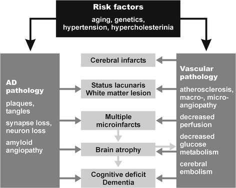
“The pathogenic chain of these and other deleterious effects, in a vicious circle, finally produces either structural cerebral disintegration (lacunes, infarcts, WMLs) with compromised neuronal metabolism, mitochondrial deficiency, oxidative stress, protein degradation, failure promoting cytoskeletal lesions with deposition of Aβ, and formation of neuritic lesions (e.g., neurofibrillary tangles).”
§ The overlap between vascular disease and Alzheimer’s disease - lessons from pathology > Review > Pathogenic factors
What the sources say
- Not checked yetcites [147] Jellinger KA, Attems J: Neuropathological evaluation of mixed dementia. J Neurol Sci. 2007, 257: 80-87. source
plaindirect- Asserted: passes — The authors put it forward as true.
- Checkable: passes — It can be checked against evidence.
- Load-bearing: passes — The paper’s conclusions depend on it.
- Based: passes — The paper gives a reason why it holds.
It may state two separate claims as one.
In the paper
“Microvascular changes in the aged brain and in AD induce impairment of cerebral perfusion, in particular decrease of regional blood flow, reduction of glucose transport and utilization, loss of vascular innervation with special impact on the cholinergic and transmitter deficits in AD [171], impairment of neurovascular regulation, ultrastructural changes in capillaries and basement membranes due to”
§ The overlap between vascular disease and Alzheimer’s disease - lessons from pathology > Review > Pathogenic factors
What the sources say
- Not checked yetcites [171] Roman GC, Kalaria RN: Vascular determinants of cholinergic deficits in Alzheimer disease and vascular dementia. Neurobiol Aging. 2006, 27: 1769-1785. source
plaindirect- Asserted: passes — The authors put it forward as true.
- Checkable: passes — It can be checked against evidence.
- Load-bearing: passes — The paper’s conclusions depend on it.
- Based: passes — The paper gives a reason why it holds.
It may state two separate claims as one.
In the paper
“These factors induce brain atrophy with cognitive and memory impairment (Figure 1) [147], although the complex cascade of these and other noxious factors needs further elucidation.”
§ The overlap between vascular disease and Alzheimer’s disease - lessons from pathology > Review > Pathogenic factors
What the sources say
- Not checked yetcites [147] Jellinger KA, Attems J: Neuropathological evaluation of mixed dementia. J Neurol Sci. 2007, 257: 80-87. source
plaindirect- Asserted: passes — The authors put it forward as true.
- Checkable: passes — It can be checked against evidence.
- Load-bearing: passes — The paper’s conclusions depend on it.
- Based: passes — The paper gives a reason why it holds.
It may state two separate claims as one.
In the paper
“Both hypertension and CAA are associated with an increased prevalence of CVLs [157]”
§ The overlap between vascular disease and Alzheimer’s disease - lessons from pathology > Review > Pathogenic factors
What the sources say
- Not checked yetcites [157] Jellinger KA, Attems J: Prevalence and pathogenic role of cerebrovascular lesions in Alzheimer’s disease. J Neurol Sci. 2005, 229-230: 37-41. source
plaincorrelational- Asserted: passes — The authors put it forward as true.
- Checkable: passes — It can be checked against evidence.
- Load-bearing: unclear — It is unclear whether the paper’s conclusions depend on it.
- Based: passes — The paper gives a reason why it holds.
It may state two separate claims as one.
In the paper
“CVD has been suggested to be an important cause of cognitive impairment in the elderly, both by itself or as a catalyst for the conversion of low-grade AD to overt dementia [186].”
§ The overlap between vascular disease and Alzheimer’s disease - lessons from pathology > Conclusions
What the sources say
- Not checked yetcites [186] Kalaria RN, Kenny RA, Ballard CG, Perry R, Ince P, Polvikoski T: Towards defining the neuropathological substrates of vascular dementia. J Neurol Sci. 2004, 226: 75-80. source
hedgedconditional- Asserted: passes — The authors put it forward as true.
- Checkable: passes — It can be checked against evidence.
- Load-bearing: passes — The paper’s conclusions depend on it.
- Based: passes — The paper gives a reason why it holds.
It may state two separate claims as one.
The authors state this as an assumption.
WMLs contribute to cognitive decline.
“and, thus, are assumed to contribute to cognitive decline [184].”
Claims other papers make about this one
Each is checked against this paper’s own text: does it say what the citing paper says it does?
The cited source says what the claim attributes to it.
In the citing paper
“Studies have consistently found a positive epidemiological correlation between Alzheimer’s disease (AD) and cardiometabolic traits, yet the biological mechanisms behind this correlation is not well understood [1–4].”
From Multi-trait association studies discover pleiotropic loci between Alzheimer’s disease and cardiometabolic traits (2021) · cites this paper as [1]
The cited source says what the claim attributes to it.
In the citing paper
“Studies have consistently found a positive epidemiological correlation between Alzheimer’s disease (AD) and cardiometabolic traits, yet the biological mechanisms behind this correlation is not well understood [1–4].”
From Multi-trait association studies discover pleiotropic loci between Alzheimer’s disease and cardiometabolic traits (2021) · cites this paper as [1]
In the citing paper
“Moreover, the growing recognition of overlapping pathologies (e.g., Lewy body inclusions, TDP-43 pathology, vascular lesions, etc.) among dementia patients indicates a wide range of biological heterogeneity within AD that necessitates a broader approach to scientific investigations of its pathophysiology [37–39].”
From Systems-based proteomics to resolve the biology of Alzheimer’s disease beyond amyloid and tau (2021) · cites this paper as [38]
Self-check
Percentages, sums and reported differences re-computed from the paper’s own numbers.
Each contradiction between two passages listed here was confirmed by an independent check.
Rated minor: it would not touch a result — a value no result rests on, a p-value that stays on the same side of the significance level, or a conflict that is clerical, resolved elsewhere in the paper, or outside its own findings.
In the paper
“Multiple infarcts (61 = 32.5%)”
§ The overlap between vascular disease and Alzheimer’s disease - lessons from pathology > Review > Topographical distribution of cerebrovascular lesions
Reported 61 · recomputed 58
Rated minor: it would not touch a result — a value no result rests on, a p-value that stays on the same side of the significance level, or a conflict that is clerical, resolved elsewhere in the paper, or outside its own findings.
In the paper
§ The overlap between vascular disease and Alzheimer’s disease - lessons from pathology > Review > Topographical distribution of cerebrovascular lesions
“Table: Table 3 Common lesions in AD, VaD, MIX, and aged controls (from[130])”
§ The overlap between vascular disease and Alzheimer’s disease - lessons from pathology > Review > Topographical distribution of cerebrovascular lesions
“The type and average prevalence of CVLs in AD, VaD, mixed dementia, and aged controls is shown in Table 3[147].”
Rated minor: it would not touch a result — a value no result rests on, a p-value that stays on the same side of the significance level, or a conflict that is clerical, resolved elsewhere in the paper, or outside its own findings.
In the paper
§ The overlap between vascular disease and Alzheimer’s disease - lessons from pathology > Review > Cerebrovascular and Alzheimer’s disease pathology in demented and non-demented elderly
“compared to 66.8% in age-matched controls, all showing low Braak stages (P <0.01).”
§ The overlap between vascular disease and Alzheimer’s disease - lessons from pathology > Review > Cerebrovascular and Alzheimer’s disease pathology in demented and non-demented elderly
“Minor and moderate vascular pathology in AD were about twice as frequent as in controls (26.2% vs. 12.2% and 20.9% vs. 11.3%; P <0.01). On the other hand, severe vascular pathology did not significantly differ between both groups (10.2% vs. 12.2%).”
Citations
Cites
Wardlaw, J. M., Smith, E. E., Biessels, G. J., Cordonnier, C., Fazekas, F., Frayne, R., Lindley, R. I., O'Brien, J. T., Barkhof, F., Benavente, O. R., et al. (2013) · The Lancet Neurology · doi:10.1016/s1474-4422(13)70124-8 · Cited by 5,544 · cited 1×
“SVD is a frequent cause of white matter lesions (WMLs; leukoaraiosis) that are increasingly detected by neuroimaging [118-121].”
BackgroundGorelick, P. B., Scuteri, A., Black, S. E., DeCarli, C., Greenberg, S. M., Iadecola, C., Launer, L. J., Laurent, S., Lopez, O. L., Nyenhuis, D., et al. (2011) · Stroke · doi:10.1161/str.0b013e3182299496 · Cited by 3,811 · cited 3×
“Epidemiological studies have shown that AD and CVD share common risk factors such as hypertension during midlife, diabetes mellitus, smoking, apolipoprotein E (ApoE) ε4 isoforms, hypercholesterolemia, homocysteinemia, and, in particular, age [16,30-34].”
BackgroundPantoni, L. (2010) · The Lancet Neurology · doi:10.1016/s1474-4422(10)70104-6 · Cited by 3,405 · cited 1×
“SVD affects small arteries and arterioles and refers to pathological changes similar to atherosclerosis that are termed small vessel arteriosclerosis/atherosclerosis, lipoor fibrohyalinosis, or hypertensive arteriopathy [115].”
BackgroundNeurovascular pathways to neurodegeneration in Alzheimer’s disease and other disorders
Zlokovic, B. V. (2011) · Nature Reviews Neuroscience · doi:10.1038/nrn3114 · Cited by 2,969 · cited 1×
“CVD has been suggested to contribute to AD neuropathological changes including selective brain atrophy and accumulation of abnormal proteins such as Aβ [24,35,84,85].”
BackgroundThe projected effect of risk factor reduction on Alzheimer's disease prevalence
Barnes, D. E., & Yaffe, K. S. (2011) · The Lancet Neurology · doi:10.1016/s1474-4422(11)70072-2 · Cited by 2,846
Mixed brain pathologies account for most dementia cases in community-dwelling older persons
Schneider, J. A., Arvanitakis, Z., Bang, W., & Bennett, D. A. (2007) · Neurology · doi:10.1212/01.wnl.0000271090.28148.24 · Cited by 1,992 · cited 2×
“The burden of vascular and AD-type pathologies are leading and independent causes of dementia in the elderly [4,9-15], suggesting additive or synergistic effects of both types of lesions on cognitive impairment [2,3,5,9,16-29].”
BackgroundThe Pathobiology of Vascular Dementia
Iadecola, C. (2013) · Neuron · doi:10.1016/j.neuron.2013.10.008 · Cited by 1,857 · cited 1×
“The pathophysiology of VaD has been critically reviewed recently [48,144-146].”
BackgroundCerebral microbleeds: a guide to detection and interpretation
Greenberg, S. M., Vernooij, M. W., Cordonnier, C., Viswanathan, A., Al-Shahi Salman, R., Warach, S., Launer, L. J., Van Buchem, M. A., & Breteler, M. M. (2009) · The Lancet Neurology · doi:10.1016/s1474-4422(09)70013-4 · Cited by 1,798
Brain Infarction and the Clinical Expression of Alzheimer Disease<subtitle>The Nun Study</subtitle>
Snowdon, D. A. (1997) · JAMA: The Journal of the American Medical Association · doi:10.1001/jama.1997.03540340047031 · Cited by 1,590
Silent brain infarcts: a systematic review
Vermeer, S. E., Longstreth, W. T., & Koudstaal, P. J. (2007) · The Lancet Neurology · doi:10.1016/s1474-4422(07)70170-9 · Cited by 1,221 · cited 1×
““Silent” lacunar infarcts are frequently detected by MRI or CCT and are not accompanied by any overt clinical symptoms, but double the risk of subsequent stroke and dementia [133].”
Background
Research works printed but not linked: 2
- Crystal H, Dickson D: Cerebral infarcts in patients with autopsy proven Alzheimer’s disease. Neurobiol Aging. 2002, 23: 207.
- Jentoft M, Parisi J, Dickson D, Johnson K, Boeve B, Knopman D, Petersen R: Neuropathologic findings in 32 nondemented elderly subjects (abs.). J Neuropathol Exp Neurol. 2011, 70: 531.
Cited by
Sorted by citations
Items 101–200 of 736
Guerriero, F., & Ricevuti, G. (2016) · Neural Regeneration Research · doi:10.4103/1673-5374.195277 · Cited by 69
“All these components strengthen the possibility that the ‘major pathological role of amyloid in AD may be to inflict vascular damage’ and hence, impair BBB function (Franzblau et al., 2013; Attems and Jellinger, 2014).”
Button, E. B., Boyce, G. K., Wilkinson, A., Stukas, S. K., Hayat, A., Fan, J., Wadsworth, B. J., Robert, J., Martens, K. M., & Wellington, C. L. (2019) · Alzheimer s Research & Therapy · doi:10.1186/s13195-019-0497-9 · Cited by 73
“The majority of AD patients have co-morbid vascular diseases and cerebrovascular pathologies including microinfarcts, cerebral atherosclerosis, and arteriolosclerosis [3, 4].”
BackgroundSong, R., Pan, K., Xu, H., Qi, X., Buchman, A. S., Bennett, D. A., & Xu, W. (2021) · Alzheimer s & Dementia · doi:10.1002/alz.12343 · Cited by 75
GLP-1 Receptor Agonists in Neurodegeneration: Neurovascular Unit in the Spotlight
Monti, G., Moreira, D. G., Richner, M., Mutsaers, H. A. M., Ferreira, N., & Jan, A. (2022) · Cells · doi:10.3390/cells11132023 · Cited by 72
Endothelial Nitric Oxide Synthase–Deficient Mice
Liao, F., Lin, G., Chen, X., Chen, L., Zheng, W., Raghow, R., Zhou, F.-M., Shih, A. Y., & Tan, X.-L. (2021) · American Journal Of Pathology · doi:10.1016/j.ajpath.2021.02.022 · Cited by 68
Georgakis, M. K., Dimitriou, N. G., Karalexi, M. A., Mihas, C. C., Nasothimiou, E. G., Tousoulis, D. M., Tsivgoulis, G. K., & Petridou, E. T. (2017) · Journal of the American Geriatrics Society · doi:10.1111/jgs.14750 · Cited by 67
“Cerebral microvascular disease is associated with cognitive deterioration, and has been implicated in the pathogenesis of both vascular dementia (VaD), and Alzheimer’s disease (AD).(3,4) Because small vessel disease is considered a systemic disorder,(5) damage of other end-organs could be accompanied by microcirculato…”
BackgroundPrion-like mechanisms in Alzheimer disease
Walker, L. C. (2018) · Handbook of clinical neurology · doi:10.1016/b978-0-444-63945-5.00016-7 · Cited by 67
“However, some degree of Aβ-CAA is almost always present in AD (Attems and Jellinger, 2014; Vinters, 2015; Kapasi and Schneider, 2016), and it is severe in around 25% of cases (Charidimou et al.”
BackgroundBos, I., Vos, S. J. B., Schindler, S. E., Hassenstab, J. J., Xiong, C., Grant, E., Verhey, F. R. J., Morris, J. C., Visser, P. J., & Fagan, A. M. (2019) · Alzheimer s & Dementia · doi:10.1016/j.jalz.2019.04.015 · Cited by 68
“This is compatible with studies showing that AD pathology and vascular disorders, as well as concomitant cerebral vascular pathology, often coexist in late-onset AD [34,35].”
ResultInfluence of cerebrovascular disease on brain networks in prodromal and clinical Alzheimer’s disease
Chong, J. S. X., Liu, S., Loke, Y. M., Hilal, S., Ikram, M. K., Xu, X., Tan, B. Y., Venketasubramanian, N., Chen, C., & Zhou, J. H. (2017) · Brain · doi:10.1093/brain/awx224 · Cited by 64
“Together, Alzheimer’s disease and cerebrovascular disease are proposed to have additive effects on cognitive decline (Zekry et al., 2002; Iadecola, 2010; Attems and Jellinger, 2014; Kalheim et al., 2017).”
BackgroundResultSharafi, S. M., Sylvestre, J., Chevrefils, C., Soucy, J., Beaulieu, S., Pascoal, T. A., Arbour, J. D., Rhéaume, M., Robillard, A., Chayer, C., et al. (2019) · Alzheimer s & Dementia Translational Research & Clinical Interventions · doi:10.1016/j.trci.2019.09.006 · Cited by 65
“studies have further shown a relationship between RVT and AD [33,35,46,47].”
ResultPaoletti, F. P., Simoni, S., Parnetti, L., & Gaetani, L. (2021) · International Journal of Molecular Sciences · doi:10.3390/ijms22094958 · Cited by 65
“Mixed AD/SVD pathology is frequently observed in AD patients, and this tren increases with age [47].”
BackgroundIshii, M., & Iadecola, C. (2015) · Biochimica et Biophysica Acta (BBA) - Molecular Basis of Disease · doi:10.1016/j.bbadis.2015.10.029 · Cited by 63
“Dementia due to vascular causes is likely to represent one end of a spectrum of age-related dementia, while dementia due to AD pathology may represent another end of the spectrum, with many cases having contributions from both [4-6].”
The road to precision medicine: Eliminating the “One Size Fits All” approach in Alzheimer’s disease
Behl, T., Kaur, I., Sehgal, A., Singh, S., Albarrati, A., Albratty, M., Najmi, A., Meraya, A. M., & BUNGAU, S. G. (2022) · Biomedicine & Pharmacotherapy · doi:10.1016/j.biopha.2022.113337 · Cited by 67
Ferroptosis of Microglia in Aging Human White Matter Injury
Adeniyi, P. A., Gong, X., MacGregor, E., Degener-O’Brien, K., McClendon, E., Garcia, M., Romero, O., Russell, J., Srivastava, T., Miller, J. A., et al. (2023) · Annals of Neurology · doi:10.1002/ana.26770 · Cited by 62
Chai, Y. L., Hilal, S., Chong, J. P. C., Ng, Y. X., Liew, O. W., Xu, X., Ikram, M. K., Venketasubramanian, N., Richards, M., Lai, M. K. P., et al. (2016) · Medicine · doi:10.1097/md.0000000000004566 · Cited by 62
“YLC and SH contributed equally to the article.”
BackgroundSerum IL‐8 is a marker of white‐matter hyperintensities in patients with Alzheimer's disease
Zhu, Y., Chai, Y. L., Hilal, S., Ikram, M. K., Venketasubramanian, N., Wong, B., Chen, C., & Lai, M. K. P. (2017) · Alzheimer s & Dementia Diagnosis Assessment & Disease Monitoring · doi:10.1016/j.dadm.2017.01.001 · Cited by 61
“Several neuroimaging markers of cerebrovascular disease (CeVD), such as white-matter hyperintensities (WMHs), cortical infarct, and lacunes, have been shown to be associated with AD [20], and may exacerbate the severity of dementia [21,22].”
Giacomelli, C., Daniele, S., & Martini, C. (2017) · Biochemical Pharmacology · doi:10.1016/j.bcp.2017.01.017 · Cited by 71
Bos, I., Verhey, F. R. J., Ramakers, I. H. G. B., Jacobs, H. I. L., Soininen, H. S., Freund‐Levi, Y., Hampel, H. J., Tsolaki, M., Wallin, Å. K., van Buchem, M. A., et al. (2017) · Alzheimer s Research & Therapy · doi:10.1186/s13195-017-0328-9 · Cited by 62
“Background Cerebrovascular disease (CVD) often coexists with Alzheimer’s disease (AD), and both conditions add to cognitive decline [1, 2].”
BackgroundPrevention and Treatment of Alzheimer’s Disease: Biological Mechanisms of Exercise
McGurran, H., Glenn, J. M., Madero, E. N., & Bott, N. T. (2019) · Journal of Alzheimer s Disease · doi:10.3233/jad-180958 · Cited by 63
“This can lessen the threshold for clinically relevant dementia due the additive effect of non-AD specific pathologies [203].”
Positive Feedback Loops in Alzheimer’s Disease: The Alzheimer’s Feedback Hypothesis
Doig, A. J. (2018) · Journal of Alzheimer s Disease · doi:10.3233/jad-180583 · Cited by 67
“Multiple stresses, such as an ApoE4 genotype [103], age, inefficient protein degradation [104], autophagic/lysosomal dysfunction [105], iron dysregulation [27], deficiencies in blood coagulation via bacterial lipopolysaccharides [106, 107], vascular conditions [108], mitochondrial dysfunction [17], viral infection [10…”
BackgroundRivera‐Rivera, L. A., Cody, K. A., Eisenmenger, L. B., Cary, P., Rowley, H. A., Carlsson, C. M., Johnson, S. C., & Johnson, K. M. (2020) · Journal of Cerebral Blood Flow & Metabolism · doi:10.1177/0271678x20910302 · Cited by 62
The Role of Cardiovascular Risk Factors and Stroke in Familial Alzheimer Disease
Tosto, G., Bird, T. D., Bennett, D. A., Boeve, B. F., Brickman, A. M., Cruchaga, C., Faber, K. M., Foroud, T., Farlow, M. R., Goate, A., et al. (2016) · JAMA Neurology · doi:10.1001/jamaneurol.2016.2539 · Cited by 62
Glushakova, O., Glushakov, A. V., Glushakov, A. O., Wijesinghe, D. S., Valadka, A. B., & Hayes, R. (2017) · Brain Circulation · doi:10.4103/bc.bc_27_16 · Cited by 65
“[112,121,122] Thus, tau isoforms are considered to be one of the earliest biomarkers of AD.”
BackgroundInfluentialExposure to ambient air pollutants and the onset of dementia in Québec, Canada
Smargiassi, A., Sidi, E. A. L., Robert, L., Plante, C., Haddad, M., Gamache, P., Burnett, R. T., Goudreau, S., Liu, L., Fournier, M., et al. (2020) · Environmental Research · doi:10.1016/j.envres.2020.109870 · Cited by 63
Chen, X., Chen, L., Lin, G., Wang, Z., Kodali, M. C., Li, M., Chen, H., Lebovitz, S. G., Ortyl, T. C., Li, L., et al. (2022) · Molecular Psychiatry · doi:10.1038/s41380-022-01701-9 · Cited by 66
Carey, A. M., & Fossati, S. (2022) · Alzheimer s & Dementia · doi:10.1002/alz.12871 · Cited by 62
Chung, C., Lee, H.-Y., Lin, P.-C., & Wang, P. (2017) · Journal of Alzheimer s Disease · doi:10.3233/jad-170349 · Cited by 63
“Cerebral microvascular pathologies, including cerebral amyloid angiopathy, arteriosclerosis, or lipohyalinosis, have been revealed in a large proportion of patients with AD [27, 28].”
BackgroundAntidepressant medications in dementia: evidence and potential mechanisms of treatment-resistance
Costello, H., Roiser, J. P., & Howard, R. (2023) · Psychological Medicine · doi:10.1017/s003329172200397x · Cited by 60
“However, the overlap of vascular dementia with AD pathology (Attems & Jellinger, 2014) and evidence that higher vascular risk factor scores predict reduced response to antidepressants in late life depression (Sheline et al., 2010) does not suggest this patient group will buck the trend of a…”
A mechanistic survey of Alzheimer's disease
Tang, Y., Zhang, D., Gong, X., & Zheng, J. (2021) · Biophysical Chemistry · doi:10.1016/j.bpc.2021.106735 · Cited by 66
Shang, X., Zhu, Z., Zhang, X., Huang, Y., Zhang, X., Liu, J., Wang, W., Tang, S., Yu, H., Ge, Z., et al. (2022) · EClinicalMedicine · doi:10.1016/j.eclinm.2022.101335 · Cited by 60
Ferreira, S. T. (2021) · Journal of Neuroendocrinology · doi:10.1111/jne.12959 · Cited by 59
Association between atherosclerosis and Alzheimer's disease: A systematic review and meta‐analysis
Xie, B., Shi, X., Xing, Y., & Tang, Y. (2020) · Brain and Behavior · doi:10.1002/brb3.1601 · Cited by 62
Glymphatic system dysfunction in neurodegenerative diseases
Beschörner, N., & Nedergaard, M. (2024) · Current Opinion in Neurology · doi:10.1097/wco.0000000000001252 · Cited by 60
Zhou, B., Li, L., Qiu, X., Wu, J., Xu, L., & Shao, W. (2020) · Molecular Medicine Reports · doi:10.3892/mmr.2020.11203 · Cited by 58
RetractedCerebrovascular contributions to aging and Alzheimer's disease in Down syndrome
Wilcock, D. M., Schmitt, F. A., & Head, E. (2016) · Biochimica et Biophysica Acta (BBA) - Molecular Basis of Disease · doi:10.1016/j.bbadis.2015.11.007 · Cited by 56
“As mentioned previously, the CVD contribution to AD is increasingly being recognized as a critical comorbidity that accelerates the age of onset of dementia and also leads to a faster progression of the disease [2,5,28,41].”
BackgroundPET imaging of the neurovascular interface in cerebrovascular disease
Evans, N. R., Tarkin, J. M., Buscombe, J. R., Markus, H. S., Rudd, J. H. F., & Warburton, E. A. (2017) · Nature Reviews Neurology · doi:10.1038/nrneurol.2017.129 · Cited by 56
A Critical Review of the Study of Neuroprotective Diets to Reduce Cognitive Decline
Duplantier, S. C., & Gardner, C. D. (2021) · Nutrients · doi:10.3390/nu13072264 · Cited by 55
“In particular, vascular disease risk factors, especially in mid-life, are associated with the build-up of beta-amyloid in the brain [26,27].”
BackgroundKelly, P., Denver, P., Satchell, S. C., Ackermann, M., Konerding, M. A., & Mitchell, C. A. (2017) · Angiogenesis · doi:10.1007/s10456-017-9568-3 · Cited by 56
“Pathological alterations to the integrity of the cerebrovasculature may critically alter the function of the neurovascular unit (NVU) and cause neuronal injury via an impairment of cerebral perfusion, an unrestricted brain entry of circulatory compounds, reduced cerebral amyloid clearance and vascular and parenchymal…”
BackgroundNetwork Localisation of White Matter Damage in Cerebral Small Vessel Disease
Petersen, M., Frey, B. M., Schlemm, E., Mayer, C., Hanning, U. M. P., Engelke, K., Fiehler, J., Borof, K., Jagodzinski, A., Gerloff, C. P., et al. (2020) · Scientific Reports · doi:10.1038/s41598-020-66013-w · Cited by 53
HDL Proteome and Alzheimer’s Disease: Evidence of a Link
Marsillach, J., Adorni, M. P., Zimetti, F., Papotti, B., Zuliani, G., & Cervellati, C. (2020) · Antioxidants · doi:10.3390/antiox9121224 · Cited by 62
“Additional frequent abnormalities are represented by the presence of neuroinflammation and cerebral vessel disease, with the latter affecting 60–90% of AD patients [3].”
BackgroundPlasma GSH levels and Alzheimer's disease. A prospective approach.: Results from the HELIAD study
Charisis, S., Ntanasi, E., Yannakoulia, M., Anastasiou, C. A., Kosmidis, M. H., Dardiotis, E., Hadjigeorgiou, G. M., Sakka, P., Veskoukis, A. S., Kouretas, D., et al. (2020) · Free Radical Biology and Medicine · doi:10.1016/j.freeradbiomed.2020.10.027 · Cited by 56
Mancini, S., Minniti, S., Gregori, M., Sancini, G., Cagnotto, A., Couraud, P., Ordóñez‐Gutiérrez, L., Wandosell, F. G., Salmona, M., & Re, F. (2015) · Nanomedicine Nanotechnology Biology and Medicine · doi:10.1016/j.nano.2015.09.003 · Cited by 54
“Brain accumulation of amyloid-β (Aβ) peptides, eventually deposited as plaques, is one of the pathological hallmarks of AD.1 Aβ accumulation has been hypothesized to result from an imbalance between Aβ production and clearance; indeed, Aβ clearance seems to be impaired in both early and late forms of AD and may contri…”
BackgroundInfluentialSegundo‐Acosta, P. S., Montero‐Calle, A., Fuentes, M., Rábano, A., Villalba, M., & Barderas, R. (2019) · Journal of Proteome Research · doi:10.1021/acs.jproteome.9b00258 · Cited by 54
Vitamin K2 Holds Promise for Alzheimer’s Prevention and Treatment
Popescu, A., & German, M. (2021) · Nutrients · doi:10.3390/nu13072206 · Cited by 57
“In addition, pathological data have suggested that vascular and neurodegenerative processes can coexist in AD [100].”
BackgroundEpidemiologic Trends, Social Determinants, and Brain Health: The Role of Life Course Inequalities
Hilal, S., & Brayne, C. (2022) · Stroke · doi:10.1161/strokeaha.121.032609 · Cited by 56
Shi, H., Koronyo, Y., Fuchs, D.-T. S., Sheyn, J., Jallow, O., Mandalia, K., Graham, S. L., Gupta, V., Mirzaei, M., Kramerov, A. A., et al. (2023) · Alzheimer s & Dementia · doi:10.1002/alz.13086 · Cited by 52
Aquaporin-4 and Cognitive Disorders
Wang, Y., Huang, C., Guo, Q., & Chu, H. (2022) · Aging and disease · doi:10.14336/ad.2021.0731 · Cited by 55
“With the in-depth study of the glymphatic system and AQP4, a close relationship between AQP4 and the main pathological features of AD has emerged, including abnormal deposition of extracellular Aβ, neurofibrillary tangles caused by tau protein accumulation and synaptic damage [9].”
BackgroundThe Role of Basement Membranes in Cerebral Amyloid Angiopathy
Howe, M. D., McCullough, L. D., & Urayama, A. (2020) · Frontiers in Physiology · doi:10.3389/fphys.2020.601320 · Cited by 50
Saeed, U., Mirza, S. S., MacIntosh, B. J., Herrmann, N., Keith, J., Ramirez, J., Nestor, S. M., Yu, Q., Knight, J., Swardfager, W., et al. (2018) · Alzheimer's & Dementia · doi:10.1016/j.jalz.2018.04.005 · Cited by 48
Aquaporin-4 and Alzheimer’s Disease
Yang, C., Huang, X., Huang, X., Mai, H., Li, J., Jiang, T., Wang, X., & Lü, T. (2016) · Journal of Alzheimer s Disease · doi:10.3233/jad-150949 · Cited by 53
Jandke, S., Garz, C., Schwanke, D., Sendtner, M., Heinze, H., Carare, R. O., & Schreiber, S. (2018) · Brain Pathology · doi:10.1111/bpa.12629 · Cited by 46
“006), or (3) a combination of (1) and (2) (n = 11 [52%] vs.”
BackgroundInfluentialRibeiro, V. T., de Souza, L. C., & Silva, A. C. S. E. (2019) · Protein and Peptide Letters · doi:10.2174/0929866527666191230103739 · Cited by 50
Elevated circulating amyloid concentrations in obesity and diabetes promote vascular dysfunction
Meakin, P. J., Coull, B. M., Tuharska, Z., McCaffery, C., Akoumianakis, I., Antoniades, C., Brown, J., Griffin, K. J., Platt, F., Ozber, C. H., et al. (2020) · Journal of Clinical Investigation · doi:10.1172/jci122237 · Cited by 22
“Indeed, cognitive decline and AD are associated with cerebrovascular and coronary atherosclerosis in humans, and people with AD exhibit pathological signs of vascular impairment in cerebral and peripheral vessels, indicating some commonality between peripheral and central disease mechanisms (2).”
Association of midlife vascular risk and AD biomarkers with subsequent cognitive decline
Pettigrew, C. A., Soldan, A., Wang, J., Wang, M., Arthur, K. C., Moghekar, A., Gottesman, R. F., & Albert, M. (2020) · Neurology · doi:10.1212/wnl.0000000000010946 · Cited by 52
“As reviewed elsewhere, there are a number of ways by which AD pathology andmeasures related to vascular risk may combine on a pathophysiologic level to exert additive effects on cognitive outcomes, which may include downstream effects on neuronal function, cerebral perfusion, and cortical atrophy.(48,49) More work is…”
BackgroundIschemia as a common trigger for Alzheimer’s disease
Elman-Shina, K., & Efrati, S. (2022) · Frontiers in Aging Neuroscience · doi:10.3389/fnagi.2022.1012779 · Cited by 48
“of vascular pathology are recognized in AD: small vessel disease and cerebral amyloid angiopathy (CAA) (Attems and Jellinger, 2014).”
BackgroundInfluentialEstrogen, menopause, and Alzheimer’s disease: understanding the link to cognitive decline in women
Mervosh, N. L., & Devi, G. (2025) · Frontiers in Molecular Biosciences · doi:10.3389/fmolb.2025.1634302 · Cited by 62
“For example, animal models of ovariectomy show increased cerebrovascular inflammation, reduced perfusion, and enhanced Aβ deposition—effects partially reversed by estrogen replacement (Hodis et al., 2016; Simpkins et al., 2009; Attems et al., 2014).”
Is Beta-Amyloid Accumulation a Cause or Consequence of Alzheimer's Disease?
Wang, S., Mims, P. N., Roman, R. J., & Fan, F. (2016) · PubMed · Cited by 48
Hickman, R. A., Flowers, X. E., & Wısnıewskı, T. (2020) · Current Neurology and Neuroscience Reports · doi:10.1007/s11910-020-01063-1 · Cited by 48
Mitochondrial dysfunction in Alzheimer’s disease - a proteomics perspective
Abyadeh, M., Gupta, V., Chitranshi, N., Gupta, V., Wu, Y., Saks, D., Wall, R. V., Fitzhenry, M. J., Basavarajappa, D. K., You, Y., et al. (2021) · Expert Review of Proteomics · doi:10.1080/14789450.2021.1918550 · Cited by 51
Energy and the Alzheimer brain
Mamelak, M. (2017) · Neuroscience & Biobehavioral Reviews · doi:10.1016/j.neubiorev.2017.02.001 · Cited by 47
“Pathology engages the Alzheimer brain’s vascular system at ll levels (Attems and Jellinger, 2014; Kelleher and Soiza, 2013). educed cerebral blood flow and/or cerebral blood flow dysreglation occurs in elderly individuals at high risk for AD even efore cognitive decline, brain atrophy or the…”
BackgroundAre the brain's vascular and Alzheimer pathologies additive or interactive?
Koncz, R., & Sachdev, P. S. (2017) · Current Opinion in Psychiatry · doi:10.1097/yco.0000000000000395 · Cited by 47
“Although the pathology underlying WMH is somewhat heterogeneous, WMH are widely used as a proxy for SVD with this caveat in mind [3].”
BackgroundTraumatic brain injury history and progression from mild cognitive impairment to Alzheimer disease.
LoBue, C., Woon, F. L. M., Rossetti, H. C., Hynan, L. S., Hart, J., & Cullum, C. M. (2018) · Neuropsychology · doi:10.1037/neu0000431 · Cited by 49
The effect of white matter hyperintensities on verbal memory
Swardfager, W., Cogo‐Moreira, H., Masellis, M., Ramirez, J., Herrmann, N., Edwards, J. D., Saleem, M., Chan, P., Yu, D., Nestor, S. M., et al. (2018) · Neurology · doi:10.1212/wnl.0000000000004983 · Cited by 47
“In the first sample, cortical stroke was excluded to infer specific effects of subcortical small vessel disease, a common co-pathology in AD that lowers the dementia threshold.(28) The effect of WMH was confirmed in an unselected, heterogeneous, consecutive, real-world case series, including patients across spectra of…”
BackgroundAlbuminuria and Cerebral Small Vessel Disease: A Systematic Review and Meta‐Analysis
Georgakis, M. K., Chatzopoulou, D., Tsivgoulis, G. K., & Petridou, E. T. (2018) · Journal of the American Geriatrics Society · doi:10.1111/jgs.15240 · Cited by 50
“Widespread vascular injury in the brain and the ensuing effect on structural and functional connectivity are main contributors to neurocognitive dysfunction.(1) Cerebral small vessel disease (SVD) is responsible for more than 20% of strokes, which are the major contributor to vascular dementia and a potential pathogen…”
BackgroundMRI load of cerebral microvascular lesions and neurodegeneration, cognitive decline, and dementia
Wang, R., Laveskog, A., Laukka, E. J., Kalpouzos, G., Bäckman, L., Fratiglioni, L., & Qiu, C. (2018) · Neurology · doi:10.1212/wnl.0000000000006355 · Cited by 47
Dysfunctional effort-based decision-making underlies apathy in genetic cerebral small vessel disease
Heron, C. J. L., Manohar, S., Plant, O., Muhammed, K., Griffanti, L., Nemeth, A. H., Douaud, G., Markus, H. S., & Husain, M. (2018) · Brain · doi:10.1093/brain/awy257 · Cited by 45
Chua, X. Y., Chai, Y. L., Chew, W. S., Chong, J. R., Ang, H. L., Xiang, P., Camara, K., Howell, A. R., Torta, F. T., Wenk, M. R., et al. (2020) · Alzheimer s Research & Therapy · doi:10.1186/s13195-020-00694-3 · Cited by 47
Sundaresan, V., Zamboni, G., Heron, C. J. L., Rothwell, P. M., Masud Husain, B., Battaglini, M., Stefano, N. D., Jenkinson, M., & Griffanti, L. (2019) · NeuroImage · doi:10.1016/j.neuroimage.2019.116056 · Cited by 46
Lee, A., Raghavan, N. S., Bhattarai, P., Siddiqui, T., Sariya, S., Reyes‐Dumeyer, D., Flowers, X. E., Cardoso, S. A. L., Jager, P. L. D., Bennett, D. A., et al. (2022) · Acta Neuropathologica · doi:10.1007/s00401-022-02431-6 · Cited by 49
“Quality control and imputation details for the ROSMAP have been reported previously [4].”
BackgroundMethodTang, A. S., Oskotsky, T., Havaldar, S., Mantyh, W. G., Bicak, M., Solsberg, C. W., Woldemariam, S., Zeng, B., Hu, Z., Oskotsky, B., et al. (2022) · Nature Communications · doi:10.1038/s41467-022-28273-0 · Cited by 45
Effects of Exercise on Type 2 Diabetes Mellitus-Related Cognitive Impairment and Dementia
Callisaya, M. L., & Nosaka, K. (2017) · Journal of Alzheimer s Disease · doi:10.3233/jad-161154 · Cited by 48
“In humans, the interaction between Alzheimer’s disease type pathology and cerebrovascular disease is likely to be additive and responsible for greater cognitive impairment [39].”
BackgroundGuglielmotto, M., Monteleone, D., Vasciaveo, V., Repetto, I. E., Manassero, G., Tabaton, M., & Tamagno, E. (2017) · Frontiers in Aging Neuroscience · doi:10.3389/fnagi.2017.00320 · Cited by 45
“Several studies show vascular pathology in 50% of the elderly population and confirm that the simultaneous presence of vascular and AD lesions is closely related to the severity of dementia (Schneider, 2009; Attems and Jellinger, 2014).”
BackgroundCognitive Reserve from the Perspective of Preclinical Alzheimer Disease
Soldan, A., Pettigrew, C. A., & Albert, M. (2019) · Clinics in Geriatric Medicine · doi:10.1016/j.cger.2019.11.006 · Cited by 46
Low, A., Prats-Sedano, M. A., Stefaniak, J. D., McKiernan, E. F., Carter, S. F., Douvani, M.-E., Mak, E., Su, L., Stupart, O., Muniz, G., et al. (2022) · Journal of Neurology, Neurosurgery & Psychiatry · doi:10.1136/jnnp-2021-327462 · Cited by 44
Cognitive reserve and midlife vascular risk: Cognitive and clinical outcomes
Soldan, A., Pettigrew, C. A., Zhu, Y., Wang, M., Gottesman, R. F., DeCarli, C. S., & Albert, M. (2020) · Annals of Clinical and Translational Neurology · doi:10.1002/acn3.51120 · Cited by 45
“5) [2-16] Years from baseline to onset of clinical symptoms of MCI – – 8.”
BackgroundVascular burden and cognition: Mediating roles of neurodegeneration and amyloid PET
Ottoy, J., Ozzoude, M., Zukotynski, K., Adamo, S. M., Scott, C. J. M., Gaudet, V., Ramirez, J., Swardfager, W., Cogo‐Moreira, H., Lam, B., et al. (2022) · Alzheimer s & Dementia · doi:10.1002/alz.12750 · Cited by 48
“…additive effects of WMH and AD pathology on cognitive decline at baseline and longitudinally [46,47], as well as a lower degree of AD pathology in the presence of vascular pathology for the same level of cognitive impairment [48] and a substantial increase in dementia risk post-stroke [49].”
Adult Astrogenesis and the Etiology of Cortical Neurodegeneration
Mohn, T. C., & Koob, A. O. (2015) · Journal of Experimental Neuroscience · doi:10.4137/jen.s25520 · Cited by 44
Post-stroke Dementia: Epidemiology, Mechanisms and Management
Hu, G., & Chen, Y.-M. (2017) · International journal of gerontology · doi:10.1016/j.ijge.2017.07.004 · Cited by 46
Cerebral Small Vessel Disease Influences Hippocampal Subfield Atrophy in Mild Cognitive Impairment
Wong, F. C. C., Yatawara, C., Low, A., Foo, H., Wong, B. Y. X., Lim, L., Wang, B. Z., Kumar, D., Ng, K. P., & Kandiah, N. (2020) · Translational Stroke Research · doi:10.1007/s12975-020-00847-4 · Cited by 43
Thal, D. R., Ronisz, A., Tousseyn, T., Upadhaya, A. R., Balakrishnan, K., Vandenberghe, R. R., Vandenbulcke, M., Arnim, C. A. F. V., Otto, M., Beach, T. G., et al. (2019) · Acta Neuropathologica Communications · doi:10.1186/s40478-019-0837-9 · Cited by 44
Pahlavian, S. H., Wang, X., Ma, S. J., Zheng, H., Casey, M., D’Orazio, L. M., Shao, X., Ringman, J. M., Chui, H. C., Wang, D. J. J., et al. (2020) · Journal of Cerebral Blood Flow & Metabolism · doi:10.1177/0271678x20927101 · Cited by 44
Kim, S. E., Kim, H. J., Jang, H., Weiner, M. W., DeCarli, C. S., Na, D. L., & Seo, S. W. (2022) · International Journal of Molecular Sciences · doi:10.3390/ijms231810490 · Cited by 42
“In fact, previous studies have shown that AD combined with vascular disease is the most prevalent mixed pathology [9,11,12].”
BackgroundMekkes, N., Groot, M., Hoekstra, E., de Boer, A., Dagkesamanskaia, E., Bouwman, S., Wehrens, S. M. T., Herbert, M. K., Wever, D. D., Rozemüller, A. J. M., et al. (2024) · Nature Medicine · doi:10.1038/s41591-024-02843-9 · Cited by 47
“Most dementias are difficult to diagnose and study due to considerable heterogeneity 3–5 , partially shared clinical and pathological features 6,7 and complex comorbidity patterns 8,9 .”
BackgroundMarongiu, R. (2019) · Frontiers in Aging Neuroscience · doi:10.3389/fnagi.2019.00242 · Cited by 47
Imaging of Tau Pathology in Neurodegenerative Diseases: An Update
Beyer, L., & Brendel, M. (2020) · Seminars in Nuclear Medicine · doi:10.1053/j.semnuclmed.2020.12.004 · Cited by 44
Program, T. V. M. V., Bone, W. P., Siewert, K. M., Jha, A., Klarin, D., Damrauer, S. M., Chang, K.-M., Tsao, P. S., Assimes, T. L., Ritchie, M. D., et al. (2021) · Alzheimer's Research & Therapy · doi:10.1186/s13195-021-00773-z · Cited by 41
Coşacak, M. İ., Bhattarai, P., Jager, P. L. D., Menon, V., Tosto, G., & Kızıl, Ç. (2022) · Cells · doi:10.3390/cells11111807 · Cited by 39
“Not only a neuronal disease, AD also includes a complex interplay of multiple cell types, such as immune cells [2,6,7], the neurovascular niche [1,8], neural stem cells [9,10], astroglia [11,12], and oligodendrocytes [13,14], among others.”
BackgroundMultimorbidity in dementia: Current perspectives and future challenges
Stirland, L., Choate, R., Zanwar, P., Zhang, P., Watermeyer, T., Valletta, M., Torso, M., Tamburin, S., Saeed, U., Ridgway, G. R., et al. (2025) · Alzheimer s & Dementia · doi:10.1002/alz.70546 · Cited by 52
“23 The relevance of this comorbid neuropathology is acknowledged by the 2024 revised criteria for the diagnosis and staging of AD.”
Hefter, D., Kaiser, M., Weyer, S. W., Papageorgiou, I. E., Both, M., Kann, O., Müller, U., & Draguhn, A. (2016) · Journal of Neuroscience · doi:10.1523/jneurosci.4130-15.2016 · Cited by 44
“This effect may constitute a link between pathological mechanisms in AD as well as in vascular disease (Attems and Jellinger, 2014).”
BackgroundThe pleiotropic vasoprotective functions of high density lipoproteins (HDL)
Boyce, G. K., Button, E. B., Sonja, S. V., & Wellington, C. L. (2017) · Journal of Biomedical Research · doi:10.7555/jbr.31.20160103 · Cited by 41
“The brain comprises only 2% of total body mass but consumes approximately 12% of total cardiac output([102]).”
BackgroundDrake, J., Initiative, A. D. N., Chambers, A. B., Ott, B. R., & Daiello, L. A. (2021) · Journal of Alzheimer s Disease · doi:10.3233/jad-200759 · Cited by 41
Low, A., Prats‐Sedano, M. A., McKiernan, E. F., Carter, S. F., Stefaniak, J. D., Nannoni, S., Su, L., Dounavi, M., Terrera, G. M., Ritchie, K. A., et al. (2022) · Alzheimer s Research & Therapy · doi:10.1186/s13195-022-01095-4 · Cited by 40
Kim, S., Sharma, C., Jung, U. J., & Kim, S. R. (2023) · Biomedicines · doi:10.3390/biomedicines11051383 · Cited by 40
Vascular contributions to cognitive impairment and dementia (VCID)
Murphy, M. P., Corriveau, R. A., & Wilcock, D. M. (2016) · Biochimica et Biophysica Acta (BBA) - Molecular Basis of Disease · doi:10.1016/j.bbadis.2016.02.010 · Cited by 39
Kelly, S. C., McKay, E. C., Beck, J. S., Collier, T. J., Dorrance, A. M., & Counts, S. (2019) · Journal of Alzheimer s Disease · doi:10.3233/jad-190090 · Cited by 44
Tank, R., Ward, J., Flegal, K. E., Smıth, D. J., Bailey, M. E. S., Cavanagh, J., & Lyall, D. M. (2021) · Neuropsychopharmacology · doi:10.1038/s41386-021-01190-4 · Cited by 42
“This could be due to vascular risk factors and diseases which tend to be associated with development of WMH, and comorbid with dementias, rather than WMH being linked to dementia directly and in isolation [20].”
BackgroundChua, X. Y., Torta, F. T., Chong, J. R., Venketasubramanian, N., Hilal, S., Wenk, M. R., Chen, C., Arumugam, T. V., Herr, D. R., & Lai, M. K. P. (2023) · Alzheimer s Research & Therapy · doi:10.1186/s13195-023-01359-7 · Cited by 44
“Both share common pathogenic factors, such as inflammation, vascular changes and apoptosis [42].”
BackgroundCSF biomarkers in neurodegenerative and vascular dementias
Llorens, F., Schmitz, M., Ferrer, I., & Zerr, I. (2016) · Progress in Neurobiology · doi:10.1016/j.pneurobio.2016.03.003 · Cited by 44
“In addition, vascular disease and AD share a large number of risk factors (Attems and Jellinger, 2014; O’Brien and Markus, 2014)”
BackgroundBlood-brain barrier and innate immunity in the pathogenesis of Alzheimer's disease
Šimić, G., Španić, E., Horvat, L. L., & Hof, P. R. (2019) · Progress in molecular biology and translational science · doi:10.1016/bs.pmbts.2019.06.003 · Cited by 40
de Montgolfier, O., Thorin‐Trescases, N., & Thorin, É. (2020) · American Journal of Hypertension · doi:10.1093/ajh/hpaa001 · Cited by 45
Metadata
- Authors
- Attems, J., & Jellinger, K. A.
- Affiliations
- Newcastle University (GB)
- Medical University of Vienna (AT)
- Corresponding author
- Attems, Johannes
- Venue
- BMC Medicine
- Published
- 2014-11-11
- Volume
- 12
- Issue
- 1
- Pages
- 206-206
- ISSN
- 1741-7015 (ISSN-L)
- PMC
- PMC4226890
- OpenAlex
- W2043522411
- Cited by
- 739
- References
- 196
- Citations by year
- 2026: 55 · 2025: 79 · 2024: 73 · 2023: 66 · 2022: 61 · 2021: 71 · 2020: 91 · 2019: 70 · 2018: 53 · 2017: 44 · 2016: 53 · 2015: 19 · 2014: 3
- FWCI
- 5.49
- Type
- journal article
- Language
- en
- Open access
- gold
- doi.org
- Locations
- BMC Medicine (publishedVersion, cc-by)
- Europe PMC (PubMed Central) (publishedVersion, cc-by)
- PubMed Central (publishedVersion, cc-by)
- PubMed (publishedVersion)
- Newcastle University ePrints (Newcastle Univesity) (cc-by)
- Funders
- Alzheimer's Society
- National Institute for Health and Care Research
- Dunhill Medical Trust (R173/1110)
- Newcastle University (G0400074)
- Newcastle upon Tyne Hospitals NHS Foundation Trust
- Medical Research Council (G0400074)
- Topics
- Health Sciences › Medicine › Psychiatry and Mental health › Dementia and Cognitive Impairment Research
- Life Sciences › Neuroscience › Neurology › Neurological Disease Mechanisms and Treatments
- Life Sciences › Neuroscience › Cellular and Molecular Neuroscience › Alzheimer's disease research and treatments
- Keywords
- Alzheimer's disease, cerebrovascular disease, cerebral amyloid angiopathy, cerebral small vessel disease, vascular dementia, mixed dementia, cognitive decline, dementia progression, subcortical infarcts
- MeSH terms
- Aging, Alzheimer Disease, Cerebrovascular Disorders, Humans
About these checks
Registry, retraction, Cites and statistics results are computed, not judged, with one exception: a printed reference that no index lists is read once by a model before it counts as a citation error. Claims are extracted by a model and kept only when their quote appears verbatim in the text; each cited source is then checked, with a model’s help, for whether it says what the claim relies on. Those verdicts can be wrong — read the claim’s quote and the cited source before relying on one.
A language model drew the claims from the paper on 2026-10-10, under version 3 of the claim definition (model deepseek-v4.1-flash, prompt extract-v3).
A second model labelled each claim on 2026-10-10 (model jev-1.13-20260917, definition revision gold-guideline-v0.2).
Statistics and arithmetic are recomputed, not judged.
Confidence bands are shown only for fields calibrated on our annotated test set. None is calibrated yet, so no band is shown.
What counts as a claim ›How we measure confidence ›How we check this ›
Are you an author, or do you see a problem with this page? Request a review
