

The DOI matches its Crossref record; 55 of the 55 references it cites were found in a registry or an index.
Nutritional immunity: the impact of metals on lung immune cells and the airway microbiome during chronic respiratory disease
Abstract
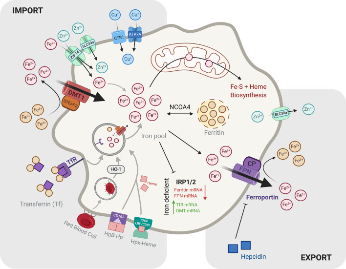
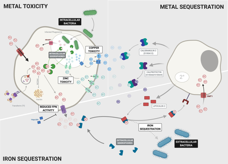
Nutritional immunity is the sequestration of bioavailable trace metals such as iron, zinc and copper by the host to limit pathogenicity by invading microorganisms. As one of the most conserved activities of the innate immune system, limiting the availability of free trace metals by cells of the immune system serves not only to conceal these vital nutrients from invading bacteria but also operates to tightly regulate host immune cell responses and function. In the setting of chronic lung disease, the regulation of trace metals by the host is often disrupted, leading to the altered availability of these nutrients to commensal and invading opportunistic pathogenic microbes. Similarly, alterations in the uptake, secretion, turnover and redox activity of these vitally important metals has significant repercussions for immune cell function including the response to and resolution of infection. This review will discuss the intricate role of nutritional immunity in host immune cells of the lung and how changes in this fundamental process as a result of chronic lung disease may alter the airway microbiome, disease progression and the response to infection.
Overview
The claims and statistics of this paper haven’t been checked yet.
Run the check — free, no account needed. It reads the open-access text and checks the sources it cites.
Integrity signals
Registry
Cites
Statistics
Figures
Verification
Claims the paper makes
The claims and statistics of this paper haven’t been checked yet.
Run the check — free, no account needed. It reads the open-access text and checks the sources it cites.
Claims other papers make about this one
Each is checked against this paper’s own text: does it say what the citing paper says it does?
In the citing paper
“In order to fight this, the host takes advantage of the toxicity and necessity of food metals by creating variables that restrict metal availability. This causes infections to either starve or accumulate too much metal to intoxicate them, a mechanism known as “nutritional immunity” (Healy et al. 2021).”
From Nutritional Immunity in Wound Infection: Unveiling the Role of Dietary Elements in Host–Pathogen Interaction (2025) · cites this paper as Healy et al. 2021
In the citing paper
“Numerous proteic factors, such as the well‐known hepcidin, lactoferrin, siderocalin, metallothionein, and calprotectin, are involved in regulating metal homeostasis in the host.”
From Nutritional Immunity in Wound Infection: Unveiling the Role of Dietary Elements in Host–Pathogen Interaction (2025) · cites this paper as Healy et al. 2021
In the citing paper
“These proteins have the dual roles of protecting host cells, transporting minerals, and preventing pathogens from accessing minerals (Healy et al. 2021).”
From Nutritional Immunity in Wound Infection: Unveiling the Role of Dietary Elements in Host–Pathogen Interaction (2025) · cites this paper as Healy et al. 2021
Self-check
Percentages, sums and reported differences re-computed from the paper’s own numbers.
The self-check needs the paper’s full text.
Citations
Cites
Gut microbiome influences efficacy of PD-1-based immunotherapy against epithelial tumors
Routy, B., Le Chatelier, E., Derosa, L., Duong, C. P. M., Alou, M. T., Daillère, R., Fluckiger, A., Messaoudene, M., Rauber, C., Roberti, M. P., et al. (2018) · Science · doi:10.1126/science.aan3706 · Cited by 5,671
Vogelmeier, C. F., Criner, G. J., Martinez, F. J., Anzueto, A., Barnes, P. J., Bourbeau, J., Celli, B. R., Chen, R., Decramer, M., Fabbri, L. M., et al. (2017) · American Journal of Respiratory and Critical Care Medicine · doi:10.1164/rccm.201701-0218pp · Cited by 3,187
Andrews, S. C., Robinson, A. K., & Rodríguez-Quiñones, F. (2003) · FEMS Microbiology Reviews · doi:10.1016/s0168-6445(03)00055-x · Cited by 2,702
Nemeth, E., Rivera, S., Gabayan, V., Keller, C., Taudorf, S., Pedersen, B. K., & Ganz, T. (2004) · Journal of Clinical Investigation · doi:10.1172/jci200420945 · Cited by 2,650
Chakarov, S., Lim, H. Y., Tan, L., Lim, S. Y., See, P., Lum, J., Zhang, X.-M., Foo, S., Nakamizo, S., Duan, K., et al. (2019) · Science · doi:10.1126/science.aau0964 · Cited by 1,114
Sindrilaru, A., Peters, T., Wieschalka, S., Baican, C., Baican, A., Peter, H., Hainzl, A., Schatz, S., Qi, Y., Schlecht, A., et al. (2011) · Journal of Clinical Investigation · doi:10.1172/jci44490 · Cited by 1,100 · cited 1×
“However, in vitro studies of macrophages isolated from chronic venous leg ulcers showed excessive erythrophagocytosis and an ensuing high intracellular iron concentration to promote M1 polarization [67].”
BackgroundBone morphogenetic protein signaling by hemojuvelin regulates hepcidin expression
Babitt, J. L., Huang, F. W., Wrighting, D. M., Xia, Y., Sidis, Y., Samad, T. A., Campagna, J. A., Chung, R. T., Schneyer, A. L., Woolf, C. J., et al. (2006) · Nature Genetics · doi:10.1038/ng1777 · Cited by 1,026
Comparison of the Respiratory Microbiome in Healthy Nonsmokers and Smokers
Morris, A., Beck, J. M., Schloss, P. D., Campbell, T. B., Crothers, K., Curtis, J. L., Flores, S. C., Fontenot, A. P., Ghedin, E., Huang, L., et al. (2013) · American Journal of Respiratory and Critical Care Medicine · doi:10.1164/rccm.201210-1913oc · Cited by 822
Acute exacerbation of idiopathic pulmonary fibrosis: incidence, risk factors and outcome
Song, J. W., Hong, S.-B., Lim, C.-M., Koh, Y., & Kim, D. S. (2011) · European Respiratory Journal · doi:10.1183/09031936.00159709 · Cited by 783
Metzler, K. D., Goosmann, C., Lubojemska, A., Zychlinsky, A., & Papayannopoulos, V. (2014) · Cell Reports · doi:10.1016/j.celrep.2014.06.044 · Cited by 741 · cited 6×
“Neutrophils Nramp‐1 [63] Lipocalin‐2 [63] Myeloperoxidase [113] Ceruloplasmin [119] Lactoferrin [110] Calprotectin (S100A8/S100A9 heterodimer) [13] Calgranulin C (S100A12) [124] Calgranulin C (S100A12) [124] ATP7A [118] ATP7B [118] Ceruloplasmin [119]”
BackgroundInfluential
Research works printed but not linked: 507
- Cornelis P, Dingemans J. Pseudomonas aeruginosa adapts its iron uptake strategies in function of the type of infections. Front Cell Infect Microbiol. 2013;3:75. · DOI
- Bachman MA, et al. Klebsiella pneumoniae yersiniabactin promotes respiratory tract infection through evasion of lipocalin 2. Infect Immun. 2011;79(8):3309–16. · DOI
- Pidcock KA, et al. Iron acquisition by Haemophilus influenzae. Infect Immun. 1988;56(4):721–5. · DOI
- Phelan JJ, et al. Modulating Iron for Metabolic Support of TB Host Defense. Front Immunol. 2018; 9(2296). · DOI
- Smith DJ, et al. Targeting iron uptake to control Pseudomonas aeruginosa infections in cystic fibrosis. Eur Respir J. 2013;42(6):1723–36. · DOI
- Babu U, Failla ML. Respiratory burst and candidacidal activity of peritoneal macrophages are impaired in copper-deficient rats. J Nutr. 1990;120(12):1692–9. · DOI
- Macomber L, Imlay JA. The iron-sulfur clusters of dehydratases are primary intracellular targets of copper toxicity. Proc Natl Acad Sci U S A. 2009;106(20):8344–9. · DOI · Paper page
- Urban CF, et al. Neutrophil extracellular traps contain calprotectin, a cytosolic protein complex involved in host defense against Candida albicans. PLoS Pathog. 2009;5(10):e1000639. · DOI
- Guillin OM, et al. Selenium, selenoproteins and viral infection. Nutrients, 2019; 11(9). · DOI
- O’Neill LA, Kishton RJ, Rathmell J. A guide to immunometabolism for immunologists. Nat Rev Immunol. 2016;16(9):553–65. · DOI · Paper page
- O’Beirne SL, et al. Alveolar macrophage immunometabolism and lung function impairment in smoking and chronic obstructive pulmonary disease. Am J Respir Crit Care Med. 2020;201(6):735–9. · DOI
- Fricker M, Gibson PG. Macrophage dysfunction in the pathogenesis and treatment of asthma. Eur Respir J. 2017;50(3):1700196. · DOI
- Bruscia EM, Bonfield TL. Cystic fibrosis lung immunity: the role of the macrophage. J Innate Immun. 2016;8(6):550–63. · DOI
- Ardain A, Marakalala MJ, Leslie A. Tissue-resident innate immunity in the lung. Immunology. 2020;159(3):245–56. · DOI
- Guilliams M, et al. Alveolar macrophages develop from fetal monocytes that differentiate into long-lived cells in the first week of life via GM-CSF. J Exp Med. 2013;210(10):1977–92. · DOI
- van de Laar L, et al. Yolk sac macrophages, fetal liver, and adult monocytes can colonize an empty niche and develop into functional tissue-resident macrophages. Immunity. 2016;44(4):755–68. · DOI
- Snelgrove RJ, et al. A critical function for CD200 in lung immune homeostasis and the severity of influenza infection. Nat Immunol. 2008;9(9):1074–83. · DOI
- Byrne AJ, et al. Pulmonary macrophages: key players in the innate defence of the airways. Thorax. 2015;70(12):1189–96. · DOI
- Neupane AS, et al. Patrolling alveolar macrophages conceal bacteria from the immune system to maintain homeostasis. Cell. 2020;183(1):110-125.e11. · DOI
- Bourdonnay E, et al. Transcellular delivery of vesicular SOCS proteins from macrophages to epithelial cells blunts inflammatory signaling. J Exp Med. 2015;212(5):729–42. · DOI
- Levine AM, et al. GM-CSF–deficient mice are susceptible to pulmonary group B streptococcal infection. J Clin Investig. 1999;103(4):563–9. · DOI
- Gonzalez-Juarrero M, et al. Disruption of granulocyte macrophage-colony stimulating factor production in the lungs severely affects the ability of mice to control Mycobacterium tuberculosis infection. J Leukoc Biol. 2005;77(6):914–22. · DOI
- Schneider C, et al. Alveolar macrophages are essential for protection from respiratory failure and associated morbidity following influenza virus infection. PLoS Pathog. 2014;10(4):e1004053. · DOI
- Yu X, et al. The cytokine TGF-β promotes the development and homeostasis of alveolar macrophages. Immunity. 2017;47(5):903-912.e4. · DOI
- Puttur F, Gregory LG, Lloyd CM. Airway macrophages as the guardians of tissue repair in the lung. Immunol Cell Biol. 2019;97(3):246–57. · DOI
- Gibbings SL, et al. Three unique interstitial macrophages in the murine lung at steady state. Am J Respir Cell Mol Biol. 2017;57(1):66–76. · DOI
- Schyns J, et al. Non-classical tissue monocytes and two functionally distinct populations of interstitial macrophages populate the mouse lung. Nat Commun. 2019;10(1):3964. · DOI
- Janssen WJ, et al. Fas determines differential fates of resident and recruited macrophages during resolution of acute lung injury. Am J Respir Crit Care Med. 2011;184(5):547–60. · DOI
- Mould KJ, et al. Cell origin dictates programming of resident versus recruited macrophages during acute lung injury. Am J Respir Cell Mol Biol. 2017;57(3):294–306. · DOI
- Machiels B, et al. A gammaherpesvirus provides protection against allergic asthma by inducing the replacement of resident alveolar macrophages with regulatory monocytes. Nat Immunol. 2017;18(12):1310–20. · DOI
- Byrne AJ, et al. Dynamics of human monocytes and airway macrophages during healthy aging and after transplant. J Exp Med, 2020; 217(3). · DOI
- Guilliams M, et al. Establishment and maintenance of the macrophage niche. Immunity. 2020;52(3):434–51. · DOI · Paper page
- Winn NC, Volk KM, Hasty AH. Regulation of tissue iron homeostasis: the macrophage “ferrostat”. JCI Insight, 2020; 5(2). · DOI
- Roth-Walter F, et al. Linking iron-deficiency with allergy: role of molecular allergens and the microbiome. Metallomics. 2017;9(12):1676–92. · DOI
- Ghio AJ, et al. DMT1 expression is increased in the lungs of hypotransferrinemic mice. Am J Physiol-Lung Cell Mol Physiol. 2003;284(6):L938–44. · DOI
- Wygrecka M, et al. Shedding of low-density lipoprotein receptor–related protein-1 in acute respiratory distress syndrome. Am J Respir Crit Care Med. 2011;184(4):438–48. · DOI
- Beker Aydemir T, et al. Zinc transporter ZIP14 functions in hepatic zinc, iron and glucose homeostasis during the innate immune response (Endotoxemia). PLoS ONE. 2012;7(10):e48679. · DOI
- Pisu D, et al. Dual RNA-Seq of Mtb-infected macrophages in vivo reveals ontologically distinct host-pathogen interactions. Cell Rep. 2020;30(2):335-350.e4. · DOI
- Wesselius LJ, Flowers CH, Skikne BS. Alveolar macrophage content of isoferritins and transferrin: comparison of nonsmokers and smokers with and without chronic airflow obstruction. Am Rev Respir Dis. 1992;145(2_pt_1):311–6. · DOI
- Neves J, et al. Disruption of the hepcidin/ferroportin regulatory system causes pulmonary iron overload and restrictive lung disease. EBioMedicine. 2017;20:230–9. · DOI
- Lim D, et al. The hepcidin-ferroportin axis controls the iron content of Salmonella-containing vacuoles in macrophages. Nat Commun. 2018;9(1):2091. · DOI · Paper page
- Nguyen NB, et al. Hepcidin expression and iron transport in alveolar macrophages. Am J Physiol Lung Cell Mol Physiol. 2006;291(3):L417–25. · DOI · Paper page
- Perez E, et al. Hepcidin is essential for alveolar macrophage function and is disrupted by smoke in a murine chronic obstructive pulmonary disease model. J Immunol. 2020;205(9):2489–98. · DOI
- Brown BL, et al. Structure of the mitochondrial aminolevulinic acid synthase, a key heme biosynthetic enzyme. Structure. 2018;26(4):580–9. · DOI
- Knutson MD, et al. Iron release from macrophages after erythrophagocytosis is up-regulated by ferroportin 1 overexpression and down-regulated by hepcidin. Proc Natl Acad Sci USA. 2005;102(5):1324–8. · DOI · Paper page
- van den Heuvel MM, et al. Regulation of CD163 on human macrophages: cross-linking of CD163 induces signaling and activation. J Leukoc Biol. 1999;66(5):858–66. · DOI
- Simões RL, et al. Proinflammatory responses of heme in alveolar macrophages: repercussion in lung hemorrhagic episodes. Mediators Inflamm. 2013;2013:946878. · DOI
- Stocker R, et al. Bilirubin is an antioxidant of possible physiological importance. Science. 1987;235(4792):1043–6. · DOI · Paper page
- Vitali SH, et al. Heme oxygenase-1 dampens the macrophage sterile inflammasome response and regulates its components in the hypoxic lung. Am J Physiol Lung Cell Mol Physiol. 2020;318(1):L125-l134. · DOI
- Oberle S, et al. The antioxidant defense protein ferritin is a novel and specific target for pentaerithrityl tetranitrate in endothelial cells. Biochem Biophys Res Commun. 1999;261(1):28–34. · DOI
- Flo TH, et al. Lipocalin 2 mediates an innate immune response to bacterial infection by sequestrating iron. Nature. 2004;432(7019):917–21. · DOI · Paper page
- Agoro R, Mura C. Inflammation-induced up-regulation of hepcidin and down-regulation of ferroportin transcription are dependent on macrophage polarization. Blood Cells Mol Dis. 2016;61:16–25. · DOI
- Poss KD, Tonegawa S. Heme oxygenase 1 is required for mammalian iron reutilization. Proc Natl Acad Sci. 1997;94(20):10919–24. · DOI · Paper page
- Agoro R, et al. Cell iron status influences macrophage polarization. PLoS ONE. 2018;13(5):e0196921. · DOI
- Artyomov MN, Sergushichev A, Schilling JD. Integrating immunometabolism and macrophage diversity. Semin Immunol. 2016;28(5):417–24. · DOI
- Russell DG, Huang L, Vanderven BC. Immunometabolism at the interface between macrophages and pathogens. Nat Rev Immunol. 2019;19(5):291–304. · DOI
- Pain D, Dancis A. Roles of Fe–S proteins: from cofactor synthesis to iron homeostasis to protein synthesis. Curr Opin Genet Dev. 2016;38:45–51. · DOI
- Canal F, et al. Regulation of the cysteine desulfurase Nfs1 and the scaffold protein IscU in macrophages stimulated with interferon-γ and lipopolysaccharide. Arch Biochem Biophys. 2007;465(1):282–92. · DOI
- Drapier JC, Hibbs JB Jr. Differentiation of murine macrophages to express nonspecific cytotoxicity for tumor cells results in L-arginine-dependent inhibition of mitochondrial iron-sulfur enzymes in the macrophage effector cells. J Immunol. 1988;140(8):2829–38. · DOI
- Pereira M, et al. Acute iron deprivation reprograms human macrophage metabolism and reduces inflammation in vivo. Cell Rep. 2019;28(2):498-511.e5. · DOI
- Salvin SB, et al. The effect of dietary zinc and prothymosin alpha on cellular immune responses of RF/J mice. Clin Immunol Immunopathol. 1987;43(3):281–8. · DOI
- Nowak JE, et al. Prophylactic zinc supplementation reduces bacterial load and improves survival in a murine model of sepsis. Pediatr Crit Care Med. 2012;13(5):e323–9. · DOI
- Wirth JJ, Fraker PJ, Kierszenbaum F. Zinc requirement for macrophage function: effect of zinc deficiency on uptake and killing of a protozoan parasite. Immunology. 1989;68(1):114–9.
- Hamon R, et al. Zinc and zinc transporters in macrophages and their roles in efferocytosis in COPD. PLoS ONE. 2014;9(10):e110056. · DOI
- Gao H, et al. Metal transporter Slc39a10 regulates susceptibility to inflammatory stimuli by controlling macrophage survival. Proc Natl Acad Sci U S A. 2017;114(49):12940–5. · DOI
- Neyrolles O, Mintz E, Catty P. Zinc and copper toxicity in host defense against pathogens: Mycobacterium tuberculosis as a model example of an emerging paradigm. Front Cell Infect Microbiol. 2013;3:89. · DOI · Paper page
- Crocker A, et al. Interaction of nutrition and infection: effect of copper deficiency on resistance to Trypanosoma lewisi. J Natl Med Assoc. 1992;84(8):697–706.
- Wagner D, et al. Elemental analysis of Mycobacterium avium-, Mycobacterium tuberculosis-, and Mycobacterium smegmatis-containing phagosomes indicates pathogen-induced microenvironments within the host cell’s endosomal system. J Immunol. 2005;174(3):1491–500. · DOI
- Shen Q, et al. Macrophage activation by IFN-γ triggers restriction of phagosomal copper from intracellular pathogens. PLoS Pathog. 2018;14(11):e1007444. · DOI
- Jabado N, et al. Natural resistance to intracellular infections: natural resistance-associated macrophage protein 1 (Nramp1) functions as a pH-dependent manganese transporter at the phagosomal membrane. J Exp Med. 2000;192(9):1237–48. · DOI · Paper page
- Davidsson L, et al. Identification of transferrin as the major plasma carrier protein for manganese introduced orally or intravenously or after in vitro addition in the rat. J Nutr. 1989;119(10):1461–4. · DOI
- Lee BC, et al. Selenoprotein MsrB1 promotes anti-inflammatory cytokine gene expression in macrophages and controls immune response in vivo. Sci Rep. 2017;7(1):5119–5119. · DOI
- Nelson SM, Lei X, Prabhu KS. Selenium levels affect the IL-4-induced expression of alternative activation markers in murine macrophages. J Nutr. 2011;141(9):1754–61. · DOI
- Carlson BA, et al. Role of selenium-containing proteins in T-cell and macrophage function. Proc Nutr Soc. 2010;69(3):300–10. · DOI
- Naik SH, et al. Development of plasmacytoid and conventional dendritic cell subtypes from single precursor cells derived in vitro and in vivo. Nat Immunol. 2007;8(11):1217–26. · DOI
- Guilliams M, Lambrecht BN, Hammad H. Division of labor between lung dendritic cells and macrophages in the defense against pulmonary infections. Mucosal Immunol. 2013;6(3):464–73. · DOI
- Sichien D, et al. Development of conventional dendritic cells: from common bone marrow progenitors to multiple subsets in peripheral tissues. Mucosal Immunol. 2017;10(4):831–44. · DOI
- Hoffmann FM, et al. Distribution and Interaction of Murine Pulmonary Phagocytes in the Naive and Allergic Lung. Front Immunol. 2018. 9. · DOI
- Cella M, et al. Plasmacytoid monocytes migrate to inflamed lymph nodes and produce large amounts of type I interferon. Nat Med. 1999;5(8):919–23. · DOI
- Van Pottelberge GR, et al. Plasmacytoid dendritic cells in pulmonary lymphoid follicles of patients with COPD. Eur Respir J. 2010;36(4):781–91. · DOI
- De Heer HJ, et al. Essential role of lung plasmacytoid dendritic cells in preventing asthmatic reactions to harmless inhaled antigen. J Exp Med. 2004;200(1):89–98. · DOI
- Verna G, et al. Iron overload mimicking conditions skews bone marrow dendritic cells differentiation into MHCII(low)CD11c(+)CD11b(+)F4/80(+) cells. Int J Mol Sci. 2020; 21(4). · DOI
- Kramer JL, et al. Differentiation of functional dendritic cells and macrophages from human peripheral blood monocyte precursors is dependent on expression of p21 (WAF1/CIP1) and requires iron. Br J Haematol. 2002;117(3):727–34. · DOI
- Kitamura H, et al. Toll-like receptor-mediated regulation of zinc homeostasis influences dendritic cell function. Nat Immunol. 2006;7(9):971–7. · DOI
- Jenne CN, et al. Neutrophils recruited to sites of infection protect from virus challenge by releasing neutrophil extracellular traps. Cell Host Microbe. 2013;13(2):169–80. · DOI
- Kumawat K, et al. LAIR-1 limits neutrophilic airway inflammation. Front Immunol. 2019;10:842. · DOI
- Casulli J, et al. CD200R deletion promotes a neutrophil niche for Francisella tularensis and increases infectious burden and mortality. Nat Commun. 2019;10(1):1–9. · DOI
- Griffiths JS, et al. Differential susceptibility of Dectin-1 isoforms to functional inactivation by neutrophil and fungal proteases. FASEB J. 2018;32(6):3385–97. · DOI
- Giacalone VD, et al. Neutrophil adaptations upon recruitment to the lung: new concepts and implications for homeostasis and disease. Int J Mol Sci. 2020; 21(3). · DOI
- Kuebler W, et al. Contribution of selectins to leucocyte sequestration in pulmonary microvessels by intravital microscopy in rabbits. J Physiol. 1997;501(Pt 2):375. · DOI
- Devi S, et al. Neutrophil mobilization via plerixafor-mediated CXCR4 inhibition arises from lung demargination and blockade of neutrophil homing to the bone marrow. J Exp Med. 2013;210(11):2321–36. · DOI
- Francis N, et al. Lactoferrin inhibits neutrophil apoptosis via blockade of proximal apoptotic signaling events. Biochim Biophys Acta. 2011;1813(10):1822–6. · DOI
- Grigorieva DV, et al. Effects of recombinant human lactoferrin on calcium signaling and functional responses of human neutrophils. Arch Biochem Biophys. 2019;675:108122. · DOI
- Maxwell PH, et al. The tumour suppressor protein VHL targets hypoxia-inducible factors for oxygen-dependent proteolysis. Nature. 1999;399(6733):271–5. · DOI · Paper page
- Sadiku P, et al. Prolyl hydroxylase 2 inactivation enhances glycogen storage and promotes excessive neutrophilic responses. J Clin Investig. 2017;127(9):3407–20. · DOI
- Mecklenburgh KI, et al. Involvement of a ferroprotein sensor in hypoxia-mediated inhibition of neutrophil apoptosis. Blood. 2002;100(8):3008–16. · DOI
- Konrad FM, et al. Heme oxygenase-1 attenuates acute pulmonary inflammation by decreasing the release of segmented neutrophils from the bone marrow. Am J Physiol Lung Cell Mol Physiol. 2014;307(9):L707–17. · DOI
- Cichon I, et al. Reduced neutrophil extracellular trap (NET) formation during systemic inflammation in mice with Menkes disease and Wilson disease: copper requirement for NET release. Front Immunol. 2019;10:3021. · DOI
- Iwata T, et al. Ceruloplasmin induces polymorphonuclear leukocyte priming in localized aggressive periodontitis. J Periodontol. 2009;80(8):1300–6. · DOI
- Hasan R, Rink L, Haase H. Chelation of free Zn2+ impairs chemotaxis, phagocytosis, oxidative burst, degranulation, and cytokine production by neutrophil granulocytes. Biol Trace Elem Res. 2016;171(1):79–88. · DOI
- Hasan R, Rink L, Haase H. Zinc signals in neutrophil granulocytes are required for the formation of neutrophil extracellular traps. Innate Immun. 2013;19(3):253–64. · DOI
- Kehl-Fie TE, et al. Nutrient metal sequestration by calprotectin inhibits bacterial superoxide defense, enhancing neutrophil killing of Staphylococcus aureus. Cell Host Microbe. 2011;10(2):158–64. · DOI · Paper page
- Isaksen B, Fagerhol MK. Calprotectin inhibits matrix metalloproteinases by sequestration of zinc. Mol Pathol: MP. 2001;54(5):289–92. · DOI
- Moroz OV, et al. Both Ca 2+ and Zn 2+ are essential for S100A12 protein oligomerization and function. BMC Biochem. 2009;10(1):1–18. · DOI
- Köse SA, Nazıroğlu M. Selenium reduces oxidative stress and calcium entry through TRPV1 channels in the neutrophils of patients with polycystic ovary syndrome. Biol Trace Elem Res. 2014;158(2):136–42. · DOI
- Kashiwakura J, et al. Pivotal advance: IgE accelerates in vitro development of mast cells and modifies their phenotype. J Leukoc Biol. 2008;84(2):357–67. · DOI
- Kirshenbaum AS, et al. Demonstration of the origin of human mast cells from CD34+ bone marrow progenitor cells. J Immunol. 1991;146(5):1410–5. · DOI
- Tsai M, et al. The rat c-kit ligand, stem cell factor, induces the development of connective tissue-type and mucosal mast cells in vivo. Analysis by anatomical distribution, histochemistry, and protease phenotype. J Exp Med. 1991;174(1):125–31. · DOI
- Suto H, et al. Mast cell-associated tnf promotes dendritic cell migration. J Immunol. 2006;176(7):4102–12. · DOI
- Levi-Schaffer F, et al. Mouse bone marrow-derived mast cells cocultured with fibroblasts. Morphology and stimulation-induced release of histamine, leukotriene B4, leukotriene C4, and prostaglandin D2. J Immunol. 1987;139(10):3431–41. · DOI
- McCurdy JD, et al. Cutting edge: distinct toll-like receptor 2 activators selectively induce different classes of mediator production from human mast cells. J Immunol. 2003;170(4):1625–9. · DOI
- Weller CL, et al. Chemotactic action of prostaglandin E2 on mouse mast cells acting via the PGE2 receptor 3. Proc Natl Acad Sci. 2007;104(28):11712–7. · DOI
- Abonia JP, et al. Constitutive homing of mast cell progenitors to the intestine depends on autologous expression of the chemokine receptor CXCR2. Blood. 2005;105(11):4308–13. · DOI
- Dvorak AM. Ultrastructural analysis of human mast cells and basophils. Chem Immunol. 1995;61:1–33.
- Kirshenbaum AS, et al. IL-3-dependent growth of basophil-like cells and mastlike cells from human bone marrow. J Immunol. 1989;142(7):2424–9. · DOI
- Varricchi G, et al. Human mast cells and basophils-How are they similar how are they different? Immunol Rev. 2018;282(1):8–34. · DOI
- Schwartz C, Eberle JU, Voehringer D. Basophils in inflammation. Eur J Pharmacol. 2016;778:90–5. · DOI
- Brunner T, Heusser CH, Dahinden CA. Human peripheral blood basophils primed by interleukin 3 (IL-3) produce IL-4 in response to immunoglobulin E receptor stimulation. J Exp Med. 1993;177(3):605–11. · DOI
- Arinobu Y, et al. Developmental checkpoints of the basophil/mast cell lineages in adult murine hematopoiesis. Proc Natl Acad Sci. 2005;102(50):18105–10. · DOI
- Cohen M, et al. Lung single-cell signaling interaction map reveals basophil role in macrophage imprinting. Cell. 2018;175(4):1031-1044.e18. · DOI
- Lamkhioued B, et al. Increased expression of eotaxin in bronchoalveolar lavage and airways of asthmatics contributes to the chemotaxis of eosinophils to the site of inflammation. J Immunol. 1997;159(9):4593–601. · DOI
- Stevens WW, et al. Detection and quantitation of eosinophils in the murine respiratory tract by flow cytometry. J Immunol Methods. 2007;327(1–2):63–74. · DOI
- Mesnil C, et al. Lung-resident eosinophils represent a distinct regulatory eosinophil subset. J Clin Investig. 2016;126(9):3279–95. · DOI
- Yi S, et al. Eosinophil recruitment is dynamically regulated by interplay among lung dendritic cell subsets after allergen challenge. Nat Commun. 2018;9(1):3879. · DOI
- Drake MG, et al. Human and mouse eosinophils have antiviral activity against parainfluenza virus. Am J Respir Cell Mol Biol. 2016;55(3):387–94. · DOI
- Palma-Carlos AG, Palma-Carlos ML, Costa AC. “Minor” hemoglobinopathies: a risk factor for asthma. Eur Ann Allergy Clin Immunol. 2005;37(5):177–82.
- Wu Z, Holwill SD, Oliveira DB. Desferrioxamine modulates chemically induced T helper 2-mediated autoimmunity in the rat. Clin Exp Immunol. 2004;135(2):194–9. · DOI
- Maazi H, et al. Iron administration reduces airway hyperreactivity and eosinophilia in a mouse model of allergic asthma. Clin Exp Immunol. 2011;166(1):80–6. · DOI
- Arnhold J, Furtmüller PG, Obinger C, Redox properties of myeloperoxidase. 2003, Redox Rep. p. 179–186. · DOI · Paper page
- Ten RM, et al. Molecular cloning of the human eosinophil peroxidase. Evidence for the existence of a peroxidase multigene family. J Exp Med. 1989;169(5):1757–69. · DOI
- Voehringer D, van Rooijen N, Locksley RM. Eosinophils develop in distinct stages and are recruited to peripheral sites by alternatively activated macrophages. J Leukoc Biol. 2007;81(6):1434–44. · DOI · Paper page
- Sumbayev VV, et al. Involvement of hypoxia-inducible factor-1 (HiF-1α) in IgE-mediated primary human basophil responses. Eur J Immunol. 2009;39(12):3511–9. · DOI
- Ünal M, et al. Serum levels of antioxidant vitamins, copper, zinc and magnesium in children with chronic rhinosinusitis. J Trace Elem Med Biol. 2004;18(2):189–92. · DOI
- Guo C-H, et al. Role of certain trace minerals in oxidative stress, inflammation, CD4/CD8 lymphocyte ratios and lung function in asthmatic patients. Ann Clin Biochem. 2011;48(4):344–51. · DOI
- Ariaee N, et al. Trace elements status in sera of patients with allergic asthma. Rep Biochem Mol Biol. 2016;5(1):20–5.
- Ho LH, et al. Labile zinc and zinc transporter ZnT4 in mast cell granules: role in regulation of caspase activation and NF-kappaB translocation. J Immunol. 2004;172(12):7750–60. · DOI
- Ugajin T, et al. Zinc-binding metallothioneins are key modulators of IL-4 production by basophils. Mol Immunol. 2015;66(2):180–8. · DOI
- Richter M, et al. Zinc status modulates bronchopulmonary eosinophil infiltration in a murine model of allergic inflammation. Chest. 2003;123(3 Suppl):446s. · DOI
- Lu H, et al. Zinc suppressed the airway inflammation in asthmatic rats: effects of zinc on generation of eotaxin, MCP-1, IL-8, IL-4, and IFN-γ. Biol Trace Elem Res. 2012;150(1–3):314–21. · DOI
- Öhrvik H, et al. Ctr2 regulates mast cell maturation by affecting the storage and expression of tryptase and proteoglycans. J Immunol. 2015;195(8):3654–64. · DOI
- Simoni Y, et al. Human innate lymphoid cell subsets possess tissue-type based heterogeneity in phenotype and frequency. Immunity. 2018;48(5):1060. · DOI
- Yudanin NA, et al. Spatial and temporal mapping of human innate lymphoid cells reveals elements of tissue specificity. Immunity. 2019;50(2):505–19. · DOI
- Kärre K, et al. Selective rejection of H-2-deficient lymphoma variants suggests alternative immune defence strategy. Nature. 1986;319(6055):675–8. · DOI
- Hart OM, et al. TLR7/8-mediated activation of human NK cells results in accessory cell-dependent IFN-γ production. J Immunol. 2005;175(3):1636–42. · DOI
- Kumar P, et al. IL-22 from conventional NK cells is epithelial regenerative and inflammation protective during influenza infection. Mucosal Immunol. 2013;6(1):69–82. · DOI
- Marquardt N, et al. Human lung natural killer cells are predominantly comprised of highly differentiated hypofunctional CD69− CD56dim cells. J Allergy Clin Immunol. 2017;139(4):1321–30. · DOI
- Fehniger TA, et al. Differential cytokine and chemokine gene expression by human NK cells following activation with IL-18 or IL-15 in combination with IL-12: implications for the innate immune response. J Immunol. 1999;162(8):4511–20. · DOI
- Yamamoto Y, et al. Lung-resident natural killer cells control pulmonary tumor growth in mice. Cancer Sci. 2018;109(9):2670–6. · DOI
- Marquardt N, et al. Unique transcriptional and protein-expression signature in human lung tissue-resident NK cells. Nat Commun. 2019;10(1):3841. · DOI
- Michel T, et al. Mouse lung and spleen natural killer cells have phenotypic and functional differences, in part influenced by macrophages. PLoS ONE. 2012;7(12):e51230. · DOI
- Cong J, Wei H. Natural killer cells in the lungs. Front Immunol. 2019;10:1416. · DOI
- Cooper GE, et al. Human CD49a(+) lung natural killer cell cytotoxicity in response to influenza a virus. Front Immunol. 2018;9:1671. · DOI
- Orange JS. Human natural killer cell deficiencies and susceptibility to infection. Microbes Infect. 2002;4(15):1545–58. · DOI
- Augustin A, Kubo RT, Sim GK. Resident pulmonary lymphocytes expressing the gamma/delta T-cell receptor. Nature. 1989;340(6230):239–41. · DOI
- Cheng M, Hu S. Lung-resident gammadelta T cells and their roles in lung diseases. Immunology. 2017;151(4):375–84. · DOI · Paper page
- Barlow JL, McKenzie ANJ. Innate lymphoid cells of the lung. Annu Rev Physiol. 2019;81:429–52. · DOI
- Trottein F, Paget C. Natural killer T cells and mucosal-associated invariant t cells in lung infections. Front Immunol. 2018;9:1750. · DOI
- Metelitsa LS, et al. Human NKT cells mediate antitumor cytotoxicity directly by recognizing target cell CD1d with bound ligand or indirectly by producing IL-2 to activate NK cells. J Immunol. 2001;167(6):3114–22. · DOI
- Lee YJ, et al. Tissue-specific distribution of iNKT cells impacts their cytokine response. Immunity. 2015;43(3):566–78. · DOI
- Thanabalasuriar A, et al. iNKT cell emigration out of the lung vasculature requires neutrophils and monocyte-derived dendritic cells in inflammation. Cell Rep. 2016;16(12):3260–72. · DOI
- Gold MC, et al. Human mucosal associated invariant T cells detect bacterially infected cells. PLoS Biol. 2010;8(6):e1000407. · DOI
- Marçais A, et al. The metabolic checkpoint kinase mTOR is essential for IL-15 signaling during the development and activation of NK cells. Nat Immunol. 2014;15(8):749–57. · DOI
- Keating SE, et al. Metabolic reprogramming supports IFN-γ production by CD56bright NK Cells. J Immunol. 2016;196(6):2552–60. · DOI
- Damiens E, et al. Effects of human lactoferrin on NK cell cytotoxicity against haematopoietic and epithelial tumour cells. Biochim Biophys Acta. 1998;1402(3):277–87. · DOI
- Hua Y, et al. Iron overload may promote alteration of NK cells and hematopoietic stem/progenitor cells by JNK and P38 pathway in myelodysplastic syndromes. Int J Hematol. 2017;106(2):248–57. · DOI
- Sottile R, et al. Iron and ferritin modulate MHC class I expression and NK cell recognition. Front Immunol. 2019;10:224. · DOI
- Kumar S, et al. Zinc-induced polymerization of killer-cell Ig-like receptor into filaments promotes its inhibitory function at cytotoxic immunological synapses. Mol Cell. 2016;62(1):21–33. · DOI
- Muzzioli M, et al. Zinc improves the development of human CD34+ cell progenitors towards NK cells and increases the expression of GATA-3 transcription factor in young and old ages. Biogerontology. 2009;10(5):593–604. · DOI
- Wessels I, Maywald M, Rink L. Zinc as a gatekeeper of immune function. Nutrients, 2017; 9(12). · DOI
- Mace EM, et al. A dual role for talin in NK cell cytotoxicity: activation of LFA-1-mediated cell adhesion and polarization of NK cells. J Immunol. 2009;182(2):948–56. · DOI
- Lv M, et al. Manganese is critical for antitumor immune responses via cGAS-STING and improves the efficacy of clinical immunotherapy. Cell Res. 2020;30(11):966–79. · DOI · Paper page
- Kiremidjian-Schumacher L, et al. Supplementation with selenium augments the functions of natural killer and lymphokine-activated killer cells. Biol Trace Elem Res. 1996;52(3):227–39. · DOI
- Brekelmans P, et al. Inhibition of proliferation and differentiation during early T cell development by anti-transferrin receptor antibody. Eur J Immunol. 1994;24(11):2896–902. · DOI
- Mincheva-Nilsson L, Hammarstrom S, Hammarstrom ML. Activated human gamma delta T lymphocytes express functional lactoferrin receptors. Scand J Immunol. 1997;46(6):609–18. · DOI
- Vargas Buonfiglio LG, et al. Airway surface liquid from smokers promotes bacterial growth and biofilm formation via iron-lactoferrin imbalance. Respir Res. 2018;19(1):42. · DOI
- Reuben A, et al. The hemochromatosis protein HFE 20 years later: an emerging role in antigen presentation and in the immune system. Immun Inflamm Dis. 2017;5(3):218–32. · DOI
- French JD, et al. Gammadelta T lymphocyte homeostasis is negatively regulated by beta2-microglobulin. J Immunol. 2009;182(4):1892–900. · DOI
- de Sousa M, et al. Iron overload in β2-microglobulin-deficient mice. Immunol Lett. 1994;39(2):105–11. · DOI
- Maia ML, et al. Invariant natural killer T cells are reduced in hereditary hemochromatosis patients. J Clin Immunol. 2015;35(1):68–74. · DOI
- Huang H, et al. Acute invariant NKT cell activation triggers an immune response that drives prominent changes in iron homeostasis. Sci Rep. 2020;10(1):21026. · DOI
- Hwang JY, Randall TD, Silva-Sanchez A. Inducible bronchus-associated lymphoid tissue: taming inflammation in the lung. Front Immunol. 2016;7:258. · DOI
- Hogg JC, et al. The nature of small-airway obstruction in chronic obstructive pulmonary disease. N Engl J Med. 2004;350(26):2645–53. · DOI
- Belz GT, et al. Tissue-resident lymphocytes: weaponized sentinels at barrier surfaces. F1000Res, 2020; 9. · DOI
- Niedzielska M, et al. Differential gene expression in human tissue resident regulatory T cells from lung, colon, and blood. Oncotarget. 2018;9(90):36166–84. · DOI
- Lu C, et al. Memory regulatory T cells home to the lung and control influenza A virus infection. Immunol Cell Biol. 2019;97(9):774–86. · DOI
- Ichikawa T, et al. CD103(hi) Treg cells constrain lung fibrosis induced by CD103(lo) tissue-resident pathogenic CD4 T cells. Nat Immunol. 2019;20(11):1469–80. · DOI
- Weiss G. Modification of iron regulation by the inflammatory response. Best Pract Res Clin Haematol. 2005;18(2):183–201. · DOI · Paper page
- Brock JH. The effect of iron and transferrin on the response of serum-free cultures of mouse lymphocytes to concanavalin A and lipopolysaccharide. Immunology. 1981;43(2):387–92.
- Mateos F, Brock JH, Perez-Arellano JL. Iron metabolism in the lower respiratory tract. Thorax. 1998;53(7):594–600. · DOI · Paper page
- Stoffel NU, et al. Iron deficiency anemia at time of vaccination predicts decreased vaccine response and iron supplementation at time of vaccination increases humoral vaccine response: a birth cohort study and a randomized trial follow-up study in Kenyan infants. Front Immunol. 2020;11:1313. · DOI · Paper page
- Frost JN, et al. Hepcidin-mediated hypoferremia disrupts immune responses to vaccination and infection. Med. 2021;2(2):164-179.e12. · DOI
- Jabara HH, et al. A missense mutation in TFRC, encoding transferrin receptor 1, causes combined immunodeficiency. Nat Genet. 2016;48(1):74–8. · DOI · Paper page
- Ehrlich R, Lemonnier FA. HFE–a novel nonclassical class I molecule that is involved in iron metabolism. Immunity. 2000;13(5):585–8. · DOI
- Roy M, et al. Supplementation with selenium and human immune cell functions. I. Effect on lymphocyte proliferation and interleukin 2 receptor expression. Biol Trace Elem Res. 1994;41(1–2):103–14. · DOI
- Elmadfa I, Meyer AL. The role of the status of selected micronutrients in shaping the immune function. Endocr Metab Immune Disord Drug Targets. 2019;19(8):1100–15. · DOI
- Budden KF, et al. Functional effects of the microbiota in chronic respiratory disease. Lancet Respir Med. 2019;7(10):907–20. · DOI
- Dickson RP, et al. Changes in the lung microbiome following lung transplantation include the emergence of two distinct Pseudomonas species with distinct clinical associations. PLoS ONE. 2014;9(5):e97214. · DOI
- Andrei A, et al. Cu Homeostasis in Bacteria: The Ins and Outs. Membranes (Basel), 2020; 10(9). · DOI · Paper page
- Dostal A, et al. Low iron availability in continuous in vitro colonic fermentations induces strong dysbiosis of the child gut microbial consortium and a decrease in main metabolites. FEMS Microbiol Ecol. 2013;83(1):161–75. · DOI · Paper page
- Tompkins GR, et al. The effects of dietary ferric iron and iron deprivation on the bacterial composition of the mouse intestine. Curr Microbiol. 2001;43(1):38–42. · DOI
200 of 507 shown
Cited by
Sorted by citations
Mineral Micronutrients in Asthma
Zajac, D. (2021) · Nutrients · doi:10.3390/nu13114001 · Cited by 33
Chen, C., Hu, X., He, D., He, X., & Shen, L. (2025) · Food Science & Nutrition · doi:10.1002/fsn3.70854 · Cited by 6
Metadata
- Authors
- Healy, C., Munoz-Wolf, N., Strydom, J., Faherty, L., Williams, N. C., Kenny, S., Donnelly, S. C., & Cloonan, S. M.
- ORCID
- Cloonan, Suzanne M.
- Venue
- Respiratory Research
- Published
- 2021-04-29
- Volume
- 22
- Issue
- 1
- ISSN
- 1465-993X
- PMC
- PMC8082489
- Cited by
- 78
- References
- 55
- Language
- en
- Funders
- Science Foundation Ireland (FRL4862</)
- Health Research Board (EIA-2019-004)
About these checks
Registry, retraction, Cites and statistics results are computed, not judged, with one exception: a printed reference that no index lists is read once by a model before it counts as a citation error. Claims are extracted by a model and kept only when their quote appears verbatim in the text; each cited source is then checked, with a model’s help, for whether it says what the claim relies on. Those verdicts can be wrong — read the claim’s quote and the cited source before relying on one.
Statistics and arithmetic are recomputed, not judged.
Confidence bands are shown only for fields calibrated on our annotated test set. None is calibrated yet, so no band is shown.
What counts as a claim ›How we measure confidence ›How we check this ›
Are you an author, or do you see a problem with this page? Request a review
How to cite
Cite CoChat for the checks on this page, and the paper for its own findings.
- This page
- CoChat. (2026). Citation check: Nutritional immunity: the impact of metals on lung immune cells and the airway microbiome during chronic respiratory disease. https://app.cochat.ai/papers/10.1186/s12931-021-01722-y
- The paper
- Healy, C., Munoz-Wolf, N., Strydom, J., Faherty, L., Williams, N. C., Kenny, S., Donnelly, S. C., & Cloonan, S. M. (2021). Nutritional immunity: the impact of metals on lung immune cells and the airway microbiome during chronic respiratory disease. Respiratory Research. https://doi.org/10.1186/s12931-021-01722-y
