Cited by 69

The DOI matches its Crossref record; 2 of the 2 references it cites were found in a registry or an index.

Exosome-sheathed porous silica nanoparticle-mediated co-delivery of 3,3′-diindolylmethane and doxorubicin attenuates cancer stem cell-driven EMT in triple negative breast cancer

Sarkar, Rupali, Biswas, Souradeep, Ghosh, Rituparna, Samanta, Priya, Pakhira, Shampa, Mondal, Mrinmoyee, Dutta Gupta, Yashaswi, Bhandary, Suman, Saha, Prosenjit, Bhowmik, Arijit, Hajra, Subhadip·2024·Journal of Nanobiotechnology

Abstract

Background: Therapeutic management of locally advanced and metastatic triple negative breast cancer (TNBC) is often limited due to resistance to conventional chemotherapy. Metastasis is responsible for more than 90% of breast cancer-associated mortality; therefore, the clinical need to prevent or target metastasis is immense. The epithelial to mesenchymal transition (EMT) of cancer stem cells (CSCs) is a crucial determinant in metastasis. Doxorubicin (DOX) is the frequently used chemotherapeutic drug against TNBC that may increase the risk of metastasis in patients. After cancer treatment, CSCs with the EMT characteristic persist, which contributes to advanced malignancy and cancer recurrence. The latest developments in nanotechnology for medicinal applications have raised the possibility of using nanomedicines to target these CSCs. Hence, we present a novel approach of combinatorial treatment of DOX with dietary indole 3,3′-diindolylmethane (DIM) which is an intriguing field of research that may target CSC mediated EMT induction in TNBC. For efficient delivery of both the compounds to the tumor niche, advance method of drug delivery based on exosomes sheathed with mesoporous silica nanoparticles may provide an attractive strategy. Results: DOX, according to our findings, was able to induce EMT in CSCs, making the breast cancer cells more aggressive and metastatic. In CSCs produced from spheres of MDAMB-231 and 4T1, overexpression of N-cadherin, Snail, Slug, and Vimentin as well as downregulation of E-cadherin by DOX treatment not only demonstrated EMT induction but also underscored the pressing need for a novel chemotherapeutic combination to counteract this detrimental effect of DOX. To reach this goal, DIM was combined with DOX and delivered to the CSCs concomitantly by loading them in mesoporous silica nanoparticles encapsulated in exosomes (e-DDMSNP). These exosomes improved the specificity, stability and better homing ability of DIM and DOX in the in vitro and in vivo CSC niche. Furthermore, after treating the CSC-enriched TNBC cell population with e-DDMSNP, a notable decrease in DOX mediated EMT induction was observed. Conclusion: Our research seeks to propose a new notion for treating TNBC by introducing this unique exosomal nano-preparation against CSC induced EMT. Graphical Abstract:

Overview

The claims and statistics of this paper haven’t been checked yet.

Run the check — free, no account needed. It reads the open-access text and checks the sources it cites.

A few free checks a day · papers already verified open instantly · results are public

Integrity signals

Registry

Crossref · 10.1186/s12951-024-02518-0

Cites

2 of 2 references verified0 citation errors · 0 not checkable

Statistics

Figures

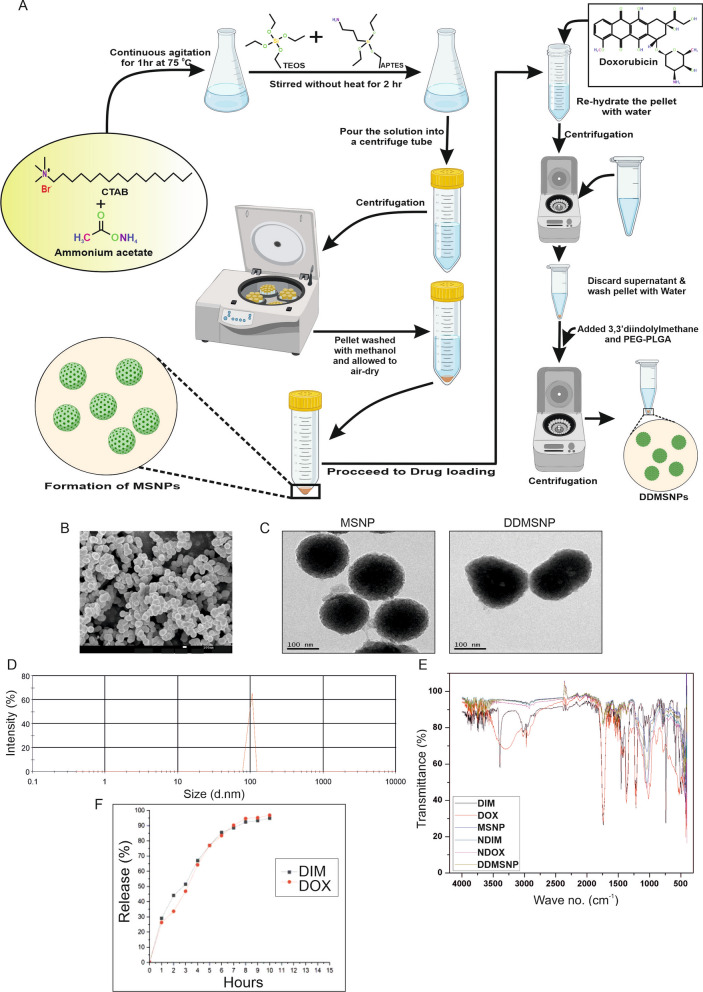
Fig. 1 Synthesis and characterization of MSNP and DDMSNP. A Modified Stӧber synthesis protocol for the preparation of MSNP and DDMSNP. B SEM image shows spherical conformations and ~ 100 nm size of synthesized MSNPs. C TEM image represents the morphological analysis of the empty MSNP as well as the loading of DIM and DOX within the MSNP. D DLS analysis shows an average size of DDMSNP to be 80–150 nm. E FTIR analysis shows comparative spectra of DIM, DOX, MSNP, NDIM, NDOX and DDMSNP to confirm successful encapsulation of DIM and DOX in the nanoparticles. F Drug release study of DDMSNP shows a prolonged release of DIM and DOX from the nanoparticle at physiological pH 7.4
Fig. 1 Synthesis and characterization of MSNP and DDMSNP. A Modified Stӧber synthesis protocol for the preparation of MSNP and DDMSNP. B SEM image shows spherical conformations and ~ 100 nm size of synthesized MSNPs. C TEM image represents the morphological analysis of the empty MSNP as well as the loading of DIM and DOX within the MSNP. D DLS analysis shows an average size of DDMSNP to be 80–150 nm. E FTIR analysis shows comparative spectra of DIM, DOX, MSNP, NDIM, NDOX and DDMSNP to confirm successful encapsulation of DIM and DOX in the nanoparticles. F Drug release study of DDMSNP shows a prolonged release of DIM and DOX from the nanoparticle at physiological pH 7.4CC BY · PubMed Central
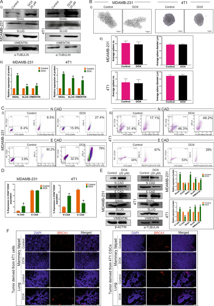
Fig. 2 Effect of DOX on TNBC cells and CSCs derived from MDAMB-231 and 4T1 cell lines. A Overexpression of Snail, Slug and Vimentin by DOX treatment. (i) Figure shows expression of Snail, Slug and Vimentin in control and 20 µM DOX treated groups of MDAMB-231 and 4T1 cells by western blot analysis. (ii) Graphical illustration denotes mean ± SD, n = 3. * indicates P ≤ 0.0002. B (i) Figure represents the sizes of CESs derived from MDAMB-231 and 4T1 cell line after DOX treatment compared to the control. (ii) Graphical representation of diameter and number of CESs of control and DOX treated groups shows no significant differences. C (i, ii) FACS analysis evaluates the expressions of N-cadherin and E-cadherin in CD44 + /CD24-/low CSC population in both cell line derived spheres of both control and 4 h DOX treated groups. D Bar diagram represents significant difference between N-cadherin expression of control and DOX treated groups, * indicates (P < 0.0001) and nonsignificant difference between E-cadherin expression of the same groups in MDAMB-231 derived CESs (left panel). The right panel denotes significant differences between N-cadherin and E-cadherin expressions of 4T1 derived CESs, * indicates (P < 0.0001). E Western blot data represents expressions of N-cadherin, E-cadherin, Snail, Slug and Vimentin in DOX treated CESs derived from MDAMB-231 and 4T1 cells. Graphical representation shows the significant differences between the expressions of these proteins in control and DOX treated groups, * indicates (P < 0.01). F Immunofluorescence study assesses BRCA1 (red) expression in mammary fatpad and metastatic lung in DOX treated and untreated groups of 4T1 cells-derived tumor-bearing BALB/c mice (n = 6 in each group) (left panel) and 4T1 CSCs- derived tumor-bearing BALB/c mice (n = 6 in each group) (right panel)
Fig. 2 Effect of DOX on TNBC cells and CSCs derived from MDAMB-231 and 4T1 cell lines. A Overexpression of Snail, Slug and Vimentin by DOX treatment. (i) Figure shows expression of Snail, Slug and Vimentin in control and 20 µM DOX treated groups of MDAMB-231 and 4T1 cells by western blot analysis. (ii) Graphical illustration denotes mean ± SD, n = 3. * indicates P ≤ 0.0002. B (i) Figure represents the sizes of CESs derived from MDAMB-231 and 4T1 cell line after DOX treatment compared to the control. (ii) Graphical representation of diameter and number of CESs of control and DOX treated groups shows no significant differences. C (i, ii) FACS analysis evaluates the expressions of N-cadherin and E-cadherin in CD44 + /CD24-/low CSC population in both cell line derived spheres of both control and 4 h DOX treated groups. D Bar diagram represents significant difference between N-cadherin expression of control and DOX treated groups, * indicates (P < 0.0001) and nonsignificant difference between E-cadherin expression of the same groups in MDAMB-231 derived CESs (left panel). The right panel denotes significant differences between N-cadherin and E-cadherin expressions of 4T1 derived CESs, * indicates (P < 0.0001). E Western blot data represents expressions of N-cadherin, E-cadherin, Snail, Slug and Vimentin in DOX treated CESs derived from MDAMB-231 and 4T1 cells. Graphical representation shows the significant differences between the expressions of these proteins in control and DOX treated groups, * indicates (P < 0.01). F Immunofluorescence study assesses BRCA1 (red) expression in mammary fatpad and metastatic lung in DOX treated and untreated groups of 4T1 cells-derived tumor-bearing BALB/c mice (n = 6 in each group) (left panel) and 4T1 CSCs- derived tumor-bearing BALB/c mice (n = 6 in each group) (right panel)CC BY · PubMed Central
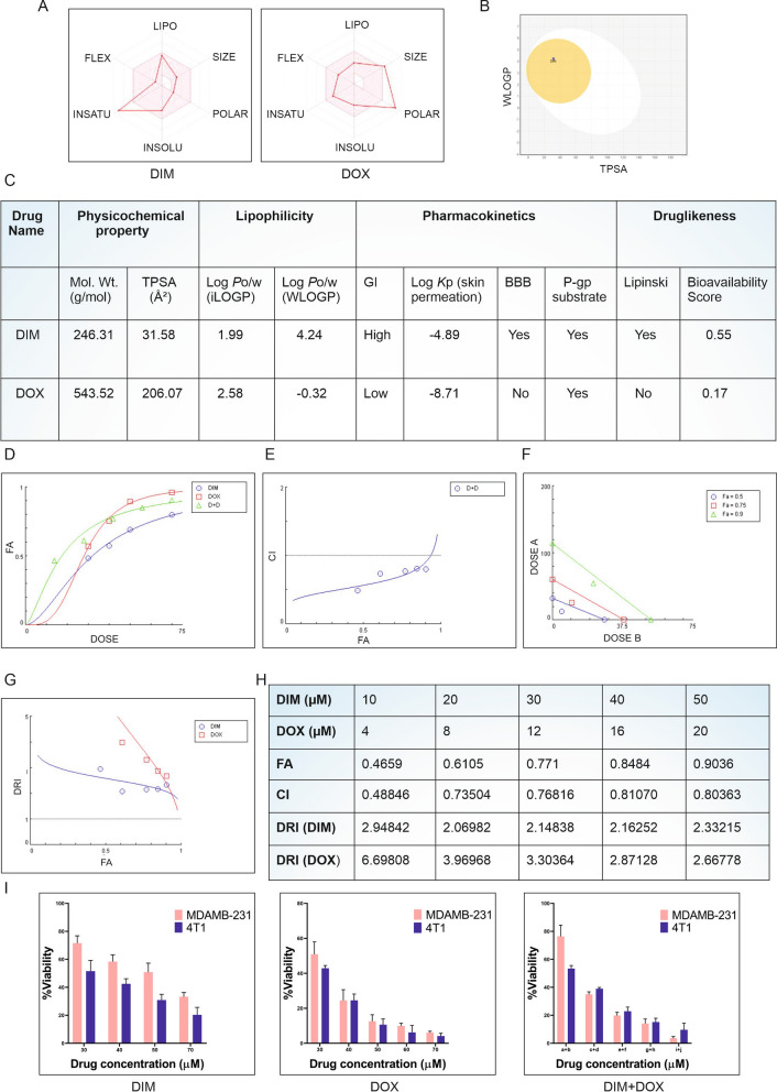
Fig. 3 Synergistic effect of DIM and DOX. A Bioavailability radar model, B boiled-egg model and C SwissADME properties of native DIM and DOX is depicted in the figure. CompuSyn analysis of DIM and DOX represents. D Dose effect plot of DIM, DOX and D + D (DIM + DOX), E plots with CI values of < 1 indicating synergism between D + D (DIM + DOX), F isobologram representing effective doses required for inhibition at 50% (Fa 0.5), 75% (Fa 0.75) and 90% (Fa 0.9) for each individual drug. Synergism is demonstrated by the dose pair plotted as a point (symbol) below their respective Fa isobole or line. G DRI of the drug combination of DIM and DOX is presented and DRI value > 1 indicates favourable drug combination. H All data are the representation of three independent experimental repeats. Fa, fraction affected; CI, combination index; DRI, dose reduction index. I MTT data demonstrates individual and the combinatorial effects of DIM and DOX in both the MDAMB-231 and 4T1 cell lines after 24 h of treatment. In this figure, a + b = DIM 10 µM + DOX 4 µM, c + d = DIM 20 µM + DOX 8 µM, e + f = DIM 30 µM + DOX 12 µM, g + h = DIM 40 µM + DOX 16 µM and i + j = DIM 50 µM + DOX 20 µM
Fig. 3 Synergistic effect of DIM and DOX. A Bioavailability radar model, B boiled-egg model and C SwissADME properties of native DIM and DOX is depicted in the figure. CompuSyn analysis of DIM and DOX represents. D Dose effect plot of DIM, DOX and D + D (DIM + DOX), E plots with CI values of < 1 indicating synergism between D + D (DIM + DOX), F isobologram representing effective doses required for inhibition at 50% (Fa 0.5), 75% (Fa 0.75) and 90% (Fa 0.9) for each individual drug. Synergism is demonstrated by the dose pair plotted as a point (symbol) below their respective Fa isobole or line. G DRI of the drug combination of DIM and DOX is presented and DRI value > 1 indicates favourable drug combination. H All data are the representation of three independent experimental repeats. Fa, fraction affected; CI, combination index; DRI, dose reduction index. I MTT data demonstrates individual and the combinatorial effects of DIM and DOX in both the MDAMB-231 and 4T1 cell lines after 24 h of treatment. In this figure, a + b = DIM 10 µM + DOX 4 µM, c + d = DIM 20 µM + DOX 8 µM, e + f = DIM 30 µM + DOX 12 µM, g + h = DIM 40 µM + DOX 16 µM and i + j = DIM 50 µM + DOX 20 µMCC BY · PubMed Central
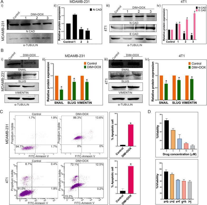
Fig. 4 Effect of DIM and DOX combination on EMT induction or apoptosis of TNBC. A (i) Expression of N-cadherin upon combinatorial treatment compared with the control. Here, “1” = DIM 10 µM + DOX 4 µM, “2” = DIM 21 µM + DOX 8 µM and “3” = DIM 30 µM + DOX 12 µM. (ii) Graphical representation shows significant change of N-cadherin expression in DIM and DOX treated MDAMB-231 cells compared to the control, * denotes P < 0.05. (iii) Immunoblotting shows the expressions of N-cadherin and E-cadherin. α-Tubulin is considered as the loading control. (iv) Bar graphs depict significant differences of N-cadherin and E-cadherin expressions between control and other groups in 4T1 cells (P < 0.001). Here, “1” = DIM 10 µM + DOX 4 µM, “2” = DIM 17 µM + DOX 7 µM and “3” = DIM 30 µM + DOX 12 µM. B (i, iii) Blots shown are the representative of expression of Snail, Slug and Vimentin in DOX treated MDAMB-231 and 4T1 cells compared to the control. α-Tubulin is used as a loading control. (ii, iv) Graphical representation illustrates significant changes in the expressions of Snail, Slug and Vimentin compared to the control (P < 0.002). C FACS analysis evaluates apoptotic cell population after 48 h of treatment with DIM and DOX in combination to MDAMB-231 and 4T1 cells. Left most panel shows control plot of untreated cells. This experiment is represented graphically in the rightmost panel (P < 0.0001). D Figure depicts the percentage of viable cells after 24 h of treatment with DOX or DIM and DOX in combination to MCF 10A cells. Here, a + b = DIM 30 µM + DOX 4 µM, c + d = DIM 40 µM + DOX 8 µM, e + f = DIM 50 µM + DOX 12 µM, g + h = DIM 60 µM + DOX 16 µM and i + j = DIM 70 µM + DOX 20 µM
Fig. 4 Effect of DIM and DOX combination on EMT induction or apoptosis of TNBC. A (i) Expression of N-cadherin upon combinatorial treatment compared with the control. Here, “1” = DIM 10 µM + DOX 4 µM, “2” = DIM 21 µM + DOX 8 µM and “3” = DIM 30 µM + DOX 12 µM. (ii) Graphical representation shows significant change of N-cadherin expression in DIM and DOX treated MDAMB-231 cells compared to the control, * denotes P < 0.05. (iii) Immunoblotting shows the expressions of N-cadherin and E-cadherin. α-Tubulin is considered as the loading control. (iv) Bar graphs depict significant differences of N-cadherin and E-cadherin expressions between control and other groups in 4T1 cells (P < 0.001). Here, “1” = DIM 10 µM + DOX 4 µM, “2” = DIM 17 µM + DOX 7 µM and “3” = DIM 30 µM + DOX 12 µM. B (i, iii) Blots shown are the representative of expression of Snail, Slug and Vimentin in DOX treated MDAMB-231 and 4T1 cells compared to the control. α-Tubulin is used as a loading control. (ii, iv) Graphical representation illustrates significant changes in the expressions of Snail, Slug and Vimentin compared to the control (P < 0.002). C FACS analysis evaluates apoptotic cell population after 48 h of treatment with DIM and DOX in combination to MDAMB-231 and 4T1 cells. Left most panel shows control plot of untreated cells. This experiment is represented graphically in the rightmost panel (P < 0.0001). D Figure depicts the percentage of viable cells after 24 h of treatment with DOX or DIM and DOX in combination to MCF 10A cells. Here, a + b = DIM 30 µM + DOX 4 µM, c + d = DIM 40 µM + DOX 8 µM, e + f = DIM 50 µM + DOX 12 µM, g + h = DIM 60 µM + DOX 16 µM and i + j = DIM 70 µM + DOX 20 µMCC BY · PubMed Central
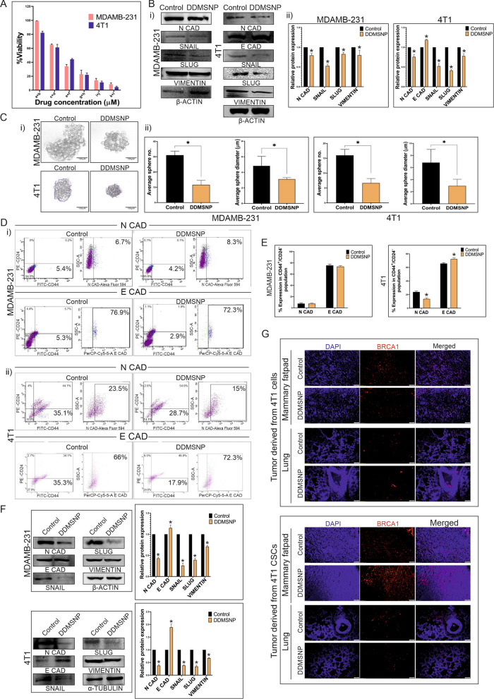
Fig. 5 Effect of DDMSNP on TNBC cells and CSCs involved in EMT. A Bar diagram represents MTT data showing the percentages of viable cells after 24 h of DDMSNP treatment encapsulating a + b = DIM 0.13 µM + DOX 0.05 µM, c + d = DIM 0.25 µM + DOX 0.1 µM, e + f = DIM 0.5 µM + DOX 0.2 µM, g + h = DIM 1 µM + DOX 0.4 µM, i + j = DIM 2 µM + DOX 0.8 µM and k + l = DIM 4 µM + DOX 1.6 µM. B (i) Immunoblotting analysis of DDMSNP (DIM 0.77 µM + DOX 0.31 µM) treated MDAMB-231 and DDMSNP (DIM 0.57 µM + DOX 0.23 µM) treated 4T1 cells shows expression of the proteins involved in EMT. (ii) Graphical representation shows significant changes in the expressions of N-cadherin, Snail, Slug and Vimentin (P < 0.05) in MDAMB-231 and E-cadherin along with the same proteins (P < 0.05) in 4T1 upon DDMSNP treatment. C (i, ii) Figures compare the number and diameter of CESs treated with DDMSNP to the control group (P < 0.01). D FACS analysis for the expressions of N-cadherin and E-cadherin in CD44 + /CD24-/low CSC population in DDMSNP treated group compared to the control in CSCs derived from MDAMB-231 and 4T1 derived CESs. E Graphical illustration denotes change in expression of EMT markers in CSCs derived from MDAMB-231 and 4T1 derived CESs. Figure shows no significant change of the markers in MDAMB-231 CSCs and significant change in 4T1 CSCs (P < 0.0001). F Western blot (left panel) and bar diagram (right panel) analysis represents the comparison between expressions of N-cadherin, E-cadherin, Snail, Slug and Vimentin in DDMSNP treated and untreated groups of CSCs from MDAMB-231 (P < 0.01) and 4T1 (P < 0.001) derived CESs. G Identification of BRCA1 (red) expression in mammary fatpad and in the metastatic lung of BALB/c mice injected with both 4T1 cells and CSCs by immunofluorescence study
Fig. 5 Effect of DDMSNP on TNBC cells and CSCs involved in EMT. A Bar diagram represents MTT data showing the percentages of viable cells after 24 h of DDMSNP treatment encapsulating a + b = DIM 0.13 µM + DOX 0.05 µM, c + d = DIM 0.25 µM + DOX 0.1 µM, e + f = DIM 0.5 µM + DOX 0.2 µM, g + h = DIM 1 µM + DOX 0.4 µM, i + j = DIM 2 µM + DOX 0.8 µM and k + l = DIM 4 µM + DOX 1.6 µM. B (i) Immunoblotting analysis of DDMSNP (DIM 0.77 µM + DOX 0.31 µM) treated MDAMB-231 and DDMSNP (DIM 0.57 µM + DOX 0.23 µM) treated 4T1 cells shows expression of the proteins involved in EMT. (ii) Graphical representation shows significant changes in the expressions of N-cadherin, Snail, Slug and Vimentin (P < 0.05) in MDAMB-231 and E-cadherin along with the same proteins (P < 0.05) in 4T1 upon DDMSNP treatment. C (i, ii) Figures compare the number and diameter of CESs treated with DDMSNP to the control group (P < 0.01). D FACS analysis for the expressions of N-cadherin and E-cadherin in CD44 + /CD24-/low CSC population in DDMSNP treated group compared to the control in CSCs derived from MDAMB-231 and 4T1 derived CESs. E Graphical illustration denotes change in expression of EMT markers in CSCs derived from MDAMB-231 and 4T1 derived CESs. Figure shows no significant change of the markers in MDAMB-231 CSCs and significant change in 4T1 CSCs (P < 0.0001). F Western blot (left panel) and bar diagram (right panel) analysis represents the comparison between expressions of N-cadherin, E-cadherin, Snail, Slug and Vimentin in DDMSNP treated and untreated groups of CSCs from MDAMB-231 (P < 0.01) and 4T1 (P < 0.001) derived CESs. G Identification of BRCA1 (red) expression in mammary fatpad and in the metastatic lung of BALB/c mice injected with both 4T1 cells and CSCs by immunofluorescence studyCC BY · PubMed Central
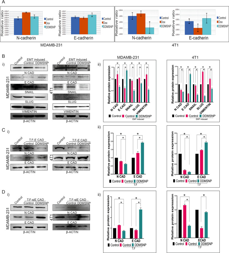
Fig. 6 Regulation of in vitro alteration of EMT markers by DDMSNP. A Total RNA extracted from DOX and DDMSNP treated and untreated MDAMB-231 and 4T1 derived CSCs are analyzed by qRT-PCR for N-cadherin and E-cadherin. B (i) Western blot data depicts the differences between the expressions of N-cadherin, E-cadherin, Snail, Slug and Vimentin in presence of DDMSNP compared to the untreated EMT induced or uninduced CSCs. β-actin is used as a loading control. (ii) The bar diagrams show relative protein expressions. * denotes P < 0.05. C (i) Western blot data from CSCs of MDAMB-231 (left panel) and 4T1 (right panel) transfected with E-cadherin expressing vectors shows differential expressions of N-cadherin and E-cadherin in non-transfected control, transfected control and DDMSNP treated transfected CSCs. (ii) Densitometric comparison for N-cadherin and E-cadherin expression among non-transfected control, transfected control and DDMSNP treated transfected CSCs is depicted by bar graphs. * denotes P < 0.05. D (i) The expressions of N-cadherin and E-cadherin in non-transfected control CSCs, transfected control CSCs and DDMSNP treated siE Cadherin (siE CAD) transfected CSCs are represented in Western blot data. (ii) Bar graphs show the densitometric comparison of N- and E-cadherin expression in non-transfected control, siE CAD transfected control, and siE CAD transfected CSCs treated with DDMSNP. * denotes P < 0.05
Fig. 6 Regulation of in vitro alteration of EMT markers by DDMSNP. A Total RNA extracted from DOX and DDMSNP treated and untreated MDAMB-231 and 4T1 derived CSCs are analyzed by qRT-PCR for N-cadherin and E-cadherin. B (i) Western blot data depicts the differences between the expressions of N-cadherin, E-cadherin, Snail, Slug and Vimentin in presence of DDMSNP compared to the untreated EMT induced or uninduced CSCs. β-actin is used as a loading control. (ii) The bar diagrams show relative protein expressions. * denotes P < 0.05. C (i) Western blot data from CSCs of MDAMB-231 (left panel) and 4T1 (right panel) transfected with E-cadherin expressing vectors shows differential expressions of N-cadherin and E-cadherin in non-transfected control, transfected control and DDMSNP treated transfected CSCs. (ii) Densitometric comparison for N-cadherin and E-cadherin expression among non-transfected control, transfected control and DDMSNP treated transfected CSCs is depicted by bar graphs. * denotes P < 0.05. D (i) The expressions of N-cadherin and E-cadherin in non-transfected control CSCs, transfected control CSCs and DDMSNP treated siE Cadherin (siE CAD) transfected CSCs are represented in Western blot data. (ii) Bar graphs show the densitometric comparison of N- and E-cadherin expression in non-transfected control, siE CAD transfected control, and siE CAD transfected CSCs treated with DDMSNP. * denotes P < 0.05CC BY · PubMed Central
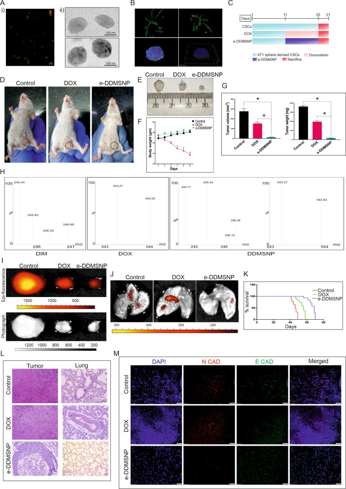
Fig. 7 Anti-metastatic effect of e-DDMSNP on TNBC-CSCs derived tumors. A (i) Encapsulation of DDMSNP within exosomes, (ii) The morphology of an empty exosome and DDMSNP loaded exosomes captured by TEM. B Images from confocal microscopy represents the uptake of e-DDMSNP in cells (upper left panel) and sphere (lower left panel). Z-stacking analysis shows internalization of e-DDMSNP in cells (upper right panel) and in the sphere (lower right panel). C Schematic representation of experimental design to study the efficacy of DOX and e-DDMSNP in 4T1 CSC derived tumor-bearing mice. D Figure represents growth inhibition of tumors derived from 4T1 CSCs in BALB/c mice after DOX and e-DDMSNP treatment. E Figure shows decreased size of tumors in treated groups compared to untreated group. F Graph represents body weight changes during treatment. Data are expressed as the mean ± SD (n = 6). G Bar diagram represents tumor volume and tumor weight of control mice and mice receiving either DOX or e-DDMSNP (n = 6, mean ± SD), * denotes P < 0.0001. H Mass spectrometric analysis shows internalization of e-DDMSNP in tumors. Highest intensity peak in graphical representation denotes molecular mass of native DIM and native DOX. Graph shows same peak with highest intensity defining exact molecular mass of DIM and DOX in tissue extracts of e-DDMSNP treated tumors. I Fluorescence and bright field imaging represents tumor tissues of control, DOX and e-DDMSNP treated groups. J Fluorescence imaging of lung of treated and untreated groups indicates lung metastasis. K Effect of DOX and e-DDMSNP treatment on survivability of RFP expressing 4T1 CSCs derived solid tumor-bearing mice. Data are analyzed using Kaplan–Meier method. L Images denote H&E staining (× 20 × magnification) of tumors and lungs of treated and untreated groups. M Immunofluorescence assay represents the expressions of N-cadherin (red) and E-cadherin (green) in untreated control and DOX or e-DDMSNP treated 4T1 CSC derived solid tumors
Fig. 7 Anti-metastatic effect of e-DDMSNP on TNBC-CSCs derived tumors. A (i) Encapsulation of DDMSNP within exosomes, (ii) The morphology of an empty exosome and DDMSNP loaded exosomes captured by TEM. B Images from confocal microscopy represents the uptake of e-DDMSNP in cells (upper left panel) and sphere (lower left panel). Z-stacking analysis shows internalization of e-DDMSNP in cells (upper right panel) and in the sphere (lower right panel). C Schematic representation of experimental design to study the efficacy of DOX and e-DDMSNP in 4T1 CSC derived tumor-bearing mice. D Figure represents growth inhibition of tumors derived from 4T1 CSCs in BALB/c mice after DOX and e-DDMSNP treatment. E Figure shows decreased size of tumors in treated groups compared to untreated group. F Graph represents body weight changes during treatment. Data are expressed as the mean ± SD (n = 6). G Bar diagram represents tumor volume and tumor weight of control mice and mice receiving either DOX or e-DDMSNP (n = 6, mean ± SD), * denotes P < 0.0001. H Mass spectrometric analysis shows internalization of e-DDMSNP in tumors. Highest intensity peak in graphical representation denotes molecular mass of native DIM and native DOX. Graph shows same peak with highest intensity defining exact molecular mass of DIM and DOX in tissue extracts of e-DDMSNP treated tumors. I Fluorescence and bright field imaging represents tumor tissues of control, DOX and e-DDMSNP treated groups. J Fluorescence imaging of lung of treated and untreated groups indicates lung metastasis. K Effect of DOX and e-DDMSNP treatment on survivability of RFP expressing 4T1 CSCs derived solid tumor-bearing mice. Data are analyzed using Kaplan–Meier method. L Images denote H&E staining (× 20 × magnification) of tumors and lungs of treated and untreated groups. M Immunofluorescence assay represents the expressions of N-cadherin (red) and E-cadherin (green) in untreated control and DOX or e-DDMSNP treated 4T1 CSC derived solid tumorsCC BY · PubMed Central

Verification

Claims the paper makes

The claims and statistics of this paper haven’t been checked yet.

Run the check — free, no account needed. It reads the open-access text and checks the sources it cites.

A few free checks a day · papers already verified open instantly · results are public

Claims other papers make about this one

Each is checked against this paper’s own text: does it say what the citing paper says it does?

Self-check

Percentages, sums and reported differences re-computed from the paper’s own numbers.

The self-check needs the paper’s full text.

Citations

Cites

Research works printed but not linked: 45

  1. Yin L, Duan J-J, Bian X-W, Yu S. Triple-negative breast cancer molecular subtyping and treatment progress. Breast Cancer Res. 2020;22:61. · DOI
  2. Pantel K, Brakenhoff RH. Dissecting the metastatic cascade. Nat Rev Cancer. 2004;4:448–56. · DOI
  3. Wang Y, Zhou BP. Epithelial-mesenchymal transition—a hallmark of breast cancer metastasis. Cancer Hallm. 2013;1:38–49. · DOI
  4. Kuşoğlu A, Biray AÇ. Cancer stem cells: a brief review of the current status. Gene. 2019;681:80–5. · DOI
  5. Biddle A, Mackenzie IC. Cancer stem cells and EMT in carcinoma. Cancer Metastasis Rev. 2012. https://doi.org/10.1007/s10555-012-9345-0. · DOI
  6. Cioce M, Ciliberto G. On the connections between cancer stem cells and EMT. Cell Cycle Georget Tex. 2012;11:4301–2. · DOI
  7. Wang S-S, Jiang J, Liang X-H, Tang Y-L. Links between cancer stem cells and epithelial-mesenchymal transition. OncoTargets Ther. 2015;8:2973–80.
  8. Hajra S, Basu A, Singha Roy S, Patra AR, Bhattacharya S. Attenuation of doxorubicin-induced cardiotoxicity and genotoxicity by an indole-based natural compound 3,3′-diindolylmethane (DIM) through activation of Nrf2/ARE signaling pathways and inhibiting apoptosis. Free Radic Res. 2017;51:812–27. · DOI
  9. Chu M, Zheng C, Chen C, Song G, Hu X, Wang Z. Targeting cancer stem cells by nutraceuticals for cancer therapy. Semin Cancer Biol. 2022;85:234–45. · DOI
  10. Thekkekkara D, Basavan D, Chandna S, Nanjan MJ. A combination of resveratrol and 3,3′-diindolylmethane, a potent radioprotector. Int J Radiat Biol. 2018;94:558–68. · DOI
  11. Prasanna PL, Renu K, Valsala GA. New molecular and biochemical insights of doxorubicin-induced hepatotoxicity. Life Sci. 2020;250:117599. · DOI
  12. Ioele G, Chieffallo M, Occhiuzzi MA, De Luca M, Garofalo A, Ragno G, et al. Anticancer drugs: recent strategies to improve stability profile, pharmacokinetic and pharmacodynamic properties. Molecules. 2022;27:5436. · DOI
  13. Banerjee M, Rajeswari VD. Critical review on the different roles of exosomes in TNBC and exosomal-mediated delivery of microRNA/siRNA/lncRNA and drug targeting signalling pathways in triple-negative breast cancer. Molecules. 2023;28:1802. · DOI
  14. Kalluri R, LeBleu VS. The biology, function, and biomedical applications of exosomes. Science. 2020;367: eaau6977. · DOI · Paper page
  15. Stöber W, Fink A, Bohn E. Controlled growth of monodisperse silica spheres in the micron size range. J Colloid Interface Sci. 1968;26:62–9. · DOI
  16. Daly CS, Flemban A, Shafei M, Conway ME, Qualtrough D, Dean SJ. Hypoxia modulates the stem cell population and induces EMT in the MCF-10A breast epithelial cell line. Oncol Rep. 2018;39:483–90.
  17. Ghosh A, Bhowmik A, Bhandary S, Putatunda S, Laskar A, Biswas A, et al. Formulation and antitumorigenic activities of nanoencapsulated nifetepimine: a promising approach in treating triple negative breast carcinoma. Nanomed Nanotechnol Biol Med. 2016;12:1973–85. · DOI
  18. Alnuqaydan AM, Almutary AG, Alshehri OY, Henidi HA, Alajlan AM, Tamim AA, et al. Evaluation of the cytotoxic activity of Tamarix articulata and its anticancer potential in prostate cancer cells. J Appl Pharm Sci. 2022;12:089–108.
  19. Bhowmik A, Chakravarti S, Ghosh A, Shaw R, Bhandary S, Bhattacharyya S, et al. Anti-SSTR2 peptide based targeted delivery of potent PLGA encapsulated 3,3′-diindolylmethane nanoparticles through blood brain barrier prevents glioma progression. Oncotarget. 2017;8:65339–58. · DOI
  20. Lombaerts M, van Wezel T, Philippo K, Dierssen JWF, Zimmerman RME, Oosting J, et al. E-cadherin transcriptional downregulation by promoter methylation but not mutation is related to epithelial-to-mesenchymal transition in breast cancer cell lines. Br J Cancer. 2006;94:661–71. · DOI
  21. Schrörs B, Boegel S, Albrecht C, Bukur T, Bukur V, Holtsträter C, et al. Multi-omics characterization of the 4T1 murine mammary gland tumor model. Front Oncol. 2020;10:1195. · DOI
  22. Scully R, Livingston DM. In search of the tumour-suppressor functions of BRCA1 and BRCA2. Nature. 2000;408:429–32. · DOI · Paper page
  23. Thiery JP. Epithelial-mesenchymal transitions in tumour progression. Nat Rev Cancer. 2002;2:442–54. · DOI
  24. Meacham CE, Morrison SJ. Tumour heterogeneity and cancer cell plasticity. Nature. 2013;501:328–37. · DOI
  25. Borovski T, De Sousa E Melo FS, Vermeulen L, Medema JP. Cancer stem cell niche: the place to be. Cancer Res. 2011;71:634–9. · DOI · Paper page
  26. Plaks V, Kong N, Werb Z. The cancer stem cell niche: how essential is the niche in regulating stemness of tumor cells? Cell Stem Cell. 2015;16:225–38. · DOI · Paper page
  27. Gote V, Nookala AR, Bolla PK, Pal D. Drug resistance in metastatic breast cancer: tumor targeted nanomedicine to the rescue. Int J Mol Sci. 2021;22:4673. · DOI
  28. Dave B, Mittal V, Tan NM, Chang JC. Epithelial-mesenchymal transition, cancer stem cells and treatment resistance. Breast Cancer Res BCR. 2012;14:202. · DOI
  29. Dent R, Hanna WM, Trudeau M, Rawlinson E, Sun P, Narod SA. Pattern of metastatic spread in triple-negative breast cancer. Breast Cancer Res Treat. 2009;115:423–8. · DOI
  30. Li Y, Wang S, Yang W, Liu H. Prognostic significance of molecular subtype, metastatic site and primary tumor surgery for survival in primary metastatic breast cancer: a SEER-based study. Medicine. 2021;100: e26619. · DOI
  31. Marshall EM, Bertaut A, Desmoulins I, Darut-Jouve A, Ponnelle T, Poillot M-L, et al. Prognostic factors of survival among women with metastatic breast cancer and impact of primary or secondary nature of disease on survival: a French population-based study. Breast J. 2017;23:138–45. · DOI
  32. Costa RLB, Gradishar WJ. Triple-negative breast cancer: current practice and future directions. J Oncol Pract. 2017;13:301–3. · DOI
  33. Gradishar WJ, Moran MS, Abraham J, Aft R, Agnese D, Allison KH, et al. NCCN Guidelines® insights: breast cancer, version 4.2021. J Natl Compr Cancer Netw JNCCN. 2021;19:484–93. · DOI
  34. Ikawati M, Jenie RI, Utomo RY, Amalina ND, Ilmawati GPN, Kawaichi M, et al. Genistein enhances cytotoxic and antimigratory activities of doxorubicin on 4T1 breast cancer cells through cell cycle arrest and ROS generation. J Appl Pharm Sci. 2020;10:095–104.
  35. Pezzani R, Salehi B, Vitalini S, Iriti M, Zuñiga FA, Sharifi-Rad J, et al. Synergistic effects of plant derivatives and conventional chemotherapeutic agents: an update on the cancer perspective. Med Kaunas Lith. 2019;55:110.
  36. Castañeda AM, Meléndez CM, Uribe D, Pedroza-Díaz J. Synergistic effects of natural compounds and conventional chemotherapeutic agents: recent insights for the development of cancer treatment strategies. Heliyon. 2022;8: e09519. · DOI
  37. Safa AR. Resistance to drugs and cell death in cancer stem cells (CSCs). J Transl Sci. 2020;6:341. · DOI
  38. Barua S, Mitragotri S. Challenges associated with penetration of nanoparticles across cell and tissue barriers: a review of current status and future prospects. Nano Today. 2014;9:223–43. · DOI
  39. Nehme Z, Roehlen N, Dhawan P, Baumert TF. Tight junction protein signaling and cancer biology. Cells. 2023;12:243. · DOI
  40. Vinogradov S, Wei X. Cancer stem cells and drug resistance: the potential of nanomedicine. Nanomedicine. 2012;7:597–615. · DOI
  41. Tan S, Wu T, Zhang D, Zhang Z. Cell or cell membrane-based drug delivery systems. Theranostics. 2015;5:863–81. · DOI · Paper page
  42. Théry C, Zitvogel L, Amigorena S. Exosomes: composition, biogenesis and function. Nat Rev Immunol. 2002;2:569–79. · DOI
  43. Vader P, Mol EA, Pasterkamp G, Schiffelers RM. Extracellular vesicles for drug delivery. Adv Drug Deliv Rev. 2016;106:148–56. · DOI · Paper page
  44. Batrakova EV, Kim MS. Using exosomes, naturally-equipped nanocarriers, for drug delivery. J Control Release Off J Control Release Soc. 2015;219:396–405. · DOI · Paper page
  45. van den Boorn JG, Dassler J, Coch C, Schlee M, Hartmann G. Exosomes as nucleic acid nanocarriers. Adv Drug Deliv Rev. 2013;65:331–5. · DOI

Cited by

Sorted by citations

See all 69

Metadata

Authors
Sarkar, R., Biswas, S., Ghosh, R., Samanta, P., Pakhira, S., Mondal, M., Dutta Gupta, Y., Bhandary, S., Saha, P., Bhowmik, A., & Hajra, S.
Venue
Journal of Nanobiotechnology
Published
2024-05-25
Volume
22
Issue
1
ISSN
1477-3155
Cited by
69
References
2
Language
en
Funders
Science and Engineering Research Board (EEQ/2020/000601, CRG/2021/007813)

About these checks

Registry, retraction, Cites and statistics results are computed, not judged, with one exception: a printed reference that no index lists is read once by a model before it counts as a citation error. Claims are extracted by a model and kept only when their quote appears verbatim in the text; each cited source is then checked, with a model’s help, for whether it says what the claim relies on. Those verdicts can be wrong — read the claim’s quote and the cited source before relying on one.

Statistics and arithmetic are recomputed, not judged.

Confidence bands are shown only for fields calibrated on our annotated test set. None is calibrated yet, so no band is shown.

What counts as a claim ›How we measure confidence ›How we check this ›

Are you an author, or do you see a problem with this page? Request a review